Movie
Movie Controller

[English] 日本語
 Yorodumi
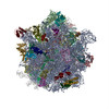
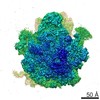
- EMDB-21031: Cryo-EM structure of the Acinetobacter baumannii Ribosome: 70S wi... -
+ Open data

- Basic information
| Entry | Database: EMDB / ID: EMD-21031 | |||||||||
|---|---|---|---|---|---|---|---|---|---|---|
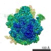
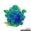
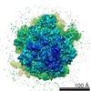
| Title | Cryo-EM structure of the Acinetobacter baumannii Ribosome: 70S with E-site tRNA | |||||||||
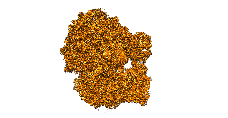
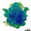
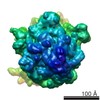
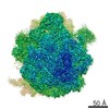
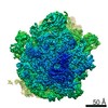
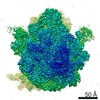
 Map data Map data | Composite EM map of 70S E-site occupied ribosome from A. baumannii created by combining three separate focused refinements (50S, 30S core and 30S head) | |||||||||
 Sample Sample |
| |||||||||
 Keywords Keywords | Ribosome / Acinetobacter baumannii | |||||||||
| Function / homology |  Function and homology information Function and homology informationassembly of large subunit precursor of preribosome / large ribosomal subunit / transferase activity / ribosomal small subunit assembly / ribosome binding / ribosome biogenesis / ribosomal small subunit biogenesis / 5S rRNA binding / ribosomal large subunit assembly / small ribosomal subunit ...assembly of large subunit precursor of preribosome / large ribosomal subunit / transferase activity / ribosomal small subunit assembly / ribosome binding / ribosome biogenesis / ribosomal small subunit biogenesis / 5S rRNA binding / ribosomal large subunit assembly / small ribosomal subunit / small ribosomal subunit rRNA binding / large ribosomal subunit rRNA binding / cytosolic small ribosomal subunit / cytosolic large ribosomal subunit / cytoplasmic translation / tRNA binding / negative regulation of translation / rRNA binding / structural constituent of ribosome / ribosome / translation / ribonucleoprotein complex / mRNA binding / RNA binding / cytoplasm / cytosol Similarity search - Function | |||||||||
| Biological species |  Acinetobacter baumannii AB0057 (bacteria) / Acinetobacter sp. CIP 51.11 (bacteria) / Acinetobacter beijerinckii ANC 3835 (bacteria) / Acinetobacter baumannii (strain AB0057) (bacteria) / Acinetobacter sp. ANC 4470 (bacteria) / Acinetobacter sp. NIPH 899 (bacteria) / Acinetobacter baumannii (bacteria) / Acinetobacter sp. 809848 (bacteria) / Acinetobacter rudis CIP 110305 (bacteria) / Acinetobacter sp. CIP 102082 (bacteria) / Acinetobacter sp. ANC 5600 (bacteria) / Acinetobacter sp. 263903-1 (bacteria) / Acinetobacter venetianus (bacteria) Acinetobacter baumannii AB0057 (bacteria) / Acinetobacter sp. CIP 51.11 (bacteria) / Acinetobacter beijerinckii ANC 3835 (bacteria) / Acinetobacter baumannii (strain AB0057) (bacteria) / Acinetobacter sp. ANC 4470 (bacteria) / Acinetobacter sp. NIPH 899 (bacteria) / Acinetobacter baumannii (bacteria) / Acinetobacter sp. 809848 (bacteria) / Acinetobacter rudis CIP 110305 (bacteria) / Acinetobacter sp. CIP 102082 (bacteria) / Acinetobacter sp. ANC 5600 (bacteria) / Acinetobacter sp. 263903-1 (bacteria) / Acinetobacter venetianus (bacteria) | |||||||||
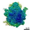
| Method | single particle reconstruction / cryo EM / Resolution: 2.82 Å | |||||||||
 Authors Authors | Morgan CE / Yu EW | |||||||||
| Funding support |  United States, 1 items United States, 1 items
| |||||||||
 Citation Citation | Journal: mBio / Year: 2020 Title: Cryo-electron Microscopy Structure of the Acinetobacter baumannii 70S Ribosome and Implications for New Antibiotic Development. Authors: Christopher E Morgan / Wei Huang / Susan D Rudin / Derek J Taylor / James E Kirby / Robert A Bonomo / Edward W Yu / Abstract: Antimicrobial resistance is a major health threat as it limits treatment options for infection. At the forefront of this serious issue is , a Gram-negative opportunistic pathogen that exhibits the ...Antimicrobial resistance is a major health threat as it limits treatment options for infection. At the forefront of this serious issue is , a Gram-negative opportunistic pathogen that exhibits the remarkable ability to resist antibiotics through multiple mechanisms. As bacterial ribosomes represent a target for multiple distinct classes of existing antimicrobial agents, we here use single-particle cryo-electron microscopy (cryo-EM) to elucidate five different structural states of the ribosome, including the 70S, 50S, and 30S forms. We also determined interparticle motions of the 70S ribosome in different tRNA bound states using three-dimensional (3D) variability analysis. Together, our structural data further our understanding of the ribosome from and other Gram-negative pathogens and will enable structure-based drug discovery to combat antibiotic-resistant bacterial infections. is a severe nosocomial threat largely due to its intrinsic antibiotic resistance and remarkable ability to acquire new resistance determinants. The bacterial ribosome serves as a major target for modern antibiotics and the design of new therapeutics. Here, we present cryo-EM structures of the 70S ribosome, revealing several unique species-specific structural features that may facilitate future drug development to combat this recalcitrant bacterial pathogen. | |||||||||
| History |
|
- Structure visualization
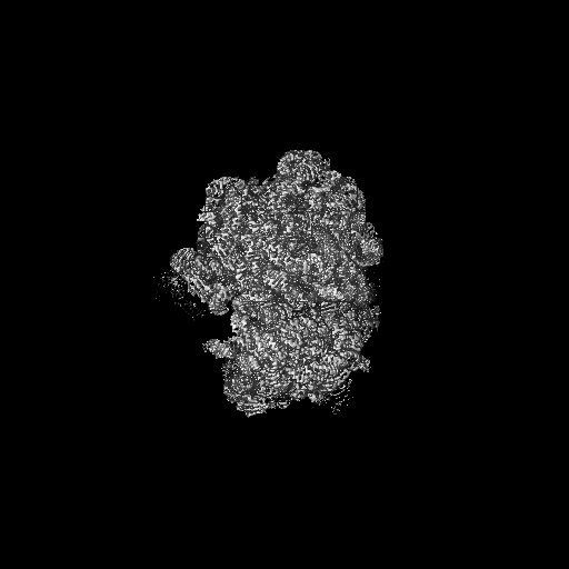
| Movie |
Movie viewer |
|---|---|
| Structure viewer | EM map: SurfViewMolmilJmol/JSmol |
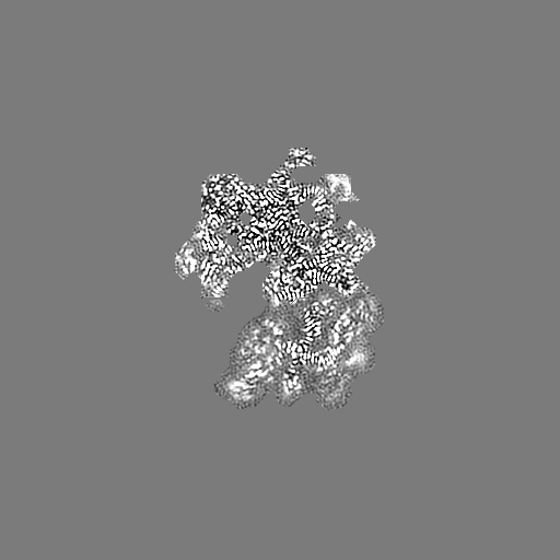
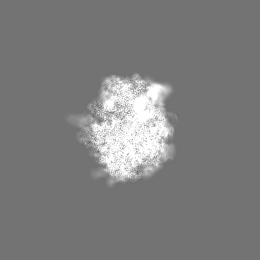
| Supplemental images |
- Downloads & links
-EMDB archive
| Map data | emd_21031.map.gz | 14.5 MB | EMDB map data format | |
|---|---|---|---|---|
| Header (meta data) | emd-21031-v30.xmlemd-21031.xml | 80.3 KB 80.3 KB | Display Display | EMDB header |
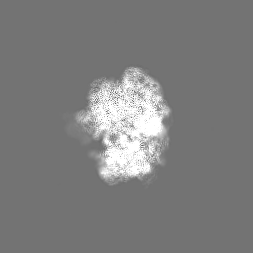
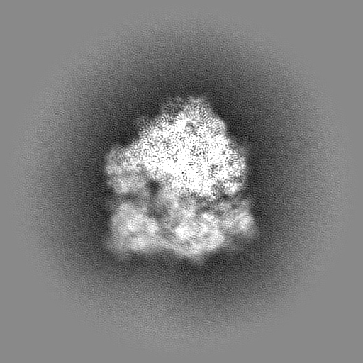
| Images |  emd_21031.png emd_21031.png | 214.6 KB | ||
| Filedesc metadata | emd-21031.cif.gz | 15.1 KB | ||
| Others | emd_21031_additional_1.map.gzemd_21031_additional_2.map.gzemd_21031_additional_3.map.gzemd_21031_additional_4.map.gz | 16.2 MB 483.8 MB 484 MB 483.5 MB | ||
| Archive directory |  http://ftp.pdbj.org/pub/emdb/structures/EMD-21031ftp://ftp.pdbj.org/pub/emdb/structures/EMD-21031 http://ftp.pdbj.org/pub/emdb/structures/EMD-21031ftp://ftp.pdbj.org/pub/emdb/structures/EMD-21031 | HTTPS FTP |
-Related structure data
| Related structure data | 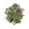 6v3aMC 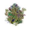 6v39C 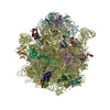 6v3bC 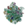 6v3dC 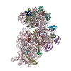 6v3eC M: atomic model generated by this map C: citing same article ( |
|---|---|
| Similar structure data |
-Links
| EMDB pages | EMDB (EBI/PDBe) / EMDataResource |
|---|---|
| Related items in Molecule of the Month |


-Map
| File | Download / File: emd_21031.map.gz / Format: CCP4 / Size: 512 MB / Type: IMAGE STORED AS FLOATING POINT NUMBER (4 BYTES) | ||||||||||||||||||||||||||||||||||||||||||||||||||||||||||||||||||||
|---|---|---|---|---|---|---|---|---|---|---|---|---|---|---|---|---|---|---|---|---|---|---|---|---|---|---|---|---|---|---|---|---|---|---|---|---|---|---|---|---|---|---|---|---|---|---|---|---|---|---|---|---|---|---|---|---|---|---|---|---|---|---|---|---|---|---|---|---|---|
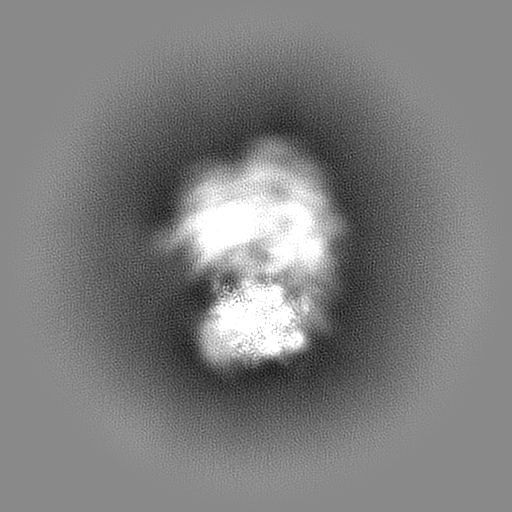
| Annotation | Composite EM map of 70S E-site occupied ribosome from A. baumannii created by combining three separate focused refinements (50S, 30S core and 30S head) | ||||||||||||||||||||||||||||||||||||||||||||||||||||||||||||||||||||
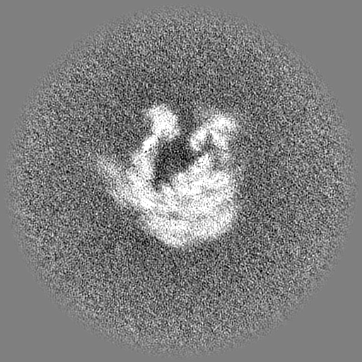
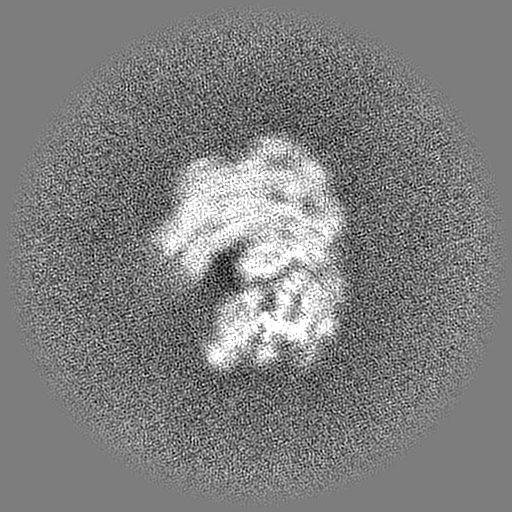
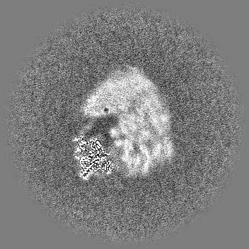
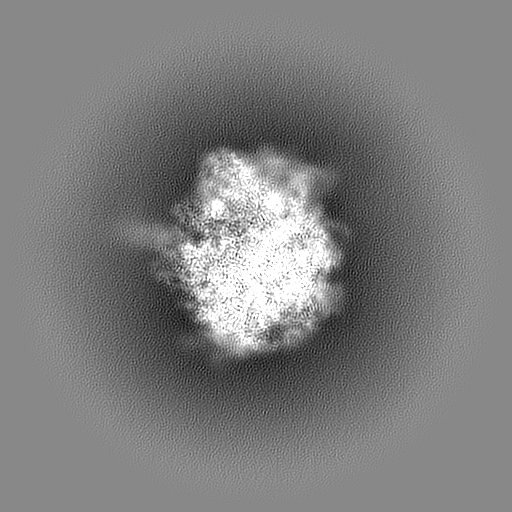
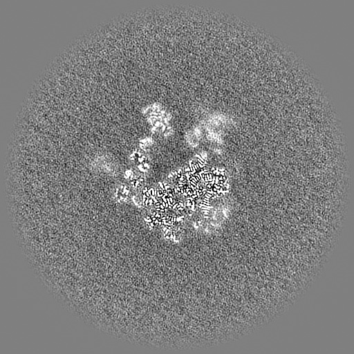
| Projections & slices | Image control
Images are generated by Spider. | ||||||||||||||||||||||||||||||||||||||||||||||||||||||||||||||||||||
| Voxel size | X=Y=Z: 1.064 Å | ||||||||||||||||||||||||||||||||||||||||||||||||||||||||||||||||||||
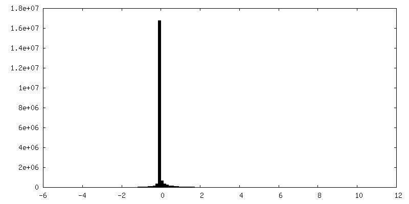
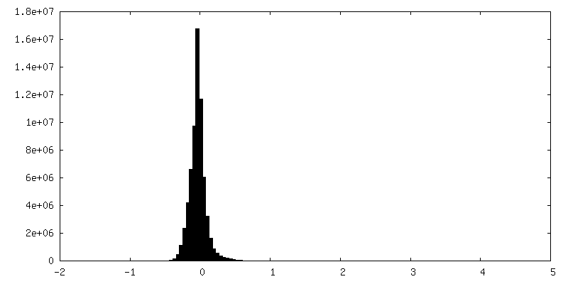
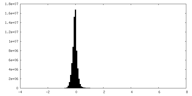
| Density |
| ||||||||||||||||||||||||||||||||||||||||||||||||||||||||||||||||||||
| Symmetry | Space group: 1 | ||||||||||||||||||||||||||||||||||||||||||||||||||||||||||||||||||||
| Details | EMDB XML:
CCP4 map header:
| ||||||||||||||||||||||||||||||||||||||||||||||||||||||||||||||||||||
 Z (Sec.)
 Y (Row.)
 X (Col.)


-Supplemental data
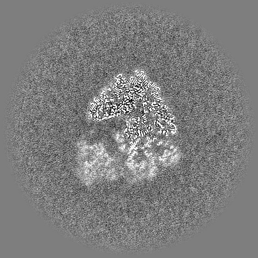
-Additional map: Composite EM map of 70S E-site occupied ribosome...
| File | emd_21031_additional_1.map | ||||||||||||
|---|---|---|---|---|---|---|---|---|---|---|---|---|---|
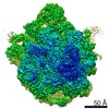
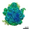
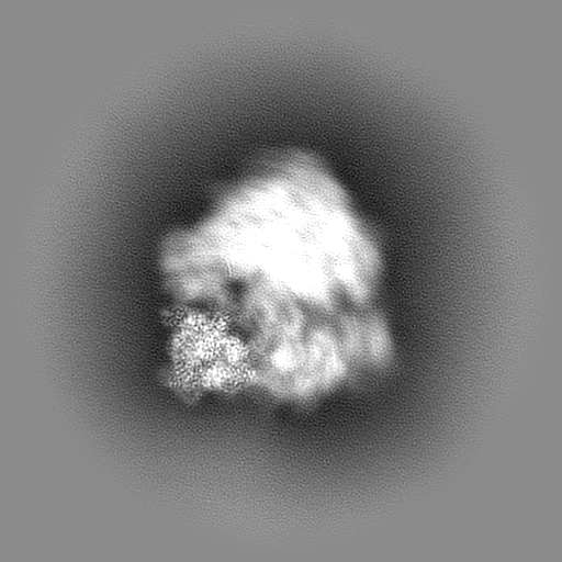
| Annotation | Composite EM map of 70S E-site occupied ribosome from A. baumannii created by combining three separate focused refinements (50S, 30S core and 30S head), local resolution | ||||||||||||
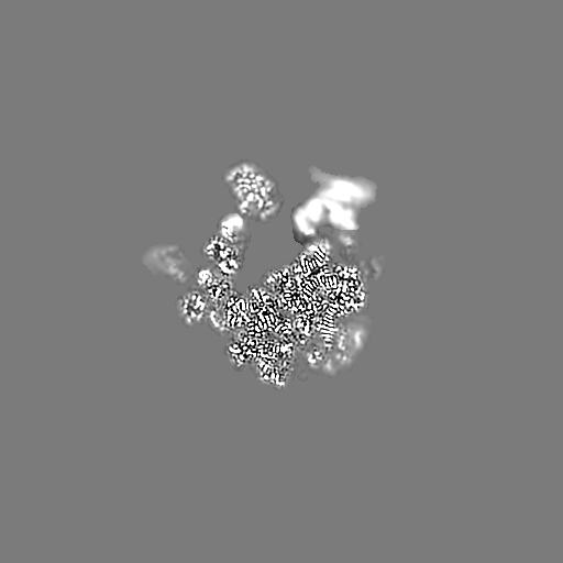
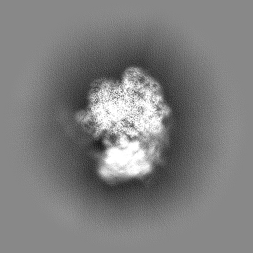
| Projections & Slices |
| ||||||||||||
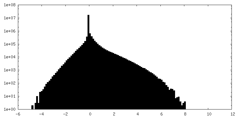
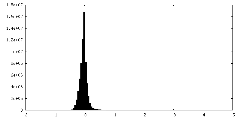
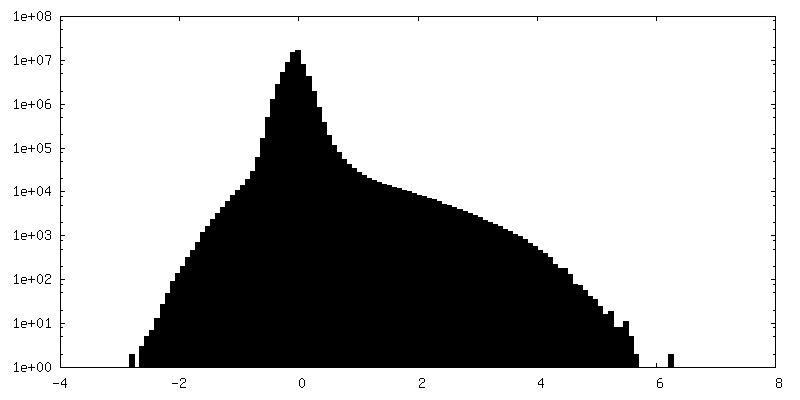
| Density Histograms |

-Additional map: Focused refinement of 30S head of 70S E-site...
| File | emd_21031_additional_2.map | ||||||||||||
|---|---|---|---|---|---|---|---|---|---|---|---|---|---|
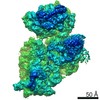
| Annotation | Focused refinement of 30S head of 70S E-site occupied ribosome from A. baumannii | ||||||||||||
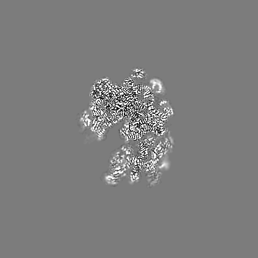
| Projections & Slices |
| ||||||||||||
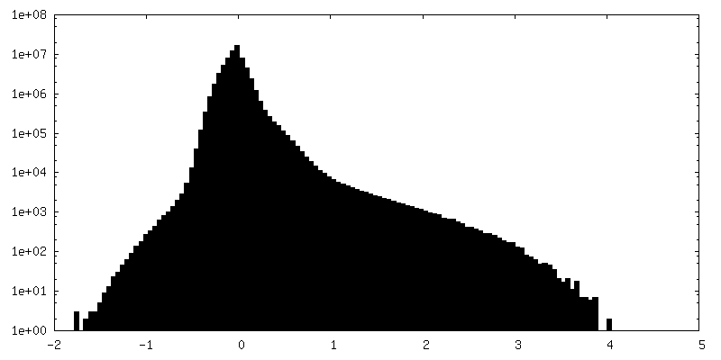
| Density Histograms |


-Additional map: Focused refinement of 30S core of 70S E-site...
| File | emd_21031_additional_3.map | ||||||||||||
|---|---|---|---|---|---|---|---|---|---|---|---|---|---|
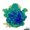
| Annotation | Focused refinement of 30S core of 70S E-site occupied ribosome from A. baumannii | ||||||||||||
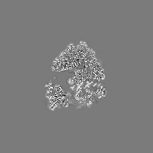
| Projections & Slices |
| ||||||||||||
| Density Histograms |


-Additional map: Focused refinement of 50S large subunit of 70S...
| File | emd_21031_additional_4.map | ||||||||||||
|---|---|---|---|---|---|---|---|---|---|---|---|---|---|
| Annotation | Focused refinement of 50S large subunit of 70S E-site occupied ribosome from A. baumannii | ||||||||||||
| Projections & Slices |
| ||||||||||||
| Density Histograms |

- Sample components
+Entire : Acinetobacter baumannii 70S ribosome with E-site tRNA bound
+Supramolecule #1: Acinetobacter baumannii 70S ribosome with E-site tRNA bound
+Macromolecule #1: 50S ribosomal protein L33
+Macromolecule #2: 50S ribosomal protein L34
+Macromolecule #3: 50S ribosomal protein L35
+Macromolecule #4: 50S ribosomal protein L36
+Macromolecule #7: 50S ribosomal protein L2
+Macromolecule #8: 50S ribosomal protein L3
+Macromolecule #9: 50S ribosomal protein L4
+Macromolecule #10: 50S ribosomal protein L5
+Macromolecule #11: 50S ribosomal protein L6
+Macromolecule #12: 50S ribosomal protein L9
+Macromolecule #13: 50S ribosomal protein L13
+Macromolecule #14: 50S ribosomal protein L14
+Macromolecule #15: 50S ribosomal protein L15
+Macromolecule #16: 50S ribosomal protein L16
+Macromolecule #17: 50S ribosomal protein L17
+Macromolecule #18: 50S ribosomal protein L18
+Macromolecule #19: 50S ribosomal protein L19
+Macromolecule #20: 50S ribosomal protein L20
+Macromolecule #21: 50S ribosomal protein L21
+Macromolecule #22: 50S ribosomal protein L22
+Macromolecule #23: 50S ribosomal protein L23
+Macromolecule #24: 50S ribosomal protein L24
+Macromolecule #25: 50S ribosomal protein L25
+Macromolecule #26: 50S ribosomal protein L27
+Macromolecule #27: 50S ribosomal protein L28
+Macromolecule #28: 50S ribosomal protein L29
+Macromolecule #29: 50S ribosomal protein L30
+Macromolecule #30: 50S ribosomal protein L32
+Macromolecule #32: 30S ribosomal protein S2
+Macromolecule #33: 30S ribosomal protein S3
+Macromolecule #34: 30S ribosomal protein S4
+Macromolecule #35: 30S ribosomal protein S5
+Macromolecule #36: 30S ribosomal protein S6
+Macromolecule #37: 30S ribosomal protein S7
+Macromolecule #38: 30S ribosomal protein S8
+Macromolecule #39: 30S ribosomal protein S9
+Macromolecule #40: 30S ribosomal protein S10
+Macromolecule #41: 30S ribosomal protein S11
+Macromolecule #42: 30S ribosomal protein S12
+Macromolecule #43: 30S ribosomal protein S13
+Macromolecule #44: 30S ribosomal protein S14
+Macromolecule #45: 30S ribosomal protein S15
+Macromolecule #46: 30S ribosomal protein S16
+Macromolecule #47: 30S ribosomal protein S17
+Macromolecule #48: 30S ribosomal protein S18
+Macromolecule #49: 30S ribosomal protein S19
+Macromolecule #50: 30S ribosomal protein S20
+Macromolecule #51: 30S ribosomal protein S21
+Macromolecule #5: 23s ribosomal RNA
+Macromolecule #6: 5s ribosomal RNA
+Macromolecule #31: 16s Ribosomal RNA
+Macromolecule #52: tRNA-met
+Macromolecule #53: mRNA
+Macromolecule #54: ZINC ION
+Macromolecule #55: MAGNESIUM ION
+Macromolecule #56: SODIUM ION
+Macromolecule #57: water

-Experimental details
-Structure determination
| Method | cryo EM |
|---|---|
 Processing Processing | single particle reconstruction |
| Aggregation state | particle |
-Sample preparation
| Buffer | pH: 7.6 |
|---|---|
| Vitrification | Cryogen name: ETHANE |
- Electron microscopy
| Microscope | FEI TITAN KRIOS |
|---|---|
| Image recording | Film or detector model: GATAN K2 SUMMIT (4k x 4k) / Detector mode: SUPER-RESOLUTION / Average electron dose: 40.0 e/Å2 |
| Electron beam | Acceleration voltage: 300 kV / Electron source:  FIELD EMISSION GUN FIELD EMISSION GUN |
| Electron optics | Illumination mode: FLOOD BEAM / Imaging mode: BRIGHT FIELD |
| Experimental equipment |  Model: Titan Krios / Image courtesy: FEI Company |