 ムービー
ムービー コントローラー


+ データを開く

- 基本情報
基本情報
| 登録情報 | データベース: EMDB / ID: EMD-21001 | |||||||||
|---|---|---|---|---|---|---|---|---|---|---|
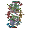
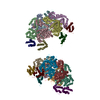
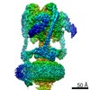
| タイトル | Cryo-EM structure of porcine ATP synthase reconstituted in small unilamellar vesicles_I | |||||||||
 マップデータ マップデータ | Cryo-EM structure of porcine ATP synthase reconstituted in small unilamellar vesicles | |||||||||
 試料 試料 |
| |||||||||
| 生物種 |  | |||||||||
| 手法 | 単粒子再構成法 / クライオ電子顕微鏡法 / 解像度: 19.0 Å | |||||||||
 データ登録者 データ登録者 | Mnatsakanyan N / Llaguno MC / Yang Y / Yan Y / Weber J / Sigworth FJ / Jonas EA | |||||||||
| 資金援助 |  米国, 2件 米国, 2件
| |||||||||
 引用 引用 | ジャーナル: Nat Commun / 年: 2019 タイトル: A mitochondrial megachannel resides in monomeric FF ATP synthase. 著者: Nelli Mnatsakanyan / Marc C Llaguno / Youshan Yang / Yangyang Yan / Joachim Weber / Fred J Sigworth / Elizabeth A Jonas / 要旨: Purified mitochondrial ATP synthase has been shown to form Ca-activated, large conductance channel activity similar to that of mitochondrial megachannel (MMC) or mitochondrial permeability transition ...Purified mitochondrial ATP synthase has been shown to form Ca-activated, large conductance channel activity similar to that of mitochondrial megachannel (MMC) or mitochondrial permeability transition pore (mPTP) but the oligomeric state required for channel formation is being debated. We reconstitute purified monomeric ATP synthase from porcine heart mitochondria into small unilamellar vesicles (SUVs) with the lipid composition of mitochondrial inner membrane and analyze its oligomeric state by electron cryomicroscopy. The cryo-EM density map reveals the presence of a single ATP synthase monomer with no density seen for a second molecule tilted at an 86 angle relative to the first. We show that this preparation of SUV-reconstituted ATP synthase monomers, when fused into giant unilamellar vesicles (GUVs), forms voltage-gated and Ca-activated channels with the key features of mPTP. Based on our findings we conclude that the ATP synthase monomer is sufficient, and dimer formation is not required, for mPTP activity. | |||||||||
| 履歴 |
|
- 構造の表示
構造の表示
| ムービー |
 ムービービューア ムービービューア |
|---|---|
| 構造ビューア | EMマップ: SurfViewMolmilJmol/JSmol |
| 添付画像 |

- ダウンロードとリンク
-EMDBアーカイブ
| マップデータ | emd_21001.map.gz | 3.1 MB | EMDBマップデータ形式 | |
|---|---|---|---|---|
| ヘッダ (付随情報) | emd-21001-v30.xmlemd-21001.xml | 14 KB 14 KB | 表示 表示 | EMDBヘッダ |
| FSC (解像度算出) | emd_21001_fsc.xml | 3.6 KB | 表示 | FSCデータファイル |
| 画像 |  emd_21001.png emd_21001.png | 113.6 KB | ||
| マスクデータ | emd_21001_msk_1.map | 3.4 MB | マスクマップ | |
| その他 | emd_21001_half_map_1.map.gzemd_21001_half_map_2.map.gz | 2.4 MB 2.4 MB | ||
| アーカイブディレクトリ |  http://ftp.pdbj.org/pub/emdb/structures/EMD-21001ftp://ftp.pdbj.org/pub/emdb/structures/EMD-21001 http://ftp.pdbj.org/pub/emdb/structures/EMD-21001ftp://ftp.pdbj.org/pub/emdb/structures/EMD-21001 | HTTPS FTP |
-検証レポート
| 文書・要旨 | emd_21001_validation.pdf.gz | 79.3 KB | 表示 | EMDB検証レポート |
|---|---|---|---|---|
| 文書・詳細版 | emd_21001_full_validation.pdf.gz | 78.4 KB | 表示 | |
| XML形式データ | emd_21001_validation.xml.gz | 493 B | 表示 | |
| アーカイブディレクトリ | https://ftp.pdbj.org/pub/emdb/validation_reports/EMD-21001ftp://ftp.pdbj.org/pub/emdb/validation_reports/EMD-21001 | HTTPS FTP |
-関連構造データ


-リンク
| EMDBのページ | EMDB (EBI/PDBe) / EMDataResource |
|---|
-マップ
| ファイル | ダウンロード / ファイル: emd_21001.map.gz / 形式: CCP4 / 大きさ: 3.4 MB / タイプ: IMAGE STORED AS FLOATING POINT NUMBER (4 BYTES) | ||||||||||||||||||||||||||||||||||||||||||||||||||||||||||||||||||||
|---|---|---|---|---|---|---|---|---|---|---|---|---|---|---|---|---|---|---|---|---|---|---|---|---|---|---|---|---|---|---|---|---|---|---|---|---|---|---|---|---|---|---|---|---|---|---|---|---|---|---|---|---|---|---|---|---|---|---|---|---|---|---|---|---|---|---|---|---|---|
| 注釈 | Cryo-EM structure of porcine ATP synthase reconstituted in small unilamellar vesicles | ||||||||||||||||||||||||||||||||||||||||||||||||||||||||||||||||||||
| 投影像・断面図 | 画像のコントロール
画像は Spider により作成 | ||||||||||||||||||||||||||||||||||||||||||||||||||||||||||||||||||||
| ボクセルのサイズ | X=Y=Z: 2.8 Å | ||||||||||||||||||||||||||||||||||||||||||||||||||||||||||||||||||||
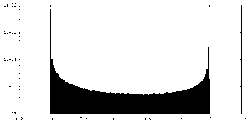
| 密度 |
| ||||||||||||||||||||||||||||||||||||||||||||||||||||||||||||||||||||
| 対称性 | 空間群: 1 | ||||||||||||||||||||||||||||||||||||||||||||||||||||||||||||||||||||
| 詳細 | EMDB XML:
CCP4マップ ヘッダ情報:
| ||||||||||||||||||||||||||||||||||||||||||||||||||||||||||||||||||||
 Z (Sec.)
 Y (Row.)
 X (Col.)


-添付データ
-マスク #1
| ファイル | emd_21001_msk_1.map | ||||||||||||
|---|---|---|---|---|---|---|---|---|---|---|---|---|---|
| 投影像・断面図 |
| ||||||||||||
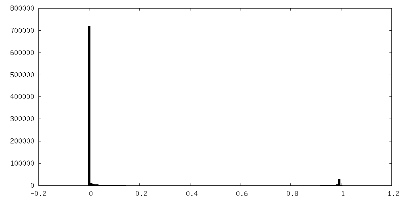
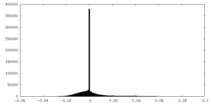
| 密度ヒストグラム |


-ハーフマップ: Cryo-EM structure of porcine ATP synthase
| ファイル | emd_21001_half_map_1.map | ||||||||||||
|---|---|---|---|---|---|---|---|---|---|---|---|---|---|
| 注釈 | Cryo-EM structure of porcine ATP synthase | ||||||||||||
| 投影像・断面図 |
| ||||||||||||
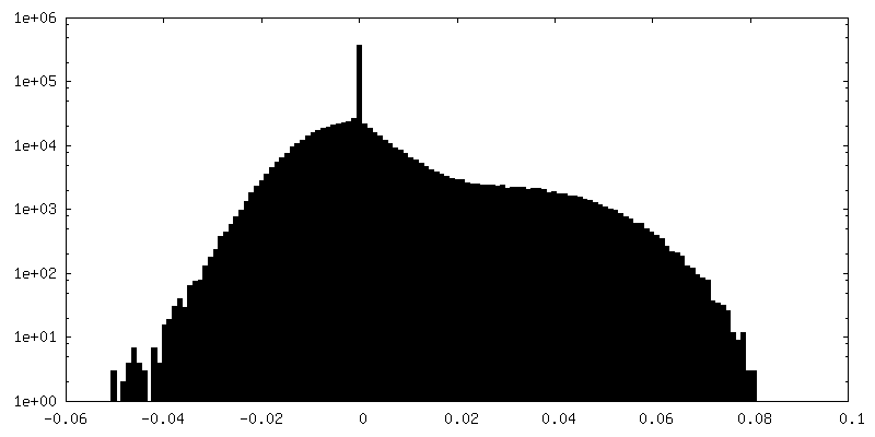
| 密度ヒストグラム |


-ハーフマップ: Cryo-EM structure of porcine ATP synthase
| ファイル | emd_21001_half_map_2.map | ||||||||||||
|---|---|---|---|---|---|---|---|---|---|---|---|---|---|
| 注釈 | Cryo-EM structure of porcine ATP synthase | ||||||||||||
| 投影像・断面図 |
| ||||||||||||
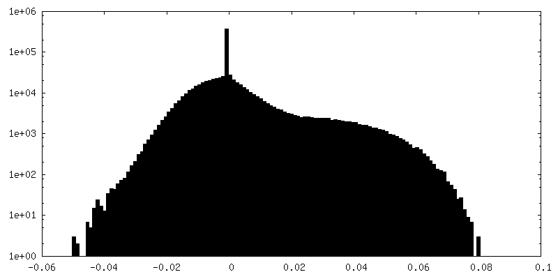
| 密度ヒストグラム |


- 試料の構成要素
-全体 : Purified ATP synthase reconstituted in liposomes
| 全体 | 名称: Purified ATP synthase reconstituted in liposomes |
|---|---|
| 要素 |
|
-超分子 #1: Purified ATP synthase reconstituted in liposomes
| 超分子 | 名称: Purified ATP synthase reconstituted in liposomes / タイプ: complex / ID: 1 / 親要素: 0 |
|---|---|
| 由来(天然) | 生物種: |
| 分子量 | 実験値: 750 KDa |
-実験情報
-構造解析
| 手法 | クライオ電子顕微鏡法 |
|---|---|
 解析 解析 | 単粒子再構成法 |
| 試料の集合状態 | particle |
-試料調製
| 濃度 | 1 mg/mL |
|---|---|
| 緩衝液 | pH: 8 / 詳細: 50 mM Tris-HCl, 150 mM NaCl, 2 mM MgSO4, 1 mM ATP |
| グリッド | モデル: C-flat-1.2/1.3 4C / 材質: GOLD / メッシュ: 300 / 支持フィルム - 材質: CARBON / 支持フィルム - トポロジー: HOLEY / 前処理 - タイプ: GLOW DISCHARGE |
| 凍結 | 凍結剤: ETHANE / チャンバー内湿度: 100 % / 装置: FEI VITROBOT MARK IV |
| 詳細 | Purified ATP synthase reconstituted in liposomes |
- 電子顕微鏡法
| 顕微鏡 | FEI TITAN KRIOS |
|---|---|
| 撮影 | フィルム・検出器のモデル: GATAN K2 QUANTUM (4k x 4k) 平均電子線量: 40.0 e/Å2 |
| 電子線 | 加速電圧: 300 kV / 電子線源:  FIELD EMISSION GUN FIELD EMISSION GUN |
| 電子光学系 | 最大 デフォーカス(補正後): 4.219 µm / 最小 デフォーカス(補正後): 1.871 µm / 倍率(補正後): 47620 / 照射モード: FLOOD BEAM / 撮影モード: BRIGHT FIELD |
| 実験機器 |  モデル: Titan Krios / 画像提供: FEI Company |
-画像解析
| 最終 再構成 | 解像度のタイプ: BY AUTHOR / 解像度: 19.0 Å / 解像度の算出法: FSC 0.143 CUT-OFF / ソフトウェア - 名称: RELION / 使用した粒子像数: 7795 |
|---|---|
| 初期 角度割当 | タイプ: MAXIMUM LIKELIHOOD |
| 最終 角度割当 | タイプ: OTHER |
| FSC曲線 (解像度の算出) | 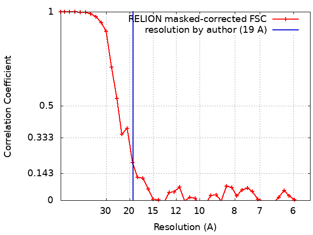 |
