 Movie
Movie Controller

[English] 日本語
 Yorodumi
- EMDB-21001: Cryo-EM structure of porcine ATP synthase reconstituted in small ... -
+ Open data

- Basic information
| Entry | Database: EMDB / ID: EMD-21001 | |||||||||
|---|---|---|---|---|---|---|---|---|---|---|
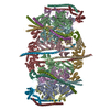
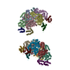
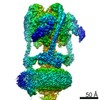
| Title | Cryo-EM structure of porcine ATP synthase reconstituted in small unilamellar vesicles_I | |||||||||
 Map data Map data | Cryo-EM structure of porcine ATP synthase reconstituted in small unilamellar vesicles | |||||||||
 Sample Sample |
| |||||||||
| Biological species |  | |||||||||
| Method | single particle reconstruction / cryo EM / Resolution: 19.0 Å | |||||||||
 Authors Authors | Mnatsakanyan N / Llaguno MC / Yang Y / Yan Y / Weber J / Sigworth FJ / Jonas EA | |||||||||
| Funding support |  United States, 2 items United States, 2 items
| |||||||||
 Citation Citation | Journal: Nat Commun / Year: 2019 Title: A mitochondrial megachannel resides in monomeric FF ATP synthase. Authors: Nelli Mnatsakanyan / Marc C Llaguno / Youshan Yang / Yangyang Yan / Joachim Weber / Fred J Sigworth / Elizabeth A Jonas / Abstract: Purified mitochondrial ATP synthase has been shown to form Ca-activated, large conductance channel activity similar to that of mitochondrial megachannel (MMC) or mitochondrial permeability transition ...Purified mitochondrial ATP synthase has been shown to form Ca-activated, large conductance channel activity similar to that of mitochondrial megachannel (MMC) or mitochondrial permeability transition pore (mPTP) but the oligomeric state required for channel formation is being debated. We reconstitute purified monomeric ATP synthase from porcine heart mitochondria into small unilamellar vesicles (SUVs) with the lipid composition of mitochondrial inner membrane and analyze its oligomeric state by electron cryomicroscopy. The cryo-EM density map reveals the presence of a single ATP synthase monomer with no density seen for a second molecule tilted at an 86 angle relative to the first. We show that this preparation of SUV-reconstituted ATP synthase monomers, when fused into giant unilamellar vesicles (GUVs), forms voltage-gated and Ca-activated channels with the key features of mPTP. Based on our findings we conclude that the ATP synthase monomer is sufficient, and dimer formation is not required, for mPTP activity. | |||||||||
| History |
|
- Structure visualization
| Movie |
 Movie viewer Movie viewer |
|---|---|
| Structure viewer | EM map: SurfViewMolmilJmol/JSmol |
| Supplemental images |

- Downloads & links
-EMDB archive
| Map data | emd_21001.map.gz | 3.1 MB | EMDB map data format | |
|---|---|---|---|---|
| Header (meta data) | emd-21001-v30.xmlemd-21001.xml | 14 KB 14 KB | Display Display | EMDB header |
| FSC (resolution estimation) | emd_21001_fsc.xml | 3.6 KB | Display | FSC data file |
| Images |  emd_21001.png emd_21001.png | 113.6 KB | ||
| Masks | emd_21001_msk_1.map | 3.4 MB | Mask map | |
| Others | emd_21001_half_map_1.map.gzemd_21001_half_map_2.map.gz | 2.4 MB 2.4 MB | ||
| Archive directory |  http://ftp.pdbj.org/pub/emdb/structures/EMD-21001ftp://ftp.pdbj.org/pub/emdb/structures/EMD-21001 http://ftp.pdbj.org/pub/emdb/structures/EMD-21001ftp://ftp.pdbj.org/pub/emdb/structures/EMD-21001 | HTTPS FTP |
-Related structure data
| Related structure data | C: citing same article ( |
|---|---|
| Similar structure data |


-Links
| EMDB pages | EMDB (EBI/PDBe) / EMDataResource |
|---|
-Map
| File | Download / File: emd_21001.map.gz / Format: CCP4 / Size: 3.4 MB / Type: IMAGE STORED AS FLOATING POINT NUMBER (4 BYTES) | ||||||||||||||||||||||||||||||||||||||||||||||||||||||||||||||||||||
|---|---|---|---|---|---|---|---|---|---|---|---|---|---|---|---|---|---|---|---|---|---|---|---|---|---|---|---|---|---|---|---|---|---|---|---|---|---|---|---|---|---|---|---|---|---|---|---|---|---|---|---|---|---|---|---|---|---|---|---|---|---|---|---|---|---|---|---|---|---|
| Annotation | Cryo-EM structure of porcine ATP synthase reconstituted in small unilamellar vesicles | ||||||||||||||||||||||||||||||||||||||||||||||||||||||||||||||||||||
| Projections & slices | Image control
Images are generated by Spider. | ||||||||||||||||||||||||||||||||||||||||||||||||||||||||||||||||||||
| Voxel size | X=Y=Z: 2.8 Å | ||||||||||||||||||||||||||||||||||||||||||||||||||||||||||||||||||||
| Density |
| ||||||||||||||||||||||||||||||||||||||||||||||||||||||||||||||||||||
| Symmetry | Space group: 1 | ||||||||||||||||||||||||||||||||||||||||||||||||||||||||||||||||||||
| Details | EMDB XML:
CCP4 map header:
| ||||||||||||||||||||||||||||||||||||||||||||||||||||||||||||||||||||
 Z (Sec.)
 Y (Row.)
 X (Col.)


-Supplemental data
-Mask #1
| File | emd_21001_msk_1.map | ||||||||||||
|---|---|---|---|---|---|---|---|---|---|---|---|---|---|
| Projections & Slices |
| ||||||||||||
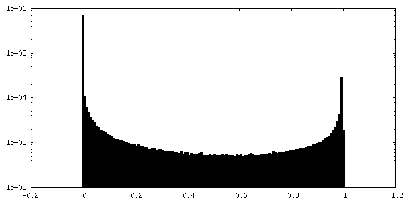
| Density Histograms |


-Half map: Cryo-EM structure of porcine ATP synthase
| File | emd_21001_half_map_1.map | ||||||||||||
|---|---|---|---|---|---|---|---|---|---|---|---|---|---|
| Annotation | Cryo-EM structure of porcine ATP synthase | ||||||||||||
| Projections & Slices |
| ||||||||||||
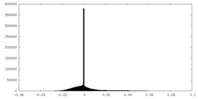
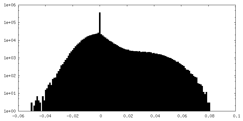
| Density Histograms |


-Half map: Cryo-EM structure of porcine ATP synthase
| File | emd_21001_half_map_2.map | ||||||||||||
|---|---|---|---|---|---|---|---|---|---|---|---|---|---|
| Annotation | Cryo-EM structure of porcine ATP synthase | ||||||||||||
| Projections & Slices |
| ||||||||||||
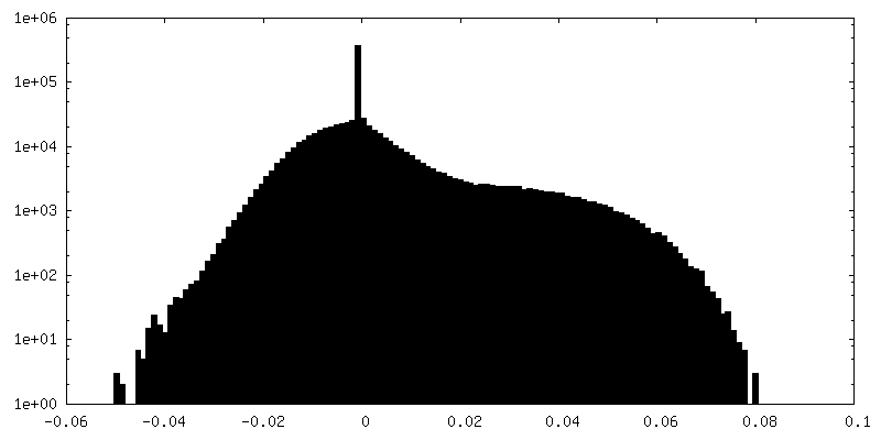
| Density Histograms |


- Sample components
-Entire : Purified ATP synthase reconstituted in liposomes
| Entire | Name: Purified ATP synthase reconstituted in liposomes |
|---|---|
| Components |
|
-Supramolecule #1: Purified ATP synthase reconstituted in liposomes
| Supramolecule | Name: Purified ATP synthase reconstituted in liposomes / type: complex / ID: 1 / Parent: 0 |
|---|---|
| Source (natural) | Organism: |
| Molecular weight | Experimental: 750 KDa |
-Experimental details
-Structure determination
| Method | cryo EM |
|---|---|
 Processing Processing | single particle reconstruction |
| Aggregation state | particle |
-Sample preparation
| Concentration | 1 mg/mL |
|---|---|
| Buffer | pH: 8 / Details: 50 mM Tris-HCl, 150 mM NaCl, 2 mM MgSO4, 1 mM ATP |
| Grid | Model: C-flat-1.2/1.3 4C / Material: GOLD / Mesh: 300 / Support film - Material: CARBON / Support film - topology: HOLEY / Pretreatment - Type: GLOW DISCHARGE |
| Vitrification | Cryogen name: ETHANE / Chamber humidity: 100 % / Instrument: FEI VITROBOT MARK IV |
| Details | Purified ATP synthase reconstituted in liposomes |
- Electron microscopy
| Microscope | FEI TITAN KRIOS |
|---|---|
| Image recording | Film or detector model: GATAN K2 QUANTUM (4k x 4k) / Average electron dose: 40.0 e/Å2 |
| Electron beam | Acceleration voltage: 300 kV / Electron source:  FIELD EMISSION GUN FIELD EMISSION GUN |
| Electron optics | Calibrated defocus max: 4.219 µm / Calibrated defocus min: 1.871 µm / Calibrated magnification: 47620 / Illumination mode: FLOOD BEAM / Imaging mode: BRIGHT FIELD |
| Experimental equipment |  Model: Titan Krios / Image courtesy: FEI Company |
-Image processing
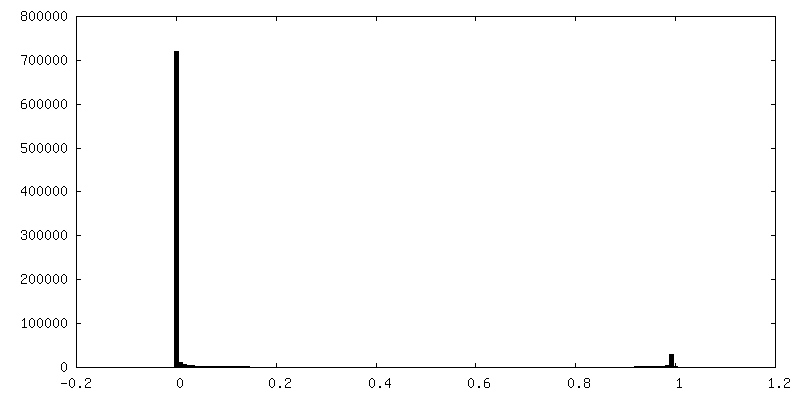
| Final reconstruction | Resolution.type: BY AUTHOR / Resolution: 19.0 Å / Resolution method: FSC 0.143 CUT-OFF / Software - Name: RELION / Number images used: 7795 |
|---|---|
| Initial angle assignment | Type: MAXIMUM LIKELIHOOD |
| Final angle assignment | Type: OTHER |
| FSC plot (resolution estimation) | 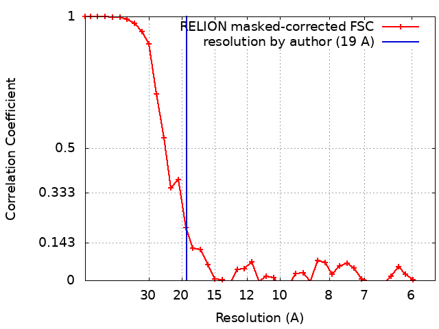 |
