[English] 日本語
 Yorodumi
- EMDB-2097: Cryo-EM structure of the F420-reducing [NiFe]-hydrogenase from a ... -
+ Open data
- Basic information
| Entry | Database: EMDB / ID: EMD-2097 | |||||||||
|---|---|---|---|---|---|---|---|---|---|---|
| Title | Cryo-EM structure of the F420-reducing [NiFe]-hydrogenase from a methanogenic archaeon with bound substrate | |||||||||
 Map data Map data | Frh complex with F420 bound | |||||||||
 Sample Sample |
| |||||||||
 Keywords Keywords | hydrogenase / [NiFe] hydrogenase / methanogenesis / F420-reducing hydrogenase | |||||||||
| Function / homology |  Function and homology information Function and homology informationcoenzyme F420 hydrogenase / coenzyme F420 hydrogenase activity / oxidoreductase activity, acting on CH or CH2 groups, with an iron-sulfur protein as acceptor / ferredoxin hydrogenase activity / nickel cation binding / iron-sulfur cluster binding / flavin adenine dinucleotide binding / 4 iron, 4 sulfur cluster binding Similarity search - Function | |||||||||
| Biological species |   Methanothermobacter marburgensis (archaea) Methanothermobacter marburgensis (archaea) | |||||||||
| Method | single particle reconstruction / cryo EM / Resolution: 4.5 Å | |||||||||
 Authors Authors | Mills DJ / Vitt S / Strauss M / Shima S / Vonck J | |||||||||
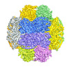
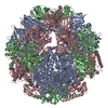
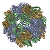
 Citation Citation |  Journal: Elife / Year: 2013 Journal: Elife / Year: 2013Title: De novo modeling of the F(420)-reducing [NiFe]-hydrogenase from a methanogenic archaeon by cryo-electron microscopy. Authors: Deryck J Mills / Stella Vitt / Mike Strauss / Seigo Shima / Janet Vonck /  Abstract: Methanogenic archaea use a [NiFe]-hydrogenase, Frh, for oxidation/reduction of F420, an important hydride carrier in the methanogenesis pathway from H2 and CO2. Frh accounts for about 1% of the ...Methanogenic archaea use a [NiFe]-hydrogenase, Frh, for oxidation/reduction of F420, an important hydride carrier in the methanogenesis pathway from H2 and CO2. Frh accounts for about 1% of the cytoplasmic protein and forms a huge complex consisting of FrhABG heterotrimers with each a [NiFe] center, four Fe-S clusters and an FAD. Here, we report the structure determined by near-atomic resolution cryo-EM of Frh with and without bound substrate F420. The polypeptide chains of FrhB, for which there was no homolog, was traced de novo from the EM map. The 1.2-MDa complex contains 12 copies of the heterotrimer, which unexpectedly form a spherical protein shell with a hollow core. The cryo-EM map reveals strong electron density of the chains of metal clusters running parallel to the protein shell, and the F420-binding site is located at the end of the chain near the outside of the spherical structure. DOI:http://dx.doi.org/10.7554/eLife.00218.001. | |||||||||
| History |
|
- Structure visualization
| Movie |
 Movie viewer Movie viewer |
|---|---|
| Structure viewer | EM map:  SurfView SurfView Molmil Molmil Jmol/JSmol Jmol/JSmol |
| Supplemental images |
- Downloads & links
-EMDB archive
| Map data |  emd_2097.map.gz emd_2097.map.gz | 84.8 MB |  EMDB map data format EMDB map data format | |
|---|---|---|---|---|
| Header (meta data) |  emd-2097-v30.xml emd-2097-v30.xml emd-2097.xml emd-2097.xml | 11 KB 11 KB | Display Display |  EMDB header EMDB header |
| Images |  EMD-2097.jpg EMD-2097.jpg | 123.3 KB | ||
| Archive directory |  http://ftp.pdbj.org/pub/emdb/structures/EMD-2097 http://ftp.pdbj.org/pub/emdb/structures/EMD-2097 ftp://ftp.pdbj.org/pub/emdb/structures/EMD-2097 ftp://ftp.pdbj.org/pub/emdb/structures/EMD-2097 | HTTPS FTP |
-Related structure data
| Related structure data |  3zfsMC  2096C C: citing same article ( M: atomic model generated by this map |
|---|---|
| Similar structure data |
- Links
Links
| EMDB pages |  EMDB (EBI/PDBe) / EMDB (EBI/PDBe) /  EMDataResource EMDataResource |
|---|---|
| Related items in Molecule of the Month |
- Map
Map
| File |  Download / File: emd_2097.map.gz / Format: CCP4 / Size: 89 MB / Type: IMAGE STORED AS FLOATING POINT NUMBER (4 BYTES) Download / File: emd_2097.map.gz / Format: CCP4 / Size: 89 MB / Type: IMAGE STORED AS FLOATING POINT NUMBER (4 BYTES) | ||||||||||||||||||||||||||||||||||||||||||||||||||||||||||||||||||||
|---|---|---|---|---|---|---|---|---|---|---|---|---|---|---|---|---|---|---|---|---|---|---|---|---|---|---|---|---|---|---|---|---|---|---|---|---|---|---|---|---|---|---|---|---|---|---|---|---|---|---|---|---|---|---|---|---|---|---|---|---|---|---|---|---|---|---|---|---|---|
| Annotation | Frh complex with F420 bound | ||||||||||||||||||||||||||||||||||||||||||||||||||||||||||||||||||||
| Projections & slices | Image control
Images are generated by Spider. | ||||||||||||||||||||||||||||||||||||||||||||||||||||||||||||||||||||
| Voxel size | X=Y=Z: 1.14 Å | ||||||||||||||||||||||||||||||||||||||||||||||||||||||||||||||||||||
| Density |
| ||||||||||||||||||||||||||||||||||||||||||||||||||||||||||||||||||||
| Symmetry | Space group: 1 | ||||||||||||||||||||||||||||||||||||||||||||||||||||||||||||||||||||
| Details | EMDB XML:
CCP4 map header:
| ||||||||||||||||||||||||||||||||||||||||||||||||||||||||||||||||||||
-Supplemental data
- Sample components
-Entire : F420-reducing [NiFe] hydrogenase Frh with bound substrate
| Entire | Name: F420-reducing [NiFe] hydrogenase Frh with bound substrate |
|---|---|
| Components |
|
-Supramolecule #1000: F420-reducing [NiFe] hydrogenase Frh with bound substrate
| Supramolecule | Name: F420-reducing [NiFe] hydrogenase Frh with bound substrate type: sample / ID: 1000 / Oligomeric state: dodecamer / Number unique components: 1 |
|---|---|
| Molecular weight | Experimental: 1.2 MDa / Theoretical: 1.2 MDa |
-Macromolecule #1: F420-reducing [NiFe] hydrogenase
| Macromolecule | Name: F420-reducing [NiFe] hydrogenase / type: protein_or_peptide / ID: 1 / Name.synonym: Frh, FrhABG Details: The Frh complex is a tetrahedral dodecamer of an FrhA, FrhB, FrhG heterotrimer. The substrate F420 is bound to FrhB Number of copies: 12 / Oligomeric state: dodecamer / Recombinant expression: No |
|---|---|
| Source (natural) | Organism:   Methanothermobacter marburgensis (archaea) / Strain: DSM 2133 / Location in cell: cytoplasm Methanothermobacter marburgensis (archaea) / Strain: DSM 2133 / Location in cell: cytoplasm |
| Molecular weight | Experimental: 1.2 MDa / Theoretical: 1.2 MDa |
| Sequence | InterPro: Coenzyme F420 hydrogenase, subunit alpha, Coenzyme F420 hydrogenase subunit beta, archaea, Coenzyme F420 hydrogenase, subunit gamma |
-Experimental details
-Structure determination
| Method | cryo EM |
|---|---|
 Processing Processing | single particle reconstruction |
| Aggregation state | particle |
- Sample preparation
| Concentration | 0.7 mg/mL |
|---|---|
| Buffer | pH: 7.6 / Details: 50mM Tris-HCl, 0.025mM FAD |
| Grid | Details: freshly glow discharged Quantifoil holey grid (1 micrometer holes) |
| Vitrification | Cryogen name: ETHANE / Chamber humidity: 70 % / Chamber temperature: 103 K / Instrument: FEI VITROBOT MARK I / Method: blotting 2.5 seconds before plunging |
- Electron microscopy
| Microscope | FEI POLARA 300 |
|---|---|
| Temperature | Min: 77 K / Max: 80 K / Average: 79 K |
| Alignment procedure | Legacy - Astigmatism: Objective lens astigmatism was corrected at 120,000 times magnification |
| Date | Feb 11, 2011 |
| Image recording | Category: FILM / Film or detector model: KODAK SO-163 FILM / Digitization - Scanner: ZEISS SCAI / Number real images: 63 / Average electron dose: 15 e/Å2 / Bits/pixel: 8 |
| Electron beam | Acceleration voltage: 200 kV / Electron source:  FIELD EMISSION GUN FIELD EMISSION GUN |
| Electron optics | Calibrated magnification: 61400 / Illumination mode: FLOOD BEAM / Imaging mode: BRIGHT FIELD / Cs: 2 mm / Nominal defocus max: 3.55 µm / Nominal defocus min: 1.65 µm / Nominal magnification: 59000 |
| Sample stage | Specimen holder model: OTHER |
| Experimental equipment |  Model: Tecnai Polara / Image courtesy: FEI Company |
- Image processing
| Details | EMAN2 |
|---|---|
| CTF correction | Details: each micrograph |
| Final reconstruction | Applied symmetry - Point group: T (tetrahedral) / Resolution.type: BY AUTHOR / Resolution: 4.5 Å / Resolution method: OTHER / Software - Name: EMAN2 / Number images used: 97000 |
| Final two d classification | Number classes: 2134 |
-Atomic model buiding 1
| Initial model | PDB ID: Chain - #0 - Chain ID: A / Chain - #1 - Chain ID: B |
|---|---|
| Software | Name: chimera, coot |
| Details | Protocol: A rigid body fit was done in Chimera and the model manually rebuilt in Coot. |
| Refinement | Space: REAL / Protocol: RIGID BODY FIT |
| Output model |  PDB-3zfs: |
 Movie
Movie Controller


 Z (Sec.)
 Y (Row.)
 X (Col.)


