 ムービー
ムービー コントローラー


+ データを開く

- 基本情報
基本情報
| 登録情報 | データベース: EMDB / ID: EMD-2091 | |||||||||
|---|---|---|---|---|---|---|---|---|---|---|
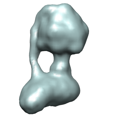
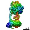
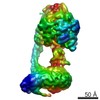
| タイトル | Electron cryomicroscopy of the bovine mitochondrial ATP synthase | |||||||||
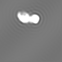
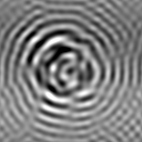
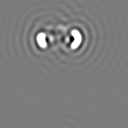
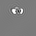
 マップデータ マップデータ | 3D map of bovine ATP synthase | |||||||||
 試料 試料 |
| |||||||||
 キーワード キーワード | bioenergetics / membrane | |||||||||
| 生物種 |  | |||||||||
| 手法 | 単粒子再構成法 / クライオ電子顕微鏡法 / 解像度: 18.0 Å | |||||||||
 データ登録者 データ登録者 | Baker LA / Watt IN / Runswick MJ / Walker JE / Rubinstein JL | |||||||||
 引用 引用 | ジャーナル: Proc Natl Acad Sci U S A / 年: 2012 タイトル: Arrangement of subunits in intact mammalian mitochondrial ATP synthase determined by cryo-EM. 著者: Lindsay A Baker / Ian N Watt / Michael J Runswick / John E Walker / John L Rubinstein /  要旨: Mitochondrial ATP synthase is responsible for the synthesis of ATP, a universal energy currency in cells. Whereas X-ray crystallography has revealed the structure of the soluble region of the complex ...Mitochondrial ATP synthase is responsible for the synthesis of ATP, a universal energy currency in cells. Whereas X-ray crystallography has revealed the structure of the soluble region of the complex and the membrane-intrinsic c-subunits, little is known about the structure of the six other proteins (a, b, f, A6L, e, and g) that comprise the membrane-bound region of the complex in animal mitochondria. Here, we present the structure of intact bovine mitochondrial ATP synthase at ∼18 Å resolution by electron cryomicroscopy of single particles in amorphous ice. The map reveals that the a-subunit and c(8)-ring of the complex interact with a small contact area and that the b-subunit spans the membrane without contacting the c(8)-ring. The e- and g-subunits extend from the a-subunit density distal to the c(8)-ring. The map was calculated from images of a preparation of the enzyme solubilized with the detergent dodecyl maltoside, which is visible in electron cryomicroscopy maps. The structure shows that the micelle surrounding the complex is curved. The observed bend in the micelle of the detergent-solubilized complex is consistent with previous electron tomography experiments and suggests that monomers of ATP synthase are sufficient to produce curvature in lipid bilayers. | |||||||||
| 履歴 |
|
- 構造の表示
構造の表示
| ムービー |
 ムービービューア ムービービューア |
|---|---|
| 構造ビューア | EMマップ: SurfViewMolmilJmol/JSmol |
| 添付画像 |
- ダウンロードとリンク
-EMDBアーカイブ
| マップデータ | emd_2091.map.gz | 7.4 MB | EMDBマップデータ形式 | |
|---|---|---|---|---|
| ヘッダ (付随情報) | emd-2091-v30.xmlemd-2091.xml | 29.9 KB 29.9 KB | 表示 表示 | EMDBヘッダ |
| 画像 |  emd_2091.png emd_2091.png | 48.2 KB | ||
| マスクデータ | emd_2091_msk_1.mapemd_2091_msk_10.mapemd_2091_msk_2.mapemd_2091_msk_3.mapemd_2091_msk_4.mapemd_2091_msk_5.mapemd_2091_msk_6.mapemd_2091_msk_7.mapemd_2091_msk_8.mapemd_2091_msk_9.map | 8 MB 8 MB 8 MB 8 MB 8 MB 8 MB 8 MB 8 MB 8 MB 8 MB | マスクマップ | |
| アーカイブディレクトリ |  http://ftp.pdbj.org/pub/emdb/structures/EMD-2091ftp://ftp.pdbj.org/pub/emdb/structures/EMD-2091 http://ftp.pdbj.org/pub/emdb/structures/EMD-2091ftp://ftp.pdbj.org/pub/emdb/structures/EMD-2091 | HTTPS FTP |
-検証レポート
| 文書・要旨 | emd_2091_validation.pdf.gz | 200.4 KB | 表示 | EMDB検証レポート |
|---|---|---|---|---|
| 文書・詳細版 | emd_2091_full_validation.pdf.gz | 199.5 KB | 表示 | |
| XML形式データ | emd_2091_validation.xml.gz | 5.6 KB | 表示 | |
| アーカイブディレクトリ | https://ftp.pdbj.org/pub/emdb/validation_reports/EMD-2091ftp://ftp.pdbj.org/pub/emdb/validation_reports/EMD-2091 | HTTPS FTP |
-関連構造データ


-リンク
| EMDBのページ | EMDB (EBI/PDBe) / EMDataResource |
|---|
-マップ
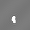
| ファイル | ダウンロード / ファイル: emd_2091.map.gz / 形式: CCP4 / 大きさ: 7.8 MB / タイプ: IMAGE STORED AS FLOATING POINT NUMBER (4 BYTES) | ||||||||||||||||||||||||||||||||||||||||||||||||||||||||||||||||||||
|---|---|---|---|---|---|---|---|---|---|---|---|---|---|---|---|---|---|---|---|---|---|---|---|---|---|---|---|---|---|---|---|---|---|---|---|---|---|---|---|---|---|---|---|---|---|---|---|---|---|---|---|---|---|---|---|---|---|---|---|---|---|---|---|---|---|---|---|---|---|
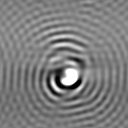
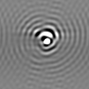
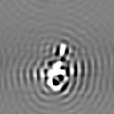
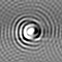
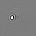
| 注釈 | 3D map of bovine ATP synthase | ||||||||||||||||||||||||||||||||||||||||||||||||||||||||||||||||||||
| 投影像・断面図 | 画像のコントロール
画像は Spider により作成 | ||||||||||||||||||||||||||||||||||||||||||||||||||||||||||||||||||||
| ボクセルのサイズ | X=Y=Z: 2.8 Å | ||||||||||||||||||||||||||||||||||||||||||||||||||||||||||||||||||||
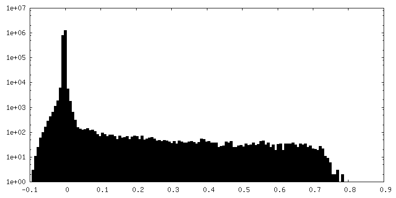
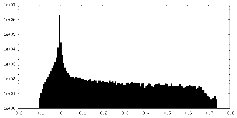
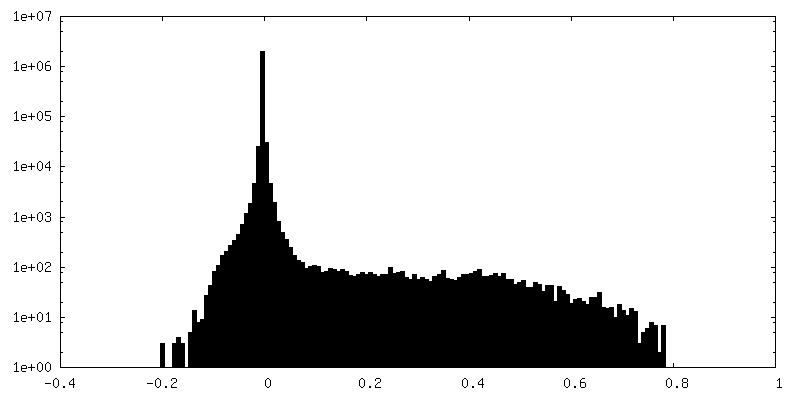
| 密度 |
| ||||||||||||||||||||||||||||||||||||||||||||||||||||||||||||||||||||
| 対称性 | 空間群: 1 | ||||||||||||||||||||||||||||||||||||||||||||||||||||||||||||||||||||
| 詳細 | EMDB XML:
CCP4マップ ヘッダ情報:
| ||||||||||||||||||||||||||||||||||||||||||||||||||||||||||||||||||||
 Z (Sec.)
 Y (Row.)
 X (Col.)


-添付データ
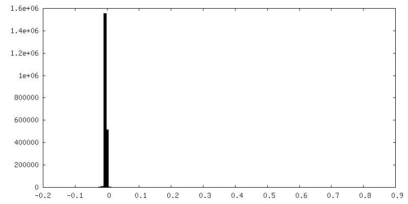
-セグメンテーションマップ: #2
| ファイル | emd_2091_msk_1.map | ||||||||||||
|---|---|---|---|---|---|---|---|---|---|---|---|---|---|
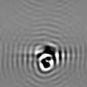
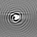
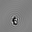
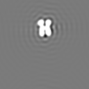
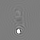
| 投影像・断面図 |
| ||||||||||||
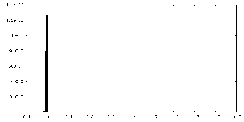
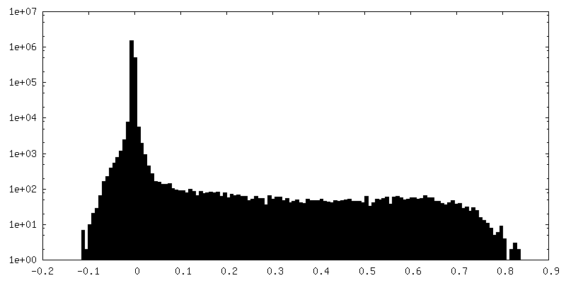
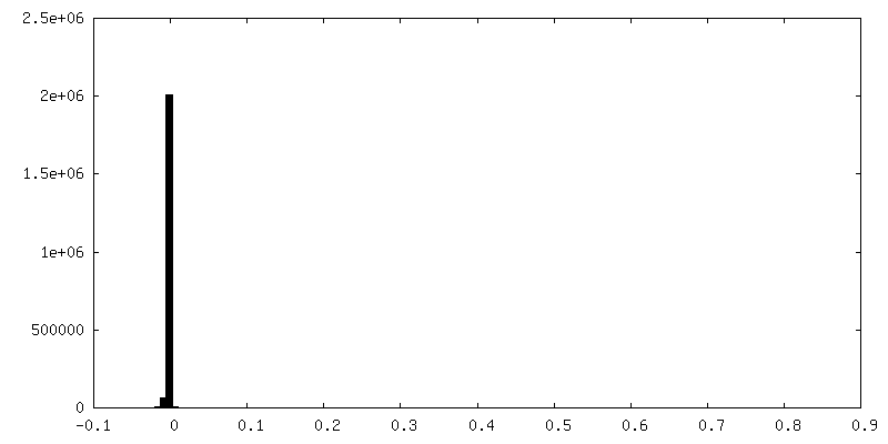
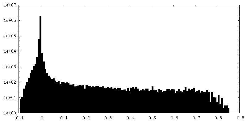
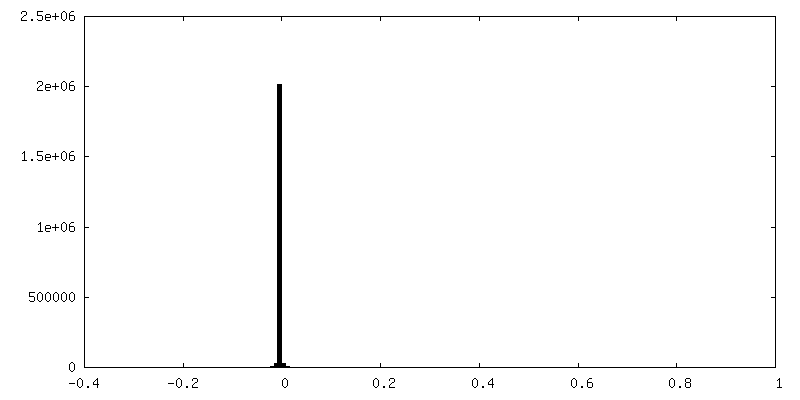
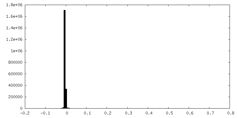
| 密度ヒストグラム |


-セグメンテーションマップ: #1
| ファイル | emd_2091_msk_10.map | ||||||||||||
|---|---|---|---|---|---|---|---|---|---|---|---|---|---|
| 投影像・断面図 |
| ||||||||||||
| 密度ヒストグラム |


-セグメンテーションマップ: #3
| ファイル | emd_2091_msk_2.map | ||||||||||||
|---|---|---|---|---|---|---|---|---|---|---|---|---|---|
| 投影像・断面図 |
| ||||||||||||
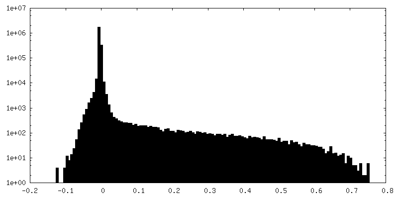
| 密度ヒストグラム |


-セグメンテーションマップ: #4
| ファイル | emd_2091_msk_3.map | ||||||||||||
|---|---|---|---|---|---|---|---|---|---|---|---|---|---|
| 投影像・断面図 |
| ||||||||||||
| 密度ヒストグラム |


-セグメンテーションマップ: #5
| ファイル | emd_2091_msk_4.map | ||||||||||||
|---|---|---|---|---|---|---|---|---|---|---|---|---|---|
| 投影像・断面図 |
| ||||||||||||
| 密度ヒストグラム |


-セグメンテーションマップ: #6
| ファイル | emd_2091_msk_5.map | ||||||||||||
|---|---|---|---|---|---|---|---|---|---|---|---|---|---|
| 投影像・断面図 |
| ||||||||||||
| 密度ヒストグラム |


-セグメンテーションマップ: #7
| ファイル | emd_2091_msk_6.map | ||||||||||||
|---|---|---|---|---|---|---|---|---|---|---|---|---|---|
| 投影像・断面図 |
| ||||||||||||
| 密度ヒストグラム |


-セグメンテーションマップ: #8
| ファイル | emd_2091_msk_7.map | ||||||||||||
|---|---|---|---|---|---|---|---|---|---|---|---|---|---|
| 投影像・断面図 |
| ||||||||||||
| 密度ヒストグラム |


-セグメンテーションマップ: #9
| ファイル | emd_2091_msk_8.map | ||||||||||||
|---|---|---|---|---|---|---|---|---|---|---|---|---|---|
| 投影像・断面図 |
| ||||||||||||
| 密度ヒストグラム |


-セグメンテーションマップ: #10
| ファイル | emd_2091_msk_9.map | ||||||||||||
|---|---|---|---|---|---|---|---|---|---|---|---|---|---|
| 投影像・断面図 |
| ||||||||||||
| 密度ヒストグラム |


- 試料の構成要素
-全体 : Bovine mitochondrial ATP synthase
| 全体 | 名称: Bovine mitochondrial ATP synthase |
|---|---|
| 要素 |
|
-超分子 #1000: Bovine mitochondrial ATP synthase
| 超分子 | 名称: Bovine mitochondrial ATP synthase / タイプ: sample / ID: 1000 詳細: The sample was maintained in a buffer containing the detergent dodecylmaltoside and phospholipids 集合状態: One hetero oligomeric ATP synthase complex / Number unique components: 1 |
|---|---|
| 分子量 | 実験値: 600 KDa / 理論値: 600 KDa / 手法: Size exclusion |
-分子 #1: ATP synthase
| 分子 | 名称: ATP synthase / タイプ: protein_or_peptide / ID: 1 / Name.synonym: ATPase, complex V / コピー数: 1 / 集合状態: monomer / 組換発現: No |
|---|---|
| 由来(天然) | 生物種: |
| 分子量 | 実験値: 600 KDa / 理論値: 600 KDa |
-実験情報
-構造解析
| 手法 | クライオ電子顕微鏡法 |
|---|---|
 解析 解析 | 単粒子再構成法 |
| 試料の集合状態 | particle |
-試料調製
| 濃度 | 2.5 mg/mL |
|---|---|
| 緩衝液 | pH: 7.8 詳細: 20 mM Tris-HCl, 100 mM NaCl, 2 mM MgSO4, 1 mM ATP, 0.02%[w/v] sodium azide, 0.1 % dodecylmaltoside, 0.1 mg/ml lipids (cardiolipin:PE:PC 3:1:1) |
| グリッド | 詳細: Quantifoil R2/2 grids |
| 凍結 | 凍結剤: ETHANE / チャンバー内湿度: 100 % / 装置: FEI VITROBOT MARK III / 手法: Equilibrate 30 s, blot 10 s |
- 電子顕微鏡法
| 顕微鏡 | FEI TECNAI F20 |
|---|---|
| アライメント法 | Legacy - 非点収差: manual astigmatism correction using image FFT |
| 日付 | 2011年1月1日 |
| 撮影 | カテゴリ: FILM / フィルム・検出器のモデル: KODAK SO-163 FILM / デジタル化 - スキャナー: ZEISS SCAI / デジタル化 - サンプリング間隔: 7 µm / 実像数: 848 / 平均電子線量: 25 e/Å2 / ビット/ピクセル: 8 |
| 電子線 | 加速電圧: 200 kV / 電子線源:  FIELD EMISSION GUN FIELD EMISSION GUN |
| 電子光学系 | 倍率(補正後): 50000 / 照射モード: FLOOD BEAM / 撮影モード: BRIGHT FIELD / Cs: 2.0 mm / 最大 デフォーカス(公称値): 7.0 µm / 最小 デフォーカス(公称値): 3.0 µm / 倍率(公称値): 50000 |
| 試料ステージ | 試料ホルダー: Gatan 626 / 試料ホルダーモデル: GATAN LIQUID NITROGEN |
| 実験機器 |  モデル: Tecnai F20 / 画像提供: FEI Company |
-画像解析
| CTF補正 | 詳細: Each particle |
|---|---|
| 最終 再構成 | 想定した対称性 - 点群: C1 (非対称) / アルゴリズム: OTHER / 解像度のタイプ: BY AUTHOR / 解像度: 18.0 Å / 解像度の算出法: FSC 0.143 CUT-OFF ソフトウェア - 名称: Spider, Rotan, Frealign, Build_fspace 使用した粒子像数: 57885 |
