National Institutes of Health/National Cancer Institute (NIH/NCI)
HSSN261200800001E
United States
National Institutes of Health/National Cancer Institute (NIH/NCI)
R01CA212403
United States
National Institutes of Health/National Human Genome Research Institute (NIH/NHGRI)
R21GM131231
United States
National Institutes of Health/National Human Genome Research Institute (NIH/NHGRI)
GM111367
United States
National Institutes of Health/National Human Genome Research Institute (NIH/NHGRI)
GM093271
United States
National Institutes of Health/National Human Genome Research Institute (NIH/NHGRI)
U24GM116788
United States
Citation
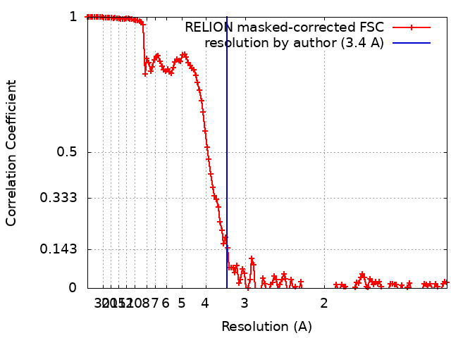
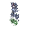
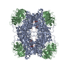
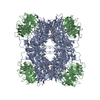
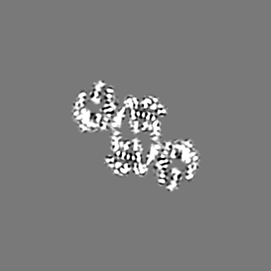
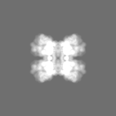
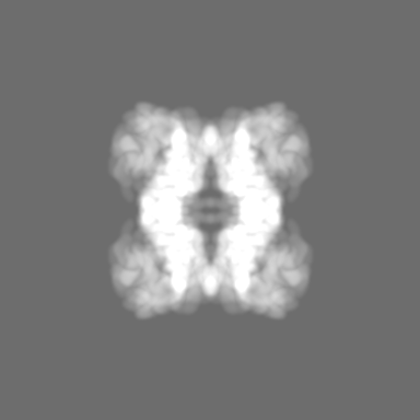
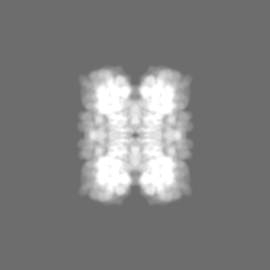
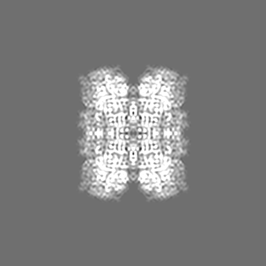
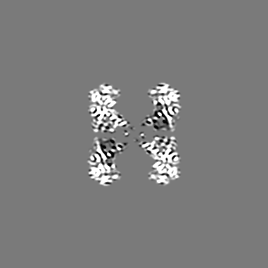
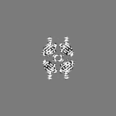
Journal: To Be Published Title: A 3.4-angstrom cryo-EM structure of the apo form of hu-man PRMT5:MEP50 complex reveals sequential catalysis of symmetric methyl transfer Authors: Zhou W / Yadav GP / Jiang Q-X / Li C
History
Deposition
Sep 26, 2019
-
Header (metadata) release
Jan 29, 2020
-
Map release
Dec 9, 2020
-
Update
Mar 20, 2024
-
Current status
Mar 20, 2024
Processing site: RCSB / Status: Released
-
Structure visualization
Movie
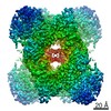
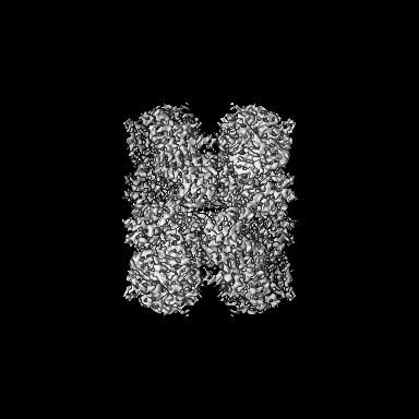
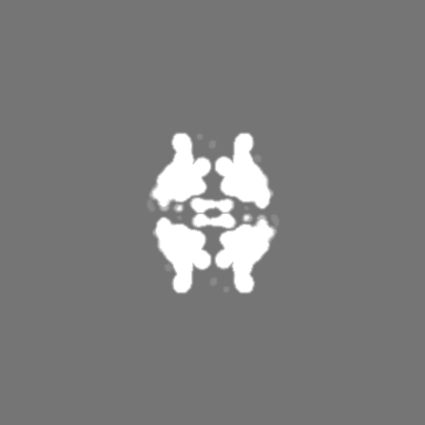
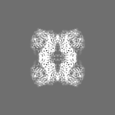
Surface view with section colored by density value
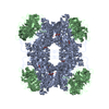
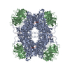
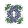
Protein or peptide: Protein arginine N-methyltransferase 5
Protein or peptide: Methylosome protein 50
-
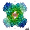
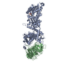
Supramolecule #1: apo PRMT5:MEP50 complex
Supramolecule
Name: apo PRMT5:MEP50 complex / type: complex / ID: 1 / Parent: 0 / Macromolecule list: all Details: the apo form of PRMT5:MEP50 heterooctomeric complex
Source (natural)
Organism: Homo sapiens (human)
Molecular weight
Theoretical: 445 KDa
-
Macromolecule #1: Protein arginine N-methyltransferase 5
Macromolecule
Name: Protein arginine N-methyltransferase 5 / type: protein_or_peptide / ID: 1 / Number of copies: 1 / Enantiomer: LEVO / EC number: type II protein arginine methyltransferase
Model: Quantifoil R1.2/1.3 / Material: GOLD / Mesh: 300 / Support film - Material: CARBON / Support film - topology: HOLEY / Support film - Film thickness: 12 / Pretreatment - Type: PLASMA CLEANING / Pretreatment - Time: 20 sec. / Pretreatment - Atmosphere: OTHER
Vitrification
Cryogen name: ETHANE / Chamber humidity: 100 % / Chamber temperature: 277 K / Instrument: FEI VITROBOT MARK II
Details
This sample was monodisperse.
-
Electron microscopy
Microscope
FEI TITAN KRIOS
Temperature
Min: 93.0 K / Max: 123.0 K
Image recording
Film or detector model: GATAN K2 SUMMIT (4k x 4k) / Detector mode: SUPER-RESOLUTION / Digitization - Frames/image: 2-39 / Average electron dose: 40.0 e/Å2
Electron beam
Acceleration voltage: 300 kV / Electron source: FIELD EMISSION GUN
Electron optics
C2 aperture diameter: 100.0 µm / Illumination mode: OTHER / Imaging mode: BRIGHT FIELD / Cs: 2.7 mm / Nominal defocus max: -4.0 µm / Nominal defocus min: -1.0 µm
In the structure databanks used in Yorodumi, some data are registered as the other names, "COVID-19 virus" and "2019-nCoV". Here are the details of the virus and the list of structure data.
Jan 31, 2019. EMDB accession codes are about to change! (news from PDBe EMDB page)
EMDB accession codes are about to change! (news from PDBe EMDB page)
The allocation of 4 digits for EMDB accession codes will soon come to an end. Whilst these codes will remain in use, new EMDB accession codes will include an additional digit and will expand incrementally as the available range of codes is exhausted. The current 4-digit format prefixed with “EMD-” (i.e. EMD-XXXX) will advance to a 5-digit format (i.e. EMD-XXXXX), and so on. It is currently estimated that the 4-digit codes will be depleted around Spring 2019, at which point the 5-digit format will come into force.
The EM Navigator/Yorodumi systems omit the EMD- prefix.
Related info.:Q: What is EMD? / ID/Accession-code notation in Yorodumi/EM Navigator
Yorodumi is a browser for structure data from EMDB, PDB, SASBDB, etc.
This page is also the successor to EM Navigator detail page, and also detail information page/front-end page for Omokage search.
The word "yorodu" (or yorozu) is an old Japanese word meaning "ten thousand". "mi" (miru) is to see.
Related info.:EMDB / PDB / SASBDB / Comparison of 3 databanks / Yorodumi Search / Aug 31, 2016. New EM Navigator & Yorodumi / Yorodumi Papers / Jmol/JSmol / Function and homology information / Changes in new EM Navigator and Yorodumi
 Movie
Movie Controller

 Yorodumi
 Open data
 Basic information
 Map data
 Sample
 Keywords
 Function and homology information
 Homo sapiens (human)
 Authors
 United States, 6 items
  Citation
 Structure visualization
 Downloads & links
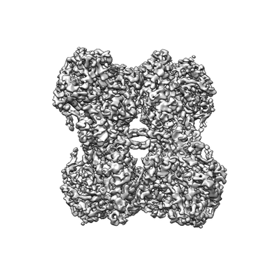 emd_20764.png
 http://ftp.pdbj.org/pub/emdb/structures/EMD-20764


 Z (Sec.)
 Y (Row.)
 X (Col.)


 Sample components
 Spodoptera frugiperda (fall armyworm)
 Processing
 Electron microscopy
 FIELD EMISSION GUN