+ データを開く
- 基本情報
基本情報
| 登録情報 | データベース: EMDB / ID: EMD-2066 | |||||||||
|---|---|---|---|---|---|---|---|---|---|---|
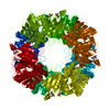
| タイトル | Electron cryo-microscopy of the L651A mutant R-peptide precursor Env of Moloney murine leukemia virus in its native state | |||||||||
 マップデータ マップデータ | Reconstruction of the L651A mutant R-peptide precursor of Moloney murine leukemia virus Env in its native state | |||||||||
 試料 試料 |
| |||||||||
 キーワード キーワード | cryo-EM / Retrovirus / spike protein / maturation cleavage / R-peptide | |||||||||
| 生物種 |  Moloney murine leukemia virus (ウイルス) Moloney murine leukemia virus (ウイルス) | |||||||||
| 手法 | 単粒子再構成法 / クライオ電子顕微鏡法 / ネガティブ染色法 / 解像度: 22.0 Å | |||||||||
 データ登録者 データ登録者 | Loving R / Wu SR / Sjoberg M / Lindqvist B / Garoff H | |||||||||
 引用 引用 |  ジャーナル: Proc Natl Acad Sci U S A / 年: 2012 ジャーナル: Proc Natl Acad Sci U S A / 年: 2012タイトル: Maturation cleavage of the murine leukemia virus Env precursor separates the transmembrane subunits to prime it for receptor triggering. 著者: Robin Löving / Shang-Rung Wu / Mathilda Sjöberg / Birgitta Lindqvist / Henrik Garoff /  要旨: The Env protein of murine leukemia virus matures by two cleavage events. First, cellular furin separates the receptor binding surface (SU) subunit from the fusion-active transmembrane (TM) subunit ...The Env protein of murine leukemia virus matures by two cleavage events. First, cellular furin separates the receptor binding surface (SU) subunit from the fusion-active transmembrane (TM) subunit and then, in the newly assembled particle, the viral protease removes a 16-residue peptide, the R-peptide from the endodomain of the TM. Both cleavage events are required to prime the Env for receptor-triggered activation. Cryoelectron microscopy (cryo-EM) analyses have shown that the mature Env forms an open cage-like structure composed of three SU-TM complexes, where the TM subunits formed separated Env legs. Here we have studied the structure of the R-peptide precursor Env by cryo-EM. TM cleavage in Moloney murine leukemia virus was inhibited by amprenavir, and the Envs were solubilized in Triton X-100 and isolated by sedimentation in a sucrose gradient. We found that the legs of the R-peptide Env were held together by trimeric interactions at the very bottom of the Env. This suggested that the R-peptide ties the TM legs together and that this prevents the activation of the TM for fusion. The model was supported by further cryo-EM studies using an R-peptide Env mutant that was fusion-competent despite an uncleaved R-peptide. The Env legs of this mutant were found to be separated, like in the mature Env. This shows that it is the TM leg separation, normally caused by R-peptide cleavage, that primes the Env for receptor triggering. | |||||||||
| 履歴 |
|
- 構造の表示
構造の表示
| ムービー |
 ムービービューア ムービービューア |
|---|---|
| 構造ビューア | EMマップ:  SurfView SurfView Molmil Molmil Jmol/JSmol Jmol/JSmol |
| 添付画像 |
- ダウンロードとリンク
-EMDBアーカイブ
| マップデータ |  emd_2066.map.gz emd_2066.map.gz | 361.2 KB |  EMDBマップデータ形式 EMDBマップデータ形式 | |
|---|---|---|---|---|
| ヘッダ (付随情報) |  emd-2066-v30.xml emd-2066-v30.xml emd-2066.xml emd-2066.xml | 10.5 KB 10.5 KB | 表示 表示 |  EMDBヘッダ EMDBヘッダ |
| 画像 |  EMD2066.png EMD2066.png | 734.4 KB | ||
| アーカイブディレクトリ |  http://ftp.pdbj.org/pub/emdb/structures/EMD-2066 http://ftp.pdbj.org/pub/emdb/structures/EMD-2066 ftp://ftp.pdbj.org/pub/emdb/structures/EMD-2066 ftp://ftp.pdbj.org/pub/emdb/structures/EMD-2066 | HTTPS FTP |
-検証レポート
| 文書・要旨 |  emd_2066_validation.pdf.gz emd_2066_validation.pdf.gz | 208.3 KB | 表示 |  EMDB検証レポート EMDB検証レポート |
|---|---|---|---|---|
| 文書・詳細版 |  emd_2066_full_validation.pdf.gz emd_2066_full_validation.pdf.gz | 207.5 KB | 表示 | |
| XML形式データ |  emd_2066_validation.xml.gz emd_2066_validation.xml.gz | 4.5 KB | 表示 | |
| アーカイブディレクトリ |  https://ftp.pdbj.org/pub/emdb/validation_reports/EMD-2066 https://ftp.pdbj.org/pub/emdb/validation_reports/EMD-2066 ftp://ftp.pdbj.org/pub/emdb/validation_reports/EMD-2066 ftp://ftp.pdbj.org/pub/emdb/validation_reports/EMD-2066 | HTTPS FTP |
-関連構造データ
- リンク
リンク
| EMDBのページ |  EMDB (EBI/PDBe) / EMDB (EBI/PDBe) /  EMDataResource EMDataResource |
|---|
- マップ
マップ
| ファイル |  ダウンロード / ファイル: emd_2066.map.gz / 形式: CCP4 / 大きさ: 422.9 KB / タイプ: IMAGE STORED AS FLOATING POINT NUMBER (4 BYTES) ダウンロード / ファイル: emd_2066.map.gz / 形式: CCP4 / 大きさ: 422.9 KB / タイプ: IMAGE STORED AS FLOATING POINT NUMBER (4 BYTES) | ||||||||||||||||||||||||||||||||||||||||||||||||||||||||||||||||||||
|---|---|---|---|---|---|---|---|---|---|---|---|---|---|---|---|---|---|---|---|---|---|---|---|---|---|---|---|---|---|---|---|---|---|---|---|---|---|---|---|---|---|---|---|---|---|---|---|---|---|---|---|---|---|---|---|---|---|---|---|---|---|---|---|---|---|---|---|---|---|
| 注釈 | Reconstruction of the L651A mutant R-peptide precursor of Moloney murine leukemia virus Env in its native state | ||||||||||||||||||||||||||||||||||||||||||||||||||||||||||||||||||||
| 投影像・断面図 | 画像のコントロール
画像は Spider により作成 | ||||||||||||||||||||||||||||||||||||||||||||||||||||||||||||||||||||
| ボクセルのサイズ | X=Y=Z: 3.5 Å | ||||||||||||||||||||||||||||||||||||||||||||||||||||||||||||||||||||
| 密度 |
| ||||||||||||||||||||||||||||||||||||||||||||||||||||||||||||||||||||
| 対称性 | 空間群: 1 | ||||||||||||||||||||||||||||||||||||||||||||||||||||||||||||||||||||
| 詳細 | EMDB XML:
CCP4マップ ヘッダ情報:
| ||||||||||||||||||||||||||||||||||||||||||||||||||||||||||||||||||||
-添付データ
- 試料の構成要素
-全体 : Native form of the L651A mutant R-peptide precursor of Moloney mu...
| 全体 | 名称: Native form of the L651A mutant R-peptide precursor of Moloney murine leukemia virus Env |
|---|---|
| 要素 |
|
-超分子 #1000: Native form of the L651A mutant R-peptide precursor of Moloney mu...
| 超分子 | 名称: Native form of the L651A mutant R-peptide precursor of Moloney murine leukemia virus Env タイプ: sample / ID: 1000 / 集合状態: trimeric / Number unique components: 1 |
|---|---|
| 分子量 | 実験値: 500 KDa / 理論値: 270 KDa / 手法: Blue native PAGE |
-分子 #1: gp70-Pr15E
| 分子 | 名称: gp70-Pr15E / タイプ: protein_or_peptide / ID: 1 / Name.synonym: (SU-TM)3; Env / 詳細: Expressed in Human Embryonic Kidney 293T cells / コピー数: 1 / 集合状態: trimeric / 組換発現: Yes |
|---|---|
| 由来(天然) | 生物種:  Moloney murine leukemia virus (ウイルス) / 別称: MO-MLV Moloney murine leukemia virus (ウイルス) / 別称: MO-MLV |
| 分子量 | 実験値: 500 KDa / 理論値: 270 KDa |
| 組換発現 | 生物種:  Homo sapiens (ヒト) / 組換プラスミド: pNCA Homo sapiens (ヒト) / 組換プラスミド: pNCA |
-実験情報
-構造解析
| 手法 | ネガティブ染色法, クライオ電子顕微鏡法 |
|---|---|
 解析 解析 | 単粒子再構成法 |
| 試料の集合状態 | particle |
- 試料調製
試料調製
| 濃度 | 0.1 mg/mL |
|---|---|
| 緩衝液 | pH: 7.4 詳細: 50 mM Hepes, 100 mM NaCl, 1.8 mM CaCl2, 0.05% Triton X-100 |
| 染色 | タイプ: NEGATIVE 詳細: The specimen was frozen in liquid ethane and transferred to liquid nitrogen for EM inspection without staining. |
| グリッド | 詳細: 400 mesh holey carbon grid. The grids were glow discharged. |
| 凍結 | 凍結剤: ETHANE / チャンバー内湿度: 99 % / チャンバー内温度: 77 K / 装置: FEI VITROBOT MARK II Timed resolved state: Vitrified 45 msec after spraying with effector 手法: Blot for 3 seconds before plunging |
- 電子顕微鏡法
| 顕微鏡 | JEOL 2100F |
|---|---|
| 温度 | 最低: 93 K / 最高: 96 K / 平均: 95 K |
| アライメント法 | Legacy - 非点収差: Objective lens astigmatism was corrected using online FFT |
| 日付 | 2011年7月1日 |
| 撮影 | カテゴリ: CCD / フィルム・検出器のモデル: GENERIC CCD / デジタル化 - サンプリング間隔: 3.5 µm / 実像数: 653 / 平均電子線量: 9 e/Å2 / ビット/ピクセル: 14 |
| Tilt angle min | 0 |
| Tilt angle max | 0 |
| 電子線 | 加速電圧: 200 kV / 電子線源:  FIELD EMISSION GUN FIELD EMISSION GUN |
| 電子光学系 | 倍率(補正後): 43200 / 照射モード: SPOT SCAN / 撮影モード: BRIGHT FIELD / Cs: 2.0 mm / 最大 デフォーカス(公称値): 4.0 µm / 最小 デフォーカス(公称値): 2.5 µm / 倍率(公称値): 43200 |
| 試料ステージ | 試料ホルダー: liquid nitrogen cooled / 試料ホルダーモデル: GATAN LIQUID NITROGEN |
- 画像解析
画像解析
| 詳細 | The particles were selected using an automatic selection program and the damaged particles were removed by visual inspection. |
|---|---|
| CTF補正 | 詳細: Each particle |
| 最終 再構成 | 想定した対称性 - 点群: C3 (3回回転対称) / アルゴリズム: OTHER / 解像度のタイプ: BY AUTHOR / 解像度: 22.0 Å / 解像度の算出法: FSC 0.5 CUT-OFF / ソフトウェア - 名称: EMAN1, EMAN2 詳細: Final maps were calculated from four averaged datasets 使用した粒子像数: 4702 |
| 最終 2次元分類 | クラス数: 131 |
 ムービー
ムービー コントローラー


 Z (Sec.)
 Y (Row.)
 X (Col.)


