ムービー
ムービー コントローラー


+ データを開く

- 基本情報
基本情報
| 登録情報 | データベース: EMDB / ID: EMD-2046 | |||||||||
|---|---|---|---|---|---|---|---|---|---|---|
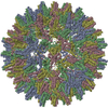
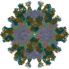
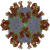
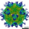
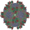
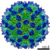
| タイトル | Cryo-electron microscopy study of hepatitis B virus decorated with the antibody E1 | |||||||||
 マップデータ マップデータ | 3D reconstruction of antibody-decorated Hepatitis B T=4 particle | |||||||||
 試料 試料 |
| |||||||||
 キーワード キーワード | Hepatitis B / core antigen / ALF / acute liver failure / antibody E1 / monoclonal / conformational epitope / fulminant hepatitis / cryo-EM | |||||||||
| 生物種 |  Homo sapiens (ヒト) / Homo sapiens (ヒト) /   Hepatitis B virus (B 型肝炎ウイルス) Hepatitis B virus (B 型肝炎ウイルス) | |||||||||
| 手法 | 単粒子再構成法 / クライオ電子顕微鏡法 / 解像度: 10.0 Å | |||||||||
 データ登録者 データ登録者 | Wu W / Chen ZC / Cheng NQ / Watts NR / Stahl SJ / Farci P / Purcell RH / Wingfield PT / Steven AC | |||||||||
 引用 引用 | ジャーナル: J Struct Biol / 年: 2013 タイトル: Specificity of an anti-capsid antibody associated with Hepatitis B Virus-related acute liver failure. 著者: Weimin Wu / Zhaochun Chen / Naiqian Cheng / Norman R Watts / Stephen J Stahl / Patrizia Farci / Robert H Purcell / Paul T Wingfield / Alasdair C Steven /  要旨: Previously, the livers of patients suffering from acute liver failure (ALF), a potentially fatal syndrome arising from infection by Hepatitis B Virus (HBV), were found to contain massive amounts of ...Previously, the livers of patients suffering from acute liver failure (ALF), a potentially fatal syndrome arising from infection by Hepatitis B Virus (HBV), were found to contain massive amounts of an antibody specific for the core antigen (HBcAg) capsid. We have used cryo-electron microscopy and molecular modeling to define its epitope. HBV capsids are icosahedral shells with 25Å-long dimeric spikes, each a 4-helix bundle, protruding from the contiguous "floor". Of the anti-HBcAg antibodies previously characterized, most bind around the spike tip while one binds to the floor. The ALF-associated antibody binds tangentially to a novel site on the side of the spike. This epitope is conformational. The Fab binds with high affinity to its principal determinants but has lower affinities for quasi-equivalent variants. The highest occupancy site is on one side of a spike, with no detectable binding to the corresponding site on the other side. Binding of one Fab per dimer was also observed by analytical ultracentrifugation. The Fab did not bind to the e-antigen dimer, a non-assembling variant of capsid protein. These findings support the propositions that antibodies with particular specificities may correlate with different clinical expressions of HBV infection and that antibodies directed to particular HBcAg epitopes may be involved in ALF pathogenesis. | |||||||||
| 履歴 |
|
- 構造の表示
構造の表示
| ムービー |
 ムービービューア ムービービューア |
|---|---|
| 構造ビューア | EMマップ: SurfViewMolmilJmol/JSmol |
| 添付画像 |
- ダウンロードとリンク
-EMDBアーカイブ
| マップデータ | emd_2046.map.gz | 21 MB | EMDBマップデータ形式 | |
|---|---|---|---|---|
| ヘッダ (付随情報) | emd-2046-v30.xmlemd-2046.xml | 11.1 KB 11.1 KB | 表示 表示 | EMDBヘッダ |
| 画像 | EMD_2046_icon.tif | 762.9 KB | ||
| アーカイブディレクトリ |  http://ftp.pdbj.org/pub/emdb/structures/EMD-2046ftp://ftp.pdbj.org/pub/emdb/structures/EMD-2046 http://ftp.pdbj.org/pub/emdb/structures/EMD-2046ftp://ftp.pdbj.org/pub/emdb/structures/EMD-2046 | HTTPS FTP |
-関連構造データ
-リンク
| EMDBのページ | EMDB (EBI/PDBe) / EMDataResource |
|---|
-マップ
| ファイル | ダウンロード / ファイル: emd_2046.map.gz / 形式: CCP4 / 大きさ: 29.8 MB / タイプ: IMAGE STORED AS FLOATING POINT NUMBER (4 BYTES) | ||||||||||||||||||||||||||||||||||||||||||||||||||||||||||||||||||||
|---|---|---|---|---|---|---|---|---|---|---|---|---|---|---|---|---|---|---|---|---|---|---|---|---|---|---|---|---|---|---|---|---|---|---|---|---|---|---|---|---|---|---|---|---|---|---|---|---|---|---|---|---|---|---|---|---|---|---|---|---|---|---|---|---|---|---|---|---|---|
| 注釈 | 3D reconstruction of antibody-decorated Hepatitis B T=4 particle | ||||||||||||||||||||||||||||||||||||||||||||||||||||||||||||||||||||
| 投影像・断面図 | 画像のコントロール
画像は Spider により作成 | ||||||||||||||||||||||||||||||||||||||||||||||||||||||||||||||||||||
| ボクセルのサイズ | X=Y=Z: 2.457 Å | ||||||||||||||||||||||||||||||||||||||||||||||||||||||||||||||||||||
| 密度 |
| ||||||||||||||||||||||||||||||||||||||||||||||||||||||||||||||||||||
| 対称性 | 空間群: 1 | ||||||||||||||||||||||||||||||||||||||||||||||||||||||||||||||||||||
| 詳細 | EMDB XML:
CCP4マップ ヘッダ情報:
| ||||||||||||||||||||||||||||||||||||||||||||||||||||||||||||||||||||
 Z (Sec.)
 Y (Row.)
 X (Col.)


-添付データ
- 試料の構成要素
-全体 : Antibody E1 to Hepatitis B core antigen
| 全体 | 名称: Antibody E1 to Hepatitis B core antigen |
|---|---|
| 要素 |
|
-超分子 #1000: Antibody E1 to Hepatitis B core antigen
| 超分子 | 名称: Antibody E1 to Hepatitis B core antigen / タイプ: sample / ID: 1000 詳細: The antibody E1 is derived from two ALF patients. T=4 hepatitis B particle is Cp149.3CA 集合状態: One antibody to one face of the dimer / Number unique components: 1 |
|---|---|
| 分子量 | 理論値: 4.06 MDa |
-超分子 #1: Hepatitis B virus
| 超分子 | 名称: Hepatitis B virus / タイプ: virus / ID: 1 / NCBI-ID: 10407 / 生物種: Hepatitis B virus / ウイルスタイプ: VIRUS-LIKE PARTICLE / ウイルス・単離状態: SPECIES / ウイルス・エンベロープ: Yes / ウイルス・中空状態: Yes |
|---|---|
| 宿主 | 生物種: Homo sapiens (ヒト) / 別称: VERTEBRATES |
-分子 #1: Antibody E1
| 分子 | 名称: Antibody E1 / タイプ: protein_or_peptide / ID: 1 / 詳細: Molecular weight is for one copy / 組換発現: Yes |
|---|---|
| 由来(天然) | 生物種: Homo sapiens (ヒト) / 別称: Human / 組織: Plasma / 細胞: B-lyphocytes |
| 分子量 | 理論値: 50 KDa |
-実験情報
-構造解析
| 手法 | クライオ電子顕微鏡法 |
|---|---|
 解析 解析 | 単粒子再構成法 |
| 試料の集合状態 | particle |
-試料調製
| 凍結 | 凍結剤: ETHANE / チャンバー内湿度: 38 % / チャンバー内温度: 100 K / 装置: OTHER / 手法: Manual plunging |
|---|
- 電子顕微鏡法
| 顕微鏡 | FEI/PHILIPS CM200FEG |
|---|---|
| 温度 | 平均: 100 K |
| アライメント法 | Legacy - 非点収差: Objective lens astigmatism was corrected at 175,000 magnification |
| 日付 | 2010年7月1日 |
| 撮影 | カテゴリ: FILM / フィルム・検出器のモデル: KODAK SO-163 FILM デジタル化 - スキャナー: NIKON SUPER COOLSCAN 9000 デジタル化 - サンプリング間隔: 6.35 µm / 実像数: 52 / 平均電子線量: 20 e/Å2 詳細: There are two kinds of particle in each micrograph. T=4 and T=3 Hepatitis B particles. ビット/ピクセル: 16 |
| 電子線 | 加速電圧: 120 kV / 電子線源:  FIELD EMISSION GUN FIELD EMISSION GUN |
| 電子光学系 | 倍率(補正後): 51689 / 照射モード: FLOOD BEAM / 撮影モード: BRIGHT FIELD / Cs: 2.0 mm / 最大 デフォーカス(公称値): 0.0018 µm / 最小 デフォーカス(公称値): 0.001 µm / 倍率(公称値): 5000 |
| 試料ステージ | 試料ホルダーモデル: GATAN LIQUID NITROGEN |
-画像解析
| 詳細 | EMAN and EMAN2 were applied to solve the structure. |
|---|---|
| CTF補正 | 詳細: Micrograph |
| 最終 再構成 | 想定した対称性 - 点群: I (正20面体型対称) / アルゴリズム: OTHER / 解像度のタイプ: BY AUTHOR / 解像度: 10.0 Å / 解像度の算出法: FSC 0.5 CUT-OFF / ソフトウェア - 名称: EMAN,EMAN2 / 使用した粒子像数: 3787 |
| 最終 角度割当 | 詳細: EMAN convention |
-原子モデル構築 1
| 初期モデル | PDB ID: Chain - #0 - Chain ID: A / Chain - #1 - Chain ID: B / Chain - #2 - Chain ID: C / Chain - #3 - Chain ID: D |
|---|---|
| ソフトウェア | 名称: Chimera,Situs |
| 詳細 | Protocol: Rigid body. The PDB structure was manually docked into the reconstruction using Chimera and was automatically fitted using Situs. |
| 精密化 | 空間: REAL / プロトコル: RIGID BODY FIT |