 Movie
Movie Controller


+ Open data

- Basic information
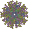
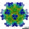
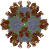
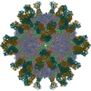
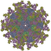
| Entry | Database: PDB / ID: 6jhq | |||||||||||||||||||||
|---|---|---|---|---|---|---|---|---|---|---|---|---|---|---|---|---|---|---|---|---|---|---|
| Title | The cryo-EM structure of HAV bound to a neutralizing antibody-F4 | |||||||||||||||||||||
 Components Components |
| |||||||||||||||||||||
 Keywords Keywords | VIRUS / Icosahedral symmetry / neutralizing antibody / HAV / complex | |||||||||||||||||||||
| Function / homology |  Function and homology information Function and homology informationhost cell mitochondrial outer membrane / symbiont-mediated suppression of host cytoplasmic pattern recognition receptor signaling pathway via inhibition of MAVS activity / picornain 3C / T=pseudo3 icosahedral viral capsid / host cell cytoplasmic vesicle membrane / host multivesicular body / ribonucleoside triphosphate phosphatase activity / nucleoside-triphosphate phosphatase / channel activity / monoatomic ion transmembrane transport ...host cell mitochondrial outer membrane / symbiont-mediated suppression of host cytoplasmic pattern recognition receptor signaling pathway via inhibition of MAVS activity / picornain 3C / T=pseudo3 icosahedral viral capsid / host cell cytoplasmic vesicle membrane / host multivesicular body / ribonucleoside triphosphate phosphatase activity / nucleoside-triphosphate phosphatase / channel activity / monoatomic ion transmembrane transport / RNA helicase activity / RNA-directed RNA polymerase / cysteine-type endopeptidase activity / viral RNA genome replication / RNA-directed RNA polymerase activity / symbiont entry into host cell / virion attachment to host cell / structural molecule activity / DNA-templated transcription / proteolysis / RNA binding / ATP binding Similarity search - Function | |||||||||||||||||||||
| Biological species |  Human hepatitis A virus Hu/Australia/HM175/1976 Human hepatitis A virus Hu/Australia/HM175/1976 | |||||||||||||||||||||
| Method | ELECTRON MICROSCOPY / single particle reconstruction / cryo EM / Resolution: 3.9 Å | |||||||||||||||||||||
 Authors Authors | Cao, L. / Liu, P. / Yang, P. / Gao, Q. / Li, H. / Sun, Y. / Zhu, L. / Lin, J. / Su, D. / Rao, Z. / Wang, X. | |||||||||||||||||||||
| Funding support |  China, 3items China, 3items
| |||||||||||||||||||||
 Citation Citation | Journal: PLoS Biol / Year: 2019 Title: Structural basis for neutralization of hepatitis A virus informs a rational design of highly potent inhibitors. Authors: Lei Cao / Pi Liu / Pan Yang / Qiang Gao / Hong Li / Yao Sun / Ling Zhu / Jianping Lin / Dan Su / Zihe Rao / Xiangxi Wang / Abstract: Hepatitis A virus (HAV), an enigmatic and ancient pathogen, is a major causative agent of acute viral hepatitis worldwide. Although there are effective vaccines, antivirals against HAV infection are ...Hepatitis A virus (HAV), an enigmatic and ancient pathogen, is a major causative agent of acute viral hepatitis worldwide. Although there are effective vaccines, antivirals against HAV infection are still required, especially during fulminant hepatitis outbreaks. A more in-depth understanding of the antigenic characteristics of HAV and the mechanisms of neutralization could aid in the development of rationally designed antiviral drugs targeting HAV. In this paper, 4 new antibodies-F4, F6, F7, and F9-are reported that potently neutralize HAV at 50% neutralizing concentration values (neut50) ranging from 0.1 nM to 0.85 nM. High-resolution cryo-electron microscopy (cryo-EM) structures of HAV bound to F4, F6, F7, and F9, together with results of our previous studies on R10 fragment of antigen binding (Fab)-HAV complex, shed light on the locations and nature of the epitopes recognized by the 5 neutralizing monoclonal antibodies (NAbs). All the epitopes locate within the same patch and are highly conserved. The key structure-activity correlates based on the antigenic sites have been established. Based on the structural data of the single conserved antigenic site and key structure-activity correlates, one promising drug candidate named golvatinib was identified by in silico docking studies. Cell-based antiviral assays confirmed that golvatinib is capable of blocking HAV infection effectively with a 50% inhibitory concentration (IC50) of approximately 1 μM. These results suggest that the single conserved antigenic site from complete HAV capsid is a good antiviral target and that golvatinib could function as a lead compound for anti-HAV drug development. | |||||||||||||||||||||
| History |
|
- Structure visualization
| Movie |
Movie viewer |
|---|---|
| Structure viewer | Molecule: MolmilJmol/JSmol |
- Downloads & links
-Download
| PDBx/mmCIF format | 6jhq.cif.gz | 210.9 KB | Display | PDBx/mmCIF format |
|---|---|---|---|---|
| PDB format | pdb6jhq.ent.gz | 166.3 KB | Display | PDB format |
| PDBx/mmJSON format | 6jhq.json.gz | Tree view | PDBx/mmJSON format | |
| Others |  Other downloads Other downloads |
-Validation report
| Arichive directory | https://data.pdbj.org/pub/pdb/validation_reports/jh/6jhqftp://data.pdbj.org/pub/pdb/validation_reports/jh/6jhq | HTTPS FTP |
|---|
-Related structure data
| Related structure data | 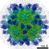 9827MC 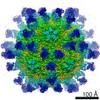 9828C 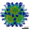 9829C 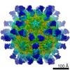 9830C  6jhrC 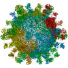 6jhsC 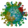 6jhtC M: map data used to model this data C: citing same article ( |
|---|---|
| Similar structure data |
-Links
 PDBj
PDBj


- Assembly
| Deposited unit | 
|
|---|---|
| 1 | x 60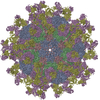
|
| 2 |
|
| 3 | x 5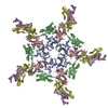
|
| 4 | x 6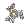
|
| 5 | 
|
| Symmetry | Point symmetry: (Schoenflies symbol: I (icosahedral)) |
-Components
| #1: Protein | Mass: 30820.629 Da / Num. of mol.: 1 / Source method: isolated from a natural source Source: (natural) Human hepatitis A virus Hu/Australia/HM175/1976References: UniProt: P08617*PLUS |
|---|---|
| #2: Protein | Mass: 24898.172 Da / Num. of mol.: 1 / Source method: isolated from a natural source Source: (natural) Human hepatitis A virus Hu/Australia/HM175/1976References: UniProt: P08617*PLUS |
| #3: Protein | Mass: 27835.693 Da / Num. of mol.: 1 / Source method: isolated from a natural source Source: (natural) Human hepatitis A virus Hu/Australia/HM175/1976References: UniProt: P08617*PLUS |
| #4: Antibody | Mass: 23939.873 Da / Num. of mol.: 1 / Source method: isolated from a natural source / Source: (natural) |
| #5: Antibody | Mass: 23592.875 Da / Num. of mol.: 1 / Source method: isolated from a natural source / Source: (natural) |
| Has protein modification | Y |
-Experimental details
-Experiment
| Experiment | Method: ELECTRON MICROSCOPY |
|---|---|
| EM experiment | Aggregation state: PARTICLE / 3D reconstruction method: single particle reconstruction |
- Sample preparation
| Component |
| ||||||||||||||||||||||||
|---|---|---|---|---|---|---|---|---|---|---|---|---|---|---|---|---|---|---|---|---|---|---|---|---|---|
| Source (natural) |
| ||||||||||||||||||||||||
| Details of virus | Empty: NO / Enveloped: NO / Isolate: STRAIN / Type: VIRION | ||||||||||||||||||||||||
| Buffer solution | pH: 7 | ||||||||||||||||||||||||
| Specimen | Embedding applied: NO / Shadowing applied: NO / Staining applied: NO / Vitrification applied: YES | ||||||||||||||||||||||||
| Vitrification | Cryogen name: ETHANE |
- Electron microscopy imaging
| Experimental equipment |  Model: Titan Krios / Image courtesy: FEI Company |
|---|---|
| Microscopy | Model: FEI TITAN KRIOS |
| Electron gun | Electron source:  FIELD EMISSION GUN / Accelerating voltage: 300 kV / Illumination mode: FLOOD BEAM FIELD EMISSION GUN / Accelerating voltage: 300 kV / Illumination mode: FLOOD BEAM |
| Electron lens | Mode: BRIGHT FIELD |
| Image recording | Electron dose: 1.3 e/Å2 / Film or detector model: GATAN K2 BASE (4k x 4k) |
- Processing
| Software | Name: PHENIX / Version: 1.11.1_2575: / Classification: refinement | ||||||||||||||||||||||||
|---|---|---|---|---|---|---|---|---|---|---|---|---|---|---|---|---|---|---|---|---|---|---|---|---|---|
| EM software |
| ||||||||||||||||||||||||
| CTF correction | Type: NONE | ||||||||||||||||||||||||
| Symmetry | Point symmetry: I (icosahedral) | ||||||||||||||||||||||||
| 3D reconstruction | Resolution: 3.9 Å / Resolution method: FSC 0.143 CUT-OFF / Num. of particles: 4536 / Symmetry type: POINT | ||||||||||||||||||||||||
| Refine LS restraints |
|
