Movie
Movie Controller


+ Open data

- Basic information
| Entry | Database: EMDB / ID: EMD-1986 | |||||||||
|---|---|---|---|---|---|---|---|---|---|---|
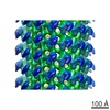
| Title | CryoEM reconstruction of the Marburg virus nucleocapsid. | |||||||||
 Map data Map data | This is a map of the Marburg virus nucleocapsid within virions. | |||||||||
 Sample Sample |
| |||||||||
 Keywords Keywords | Marburg / virus / nucleocapsid / Nucleoprotein / VP24 / VP35 | |||||||||
| Biological species |  Marburg marburgvirus Marburg marburgvirus | |||||||||
| Method | helical reconstruction / cryo EM / Resolution: 25.0 Å | |||||||||
 Authors Authors | Bharat TAM / Riches JD / Kolesnikova L / Welsch S / Kraehling V / Davey N / Parsy ML / Becker S / Briggs JAG | |||||||||
 Citation Citation | Journal: PLoS Biol / Year: 2011 Title: Cryo-electron tomography of Marburg virus particles and their morphogenesis within infected cells. Authors: Tanmay A M Bharat / James D Riches / Larissa Kolesnikova / Sonja Welsch / Verena Krähling / Norman Davey / Marie-Laure Parsy / Stephan Becker / John A G Briggs /  Abstract: Several major human pathogens, including the filoviruses, paramyxoviruses, and rhabdoviruses, package their single-stranded RNA genomes within helical nucleocapsids, which bud through the plasma ...Several major human pathogens, including the filoviruses, paramyxoviruses, and rhabdoviruses, package their single-stranded RNA genomes within helical nucleocapsids, which bud through the plasma membrane of the infected cell to release enveloped virions. The virions are often heterogeneous in shape, which makes it difficult to study their structure and assembly mechanisms. We have applied cryo-electron tomography and sub-tomogram averaging methods to derive structures of Marburg virus, a highly pathogenic filovirus, both after release and during assembly within infected cells. The data demonstrate the potential of cryo-electron tomography methods to derive detailed structural information for intermediate steps in biological pathways within intact cells. We describe the location and arrangement of the viral proteins within the virion. We show that the N-terminal domain of the nucleoprotein contains the minimal assembly determinants for a helical nucleocapsid with variable number of proteins per turn. Lobes protruding from alternate interfaces between each nucleoprotein are formed by the C-terminal domain of the nucleoprotein, together with viral proteins VP24 and VP35. Each nucleoprotein packages six RNA bases. The nucleocapsid interacts in an unusual, flexible "Velcro-like" manner with the viral matrix protein VP40. Determination of the structures of assembly intermediates showed that the nucleocapsid has a defined orientation during transport and budding. Together the data show striking architectural homology between the nucleocapsid helix of rhabdoviruses and filoviruses, but unexpected, fundamental differences in the mechanisms by which the nucleocapsids are then assembled together with matrix proteins and initiate membrane envelopment to release infectious virions, suggesting that the viruses have evolved different solutions to these conserved assembly steps. | |||||||||
| History |
|
- Structure visualization
| Movie |
 Movie viewer Movie viewer |
|---|---|
| Structure viewer | EM map: SurfViewMolmilJmol/JSmol |
| Supplemental images |

- Downloads & links
-EMDB archive
| Map data | emd_1986.map.gz | 7.3 MB | EMDB map data format | |
|---|---|---|---|---|
| Header (meta data) | emd-1986-v30.xmlemd-1986.xml | 9.5 KB 9.5 KB | Display Display | EMDB header |
| Images | 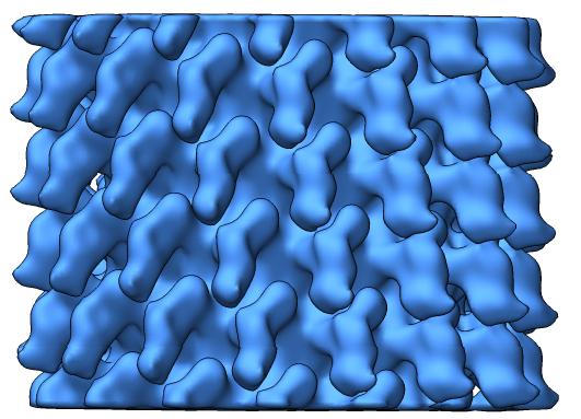 emd-1986.jpg emd-1986.jpg | 37.2 KB | ||
| Archive directory |  http://ftp.pdbj.org/pub/emdb/structures/EMD-1986ftp://ftp.pdbj.org/pub/emdb/structures/EMD-1986 http://ftp.pdbj.org/pub/emdb/structures/EMD-1986ftp://ftp.pdbj.org/pub/emdb/structures/EMD-1986 | HTTPS FTP |
-Related structure data
| Similar structure data |
|---|
-Links
| EMDB pages | EMDB (EBI/PDBe) / EMDataResource |
|---|
-Map
| File | Download / File: emd_1986.map.gz / Format: CCP4 / Size: 7.5 MB / Type: IMAGE STORED AS FLOATING POINT NUMBER (4 BYTES) | ||||||||||||||||||||||||||||||||||||||||||||||||||||||||||||||||||||
|---|---|---|---|---|---|---|---|---|---|---|---|---|---|---|---|---|---|---|---|---|---|---|---|---|---|---|---|---|---|---|---|---|---|---|---|---|---|---|---|---|---|---|---|---|---|---|---|---|---|---|---|---|---|---|---|---|---|---|---|---|---|---|---|---|---|---|---|---|---|
| Annotation | This is a map of the Marburg virus nucleocapsid within virions. | ||||||||||||||||||||||||||||||||||||||||||||||||||||||||||||||||||||
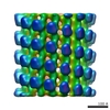
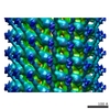
| Projections & slices | Image control
Images are generated by Spider. generated in cubic-lattice coordinate | ||||||||||||||||||||||||||||||||||||||||||||||||||||||||||||||||||||
| Voxel size | X=Y=Z: 4 Å | ||||||||||||||||||||||||||||||||||||||||||||||||||||||||||||||||||||
| Density |
| ||||||||||||||||||||||||||||||||||||||||||||||||||||||||||||||||||||
| Symmetry | Space group: 0 | ||||||||||||||||||||||||||||||||||||||||||||||||||||||||||||||||||||
| Details | EMDB XML:
CCP4 map header:
| ||||||||||||||||||||||||||||||||||||||||||||||||||||||||||||||||||||
 Z (Sec.)
 Y (Row.)
 X (Col.)


-Supplemental data
- Sample components
-Entire : Marburg virus nucleocapsid
| Entire | Name: Marburg virus nucleocapsid |
|---|---|
| Components |
|
-Supramolecule #1000: Marburg virus nucleocapsid
| Supramolecule | Name: Marburg virus nucleocapsid / type: sample / ID: 1000 / Number unique components: 1 |
|---|
-Supramolecule #1: Marburg marburgvirus
| Supramolecule | Name: Marburg marburgvirus / type: virus / ID: 1 / Name.synonym: Marburg virus / NCBI-ID: 11269 / Sci species name: Marburg marburgvirus / Database: NCBI / Virus type: VIRION / Virus isolate: STRAIN / Virus enveloped: Yes / Virus empty: No / Syn species name: Marburg virus |
|---|---|
| Host (natural) | Organism:  Homo sapiens (human) / synonym: VERTEBRATES Homo sapiens (human) / synonym: VERTEBRATES |
-Experimental details
-Structure determination
| Method | cryo EM |
|---|---|
 Processing Processing | helical reconstruction |
| Aggregation state | filament |
-Sample preparation
| Buffer | Details: PBS buffer with 4% PFA. |
|---|---|
| Vitrification | Cryogen name: ETHANE / Instrument: HOMEMADE PLUNGER / Details: Vitrification instrument: Plunge freezer / Method: Plunge freezing |
- Electron microscopy
| Microscope | FEI/PHILIPS CM120T |
|---|---|
| Image recording | Category: FILM / Film or detector model: KODAK SO-163 FILM / Digitization - Scanner: ZEISS SCAI / Digitization - Sampling interval: 14 µm / Average electron dose: 10 e/Å2 |
| Tilt angle min | 0 |
| Tilt angle max | 0 |
| Electron beam | Acceleration voltage: 120 kV / Electron source: LAB6 |
| Electron optics | Illumination mode: FLOOD BEAM / Imaging mode: BRIGHT FIELD / Cs: 6.3 mm / Nominal defocus max: 0.004 µm / Nominal defocus min: 0.0006 µm / Nominal magnification: 37000 |
| Sample stage | Specimen holder: Gatan 626 / Specimen holder model: GATAN LIQUID NITROGEN |
-Image processing
| Final reconstruction | Applied symmetry - Helical parameters - Δz: 5.0 Å Applied symmetry - Helical parameters - Δ&Phi: 24.3 ° Algorithm: OTHER / Resolution.type: BY AUTHOR / Resolution: 25.0 Å / Resolution method: FSC 0.5 CUT-OFF / Software - Name: Spider, Bsoft |
|---|---|
| CTF correction | Details: Phase flip |
-Atomic model buiding 1
| Initial model | PDB ID: |
|---|---|
| Software | Name:  Chimera Chimera |
| Details | Protocol: Rigid body. 4 monomers from the VSV Nucleoprotein structure were fitted in as a single rigid body without any corrections. |
| Refinement | Space: REAL / Protocol: RIGID BODY FIT |