 Movie
Movie Controller

[English] 日本語
 Yorodumi
- EMDB-1026: Microscopic evidence for a minus-end-directed power stroke in the... -
+ Open data

- Basic information
| Entry | Database: EMDB / ID: EMD-1026 | |||||||||
|---|---|---|---|---|---|---|---|---|---|---|
| Title | Microscopic evidence for a minus-end-directed power stroke in the kinesin motor ncd. | |||||||||
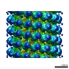
 Map data Map data | helical reconstruction of microtubule motor complex AMPPNP state | |||||||||
 Sample Sample |
| |||||||||
| Biological species |  | |||||||||
| Method | helical reconstruction / cryo EM / negative staining | |||||||||
 Authors Authors | Wendt TG | |||||||||
 Citation Citation | Journal: EMBO J / Year: 2002 Title: Microscopic evidence for a minus-end-directed power stroke in the kinesin motor ncd. Authors: Thomas G Wendt / Niels Volkmann / Georgios Skiniotis / Kenneth N Goldie / Jens Müller / Eckhard Mandelkow / Andreas Hoenger /  Abstract: We used cryo-electron microscopy and image reconstruction to investigate the structure and microtubule-binding configurations of dimeric non-claret disjunctional (ncd) motor domains under various ...We used cryo-electron microscopy and image reconstruction to investigate the structure and microtubule-binding configurations of dimeric non-claret disjunctional (ncd) motor domains under various nucleotide conditions, and applied molecular docking using ncd's dimeric X-ray structure to generate a mechanistic model for force transduction. To visualize the alpha-helical coiled-coil neck better, we engineered an SH3 domain to the N-terminal end of our ncd construct (296-700). Ncd exhibits strikingly different nucleotide-dependent three-dimensional conformations and microtubule-binding patterns from those of conventional kinesin. In the absence of nucleotide, the neck adapts a configuration close to that found in the X-ray structure with stable interactions between the neck and motor core domain. Minus-end-directed movement is based mainly on two key events: (i) the stable neck-core interactions in ncd generate a binding geometry between motor and microtubule which places the motor ahead of its cargo in the minus-end direction; and (ii) after the uptake of ATP, the two heads rearrange their position relative to each other in a way that promotes a swing of the neck in the minus-end direction. | |||||||||
| History |
|
- Structure visualization
| Movie |
 Movie viewer Movie viewer |
|---|---|
| Structure viewer | EM map: SurfViewMolmilJmol/JSmol |
| Supplemental images |
 UCSF Chimera
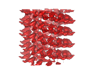
- Downloads & links
-EMDB archive
| Map data | emd_1026.map.gz | 602.2 KB | EMDB map data format | |
|---|---|---|---|---|
| Header (meta data) | emd-1026-v30.xmlemd-1026.xml | 10.3 KB 10.3 KB | Display Display | EMDB header |
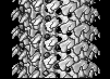
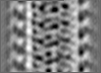
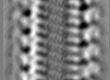
| Images |  1026.gif 1026.gif | 35.3 KB | ||
| Filedesc layerLines | emd_1026_ll.cif.gz | 12 KB | ||
| Archive directory |  http://ftp.pdbj.org/pub/emdb/structures/EMD-1026ftp://ftp.pdbj.org/pub/emdb/structures/EMD-1026 http://ftp.pdbj.org/pub/emdb/structures/EMD-1026ftp://ftp.pdbj.org/pub/emdb/structures/EMD-1026 | HTTPS FTP |
-Related structure data


-Links
| EMDB pages | EMDB (EBI/PDBe) / EMDataResource |
|---|
-Map
| File | Download / File: emd_1026.map.gz / Format: CCP4 / Size: 2.8 MB / Type: IMAGE STORED AS FLOATING POINT NUMBER (4 BYTES) | ||||||||||||||||||||||||||||||||||||||||||||||||||||||||||||
|---|---|---|---|---|---|---|---|---|---|---|---|---|---|---|---|---|---|---|---|---|---|---|---|---|---|---|---|---|---|---|---|---|---|---|---|---|---|---|---|---|---|---|---|---|---|---|---|---|---|---|---|---|---|---|---|---|---|---|---|---|---|
| Annotation | helical reconstruction of microtubule motor complex AMPPNP state | ||||||||||||||||||||||||||||||||||||||||||||||||||||||||||||
| Projections & slices | Image control
Images are generated by Spider. generated in cubic-lattice coordinate | ||||||||||||||||||||||||||||||||||||||||||||||||||||||||||||
| Voxel size | X=Y=Z: 5.53 Å | ||||||||||||||||||||||||||||||||||||||||||||||||||||||||||||
| Density |
| ||||||||||||||||||||||||||||||||||||||||||||||||||||||||||||
| Symmetry | Space group: 1 | ||||||||||||||||||||||||||||||||||||||||||||||||||||||||||||
| Details | EMDB XML:
CCP4 map header:
| ||||||||||||||||||||||||||||||||||||||||||||||||||||||||||||
 Z (Sec.)
 Y (Row.)
 X (Col.)


-Supplemental data
- Sample components
-Entire : Ncd296 motor fragment from Drosophila Melanogaster AMPPNP state
| Entire | Name: Ncd296 motor fragment from Drosophila Melanogaster AMPPNP state |
|---|---|
| Components |
|
-Supramolecule #1000: Ncd296 motor fragment from Drosophila Melanogaster AMPPNP state
| Supramolecule | Name: Ncd296 motor fragment from Drosophila Melanogaster AMPPNP state type: sample / ID: 1000 / Oligomeric state: dimer / Number unique components: 2 |
|---|
-Macromolecule #1: Ncd296 dimer
| Macromolecule | Name: Ncd296 dimer / type: protein_or_peptide / ID: 1 / Number of copies: 2 / Oligomeric state: dimer / Recombinant expression: Yes |
|---|---|
| Source (natural) | Organism: |
| Molecular weight | Experimental: 92 KDa |
| Recombinant expression | Organism:  |
-Macromolecule #2: tubulin dimer
| Macromolecule | Name: tubulin dimer / type: protein_or_peptide / ID: 2 / Name.synonym: alpha beta tubulin Details: bovine tubulin was purchased from Cytoskeleton Inc. (Denver,CO) Oligomeric state: polymer of alpha beta dimers / Recombinant expression: No / Database: NCBI |
|---|---|
| Source (natural) | Organism: |
| Molecular weight | Experimental: 100 KDa |
-Experimental details
-Structure determination
| Method | negative staining, cryo EM |
|---|---|
 Processing Processing | helical reconstruction |
| Aggregation state | filament |
-Sample preparation
| Concentration | 1.9 mg/mL |
|---|---|
| Buffer | pH: 6.8 / Details: 80mM PIPES, 2mM MgCl2, 2mM AMPPNP |
| Staining | Type: NEGATIVE / Details: plunge-frozen in liquid ethane |
| Grid | Details: quantifoil holey grids |
| Vitrification | Cryogen name: ETHANE / Chamber temperature: 93 K / Instrument: HOMEMADE PLUNGER Details: Vitrification instrument: standard guillotine. vitrification carried out at room temperature |
- Electron microscopy
| Microscope | FEI/PHILIPS CM200FEG/ST |
|---|---|
| Temperature | Average: 93 K |
| Alignment procedure | Legacy - Astigmatism: objective lens astigmatism was corrected at 150K mag |
| Details | low dose imaging |
| Image recording | Category: FILM / Film or detector model: KODAK SO-163 FILM / Digitization - Scanner: ZEISS SCAI / Digitization - Sampling interval: 21 µm / Number real images: 14 / Od range: 0.9 / Bits/pixel: 8 |
| Tilt angle min | 0 |
| Tilt angle max | 0 |
| Electron beam | Acceleration voltage: 160 kV / Electron source: TUNGSTEN HAIRPIN |
| Electron optics | Illumination mode: FLOOD BEAM / Imaging mode: BRIGHT FIELD / Cs: 1.26 mm / Nominal defocus max: 2.0 µm / Nominal defocus min: 1.5 µm / Nominal magnification: 38000 |
| Sample stage | Specimen holder: side entry / Specimen holder model: GATAN LIQUID NITROGEN |
-Image processing
| Details | 257 subunits per 137 helical turns |
|---|---|
| Final reconstruction | Applied symmetry - Helical parameters - Axial symmetry: C1 (asymmetric) Algorithm: OTHER / Software - Name: PHOELIX Details: final map was calculated from 25 datasets of near and far sides |
