 Movie
Movie Controller

[English] 日本語
 Yorodumi
- EMDB-19787: Methyl-coenzyme M reductase activation complex binding to the A2 ... -
+ Open data

- Basic information
| Entry |  | |||||||||
|---|---|---|---|---|---|---|---|---|---|---|
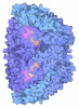
| Title | Methyl-coenzyme M reductase activation complex binding to the A2 component | |||||||||
 Map data Map data | ||||||||||
 Sample Sample |
| |||||||||
 Keywords Keywords | Methyl-coenzyme M reductase / activation complex / ATPase / Iron-sulfur clusters / OXIDOREDUCTASE | |||||||||
| Function / homology |  Function and homology information Function and homology informationorganic phosphonate catabolic process / coenzyme-B sulfoethylthiotransferase / coenzyme-B sulfoethylthiotransferase activity / methanogenesis / ATP hydrolysis activity / ATP binding / metal ion binding / cytoplasm Similarity search - Function | |||||||||
| Biological species |  Methanococcus maripaludis (archaea) Methanococcus maripaludis (archaea) | |||||||||
| Method | single particle reconstruction / cryo EM / Resolution: 2.56 Å | |||||||||
 Authors Authors | Ramirez-Amador F / Paul S / Kumar A / Schuller JM | |||||||||
| Funding support | European Union, 1 items
| |||||||||
 Citation Citation | Journal: Nature / Year: 2025 Title: Structure of the ATP-driven methyl-coenzyme M reductase activation complex. Authors: Fidel Ramírez-Amador / Sophia Paul / Anuj Kumar / Christian Lorent / Sebastian Keller / Stefan Bohn / Thinh Nguyen / Stefano Lometto / Dennis Vlegels / Jörg Kahnt / Darja Deobald / Frank ...Authors: Fidel Ramírez-Amador / Sophia Paul / Anuj Kumar / Christian Lorent / Sebastian Keller / Stefan Bohn / Thinh Nguyen / Stefano Lometto / Dennis Vlegels / Jörg Kahnt / Darja Deobald / Frank Abendroth / Olalla Vázquez / Georg Hochberg / Silvan Scheller / Sven T Stripp / Jan Michael Schuller /   Abstract: Methyl-coenzyme M reductase (MCR) is the enzyme responsible for nearly all biologically generated methane. Its active site comprises coenzyme F, a porphyrin-based cofactor with a central nickel ion ...Methyl-coenzyme M reductase (MCR) is the enzyme responsible for nearly all biologically generated methane. Its active site comprises coenzyme F, a porphyrin-based cofactor with a central nickel ion that is active exclusively in the Ni(I) state. How methanogenic archaea perform the reductive activation of F represents a major gap in our understanding of one of the most ancient bioenergetic systems in nature. Here we purified and characterized the MCR activation complex from Methanococcus maripaludis. McrC, a small subunit encoded in the mcr operon, co-purifies with the methanogenic marker proteins Mmp7, Mmp17, Mmp3 and the A2 component. We demonstrated that this complex can activate MCR in vitro in a strictly ATP-dependent manner, enabling the formation of methane. In addition, we determined the cryo-electron microscopy structure of the MCR activation complex exhibiting different functional states with local resolutions reaching 1.8-2.1 Å. Our data revealed three complex iron-sulfur clusters that formed an electron transfer pathway towards F. Topology and electron paramagnetic resonance spectroscopy analyses indicate that these clusters are similar to the [8Fe-9S-C] cluster, a maturation intermediate of the catalytic cofactor in nitrogenase. Altogether, our findings offer insights into the activation mechanism of MCR and prospects on the early evolution of nitrogenase. | |||||||||
| History |
|
- Structure visualization
| Supplemental images |
|---|

- Downloads & links
-EMDB archive
| Map data | emd_19787.map.gz | 211.7 MB | EMDB map data format | |
|---|---|---|---|---|
| Header (meta data) | emd-19787-v30.xmlemd-19787.xml | 37.5 KB 37.5 KB | Display Display | EMDB header |
| FSC (resolution estimation) | emd_19787_fsc_1.xmlemd_19787_fsc_2.xmlemd_19787_fsc_3.xmlemd_19787_fsc_4.xml | 15.8 KB 15.9 KB 15.8 KB 15.9 KB | Display Display Display Display | FSC data file |
| Images |  emd_19787.png emd_19787.png | 64.3 KB | ||
| Masks | emd_19787_msk_1.mapemd_19787_msk_2.mapemd_19787_msk_3.mapemd_19787_msk_4.mapemd_19787_msk_5.mapemd_19787_msk_6.map | 421.9 MB 421.9 MB 421.9 MB 421.9 MB 421.9 MB 421.9 MB | Mask map | |
| Filedesc metadata | emd-19787.cif.gz | 10.5 KB | ||
| Others | emd_19787_half_map_1.map.gzemd_19787_half_map_2.map.gz | 391.4 MB 391.4 MB | ||
| Archive directory |  http://ftp.pdbj.org/pub/emdb/structures/EMD-19787ftp://ftp.pdbj.org/pub/emdb/structures/EMD-19787 http://ftp.pdbj.org/pub/emdb/structures/EMD-19787ftp://ftp.pdbj.org/pub/emdb/structures/EMD-19787 | HTTPS FTP |
-Related structure data
| Related structure data |  8s7vMC  8s7xC  9h1lC M: atomic model generated by this map C: citing same article ( |
|---|---|
| Similar structure data |


-Links
| EMDB pages | EMDB (EBI/PDBe) / EMDataResource |
|---|---|
| Related items in Molecule of the Month |


-Map
| File | Download / File: emd_19787.map.gz / Format: CCP4 / Size: 421.9 MB / Type: IMAGE STORED AS FLOATING POINT NUMBER (4 BYTES) | ||||||||||||||||||||||||||||||||||||
|---|---|---|---|---|---|---|---|---|---|---|---|---|---|---|---|---|---|---|---|---|---|---|---|---|---|---|---|---|---|---|---|---|---|---|---|---|---|
| Projections & slices | Image control
Images are generated by Spider. | ||||||||||||||||||||||||||||||||||||
| Voxel size | X=Y=Z: 0.73 Å | ||||||||||||||||||||||||||||||||||||
| Density |
| ||||||||||||||||||||||||||||||||||||
| Symmetry | Space group: 1 | ||||||||||||||||||||||||||||||||||||
| Details | EMDB XML:
|
 Z (Sec.)
 Y (Row.)
 X (Col.)


-Supplemental data
-Mask #1
| File | emd_19787_msk_1.map | ||||||||||||
|---|---|---|---|---|---|---|---|---|---|---|---|---|---|
| Projections & Slices |
| ||||||||||||
| Density Histograms |


-Mask #2
| File | emd_19787_msk_2.map | ||||||||||||
|---|---|---|---|---|---|---|---|---|---|---|---|---|---|
| Projections & Slices |
| ||||||||||||
| Density Histograms |


-Mask #3
| File | emd_19787_msk_3.map | ||||||||||||
|---|---|---|---|---|---|---|---|---|---|---|---|---|---|
| Projections & Slices |
| ||||||||||||
| Density Histograms |


-Mask #4
| File | emd_19787_msk_4.map | ||||||||||||
|---|---|---|---|---|---|---|---|---|---|---|---|---|---|
| Projections & Slices |
| ||||||||||||
| Density Histograms |


-Mask #5
| File | emd_19787_msk_5.map | ||||||||||||
|---|---|---|---|---|---|---|---|---|---|---|---|---|---|
| Projections & Slices |
| ||||||||||||
| Density Histograms |


-Mask #6
| File | emd_19787_msk_6.map | ||||||||||||
|---|---|---|---|---|---|---|---|---|---|---|---|---|---|
| Projections & Slices |
| ||||||||||||
| Density Histograms |


-Half map: #2
| File | emd_19787_half_map_1.map | ||||||||||||
|---|---|---|---|---|---|---|---|---|---|---|---|---|---|
| Projections & Slices |
| ||||||||||||
| Density Histograms |


-Half map: #1
| File | emd_19787_half_map_2.map | ||||||||||||
|---|---|---|---|---|---|---|---|---|---|---|---|---|---|
| Projections & Slices |
| ||||||||||||
| Density Histograms |


- Sample components
+Entire : Methyl-coenzyme M reductase activation complex binding to A2 component
+Supramolecule #1: Methyl-coenzyme M reductase activation complex binding to A2 component
+Macromolecule #1: Methyl-coenzyme M reductase subunit gamma
+Macromolecule #2: Methyl-coenzyme M reductase subunit beta
+Macromolecule #3: Methyl-coenzyme M reductase subunit alpha
+Macromolecule #4: Methanogenesis marker protein 17
+Macromolecule #5: Methanogenesis marker protein 7
+Macromolecule #6: Methyl-coenzyme M reductase operon protein C
+Macromolecule #7: Glycine betaine/carnitine/choline transport ATP-binding protein OpuCA
+Macromolecule #8: UPF0288 protein MmarC6_0796
+Macromolecule #9: DUF2098 domain-containing protein
+Macromolecule #10: O-PHOSPHONO-N-{(2E)-7-[(2-SULFOETHYL)DITHIO]HEPT-2-ENOYL}-L-THREONINE
+Macromolecule #11: Coenzyme B
+Macromolecule #12: 1-THIOETHANESULFONIC ACID
+Macromolecule #13: FACTOR 430
+Macromolecule #14: FeFe cofactor
+Macromolecule #15: ZINC ION
+Macromolecule #16: ADENOSINE-5'-TRIPHOSPHATE

+Macromolecule #17: MAGNESIUM ION
-Experimental details
-Structure determination
| Method | cryo EM |
|---|---|
 Processing Processing | single particle reconstruction |
| Aggregation state | 3D array |
-Sample preparation
| Concentration | 1.25 mg/mL | |||||||||
|---|---|---|---|---|---|---|---|---|---|---|
| Buffer | pH: 7.6 Component:
| |||||||||
| Grid | Model: Quantifoil R1.2/1.3 / Material: COPPER / Mesh: 300 / Pretreatment - Type: GLOW DISCHARGE / Pretreatment - Time: 25 sec. / Details: 15 mA | |||||||||
| Vitrification | Cryogen name: ETHANE-PROPANE / Chamber humidity: 100 % / Chamber temperature: 277 K / Instrument: FEI VITROBOT MARK IV Details: Vitrification setup inside a Coy Lab's Vinyl Anaerobic Chamber to preserve the protein sample under strict anaerobic conditions (95% O2 / 5% H2).. |
- Electron microscopy
| Microscope | TFS KRIOS |
|---|---|
| Image recording | Film or detector model: FEI FALCON IV (4k x 4k) / Number real images: 17548 / Average electron dose: 60.0 e/Å2 Details: 7781 micrographs with a 20 deg pretilt to overcome preferred orientation problems |
| Electron beam | Acceleration voltage: 300 kV / Electron source:  FIELD EMISSION GUN FIELD EMISSION GUN |
| Electron optics | Illumination mode: FLOOD BEAM / Imaging mode: BRIGHT FIELD / Cs: 2.7 mm / Nominal defocus max: 2.0 µm / Nominal defocus min: 0.5 µm / Nominal magnification: 165000 |
| Experimental equipment |  Model: Titan Krios / Image courtesy: FEI Company |
+Image processing

-Atomic model buiding 1
| Initial model | Chain - Source name: AlphaFold / Chain - Initial model type: in silico model |
|---|---|
| Details | Rigid body fitting in Chimera and Coot. Further real-space refinement in Phenix. |
| Refinement | Space: REAL / Protocol: AB INITIO MODEL |
| Output model | PDB-8s7v: |
