 Movie
Movie Controller

[English] 日本語
 Yorodumi
- EMDB-19644: Tomographic reconstruction of the follicle cell nuclear periphery... -
+ Open data

- Basic information
| Entry | 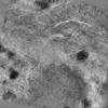 | |||||||||
|---|---|---|---|---|---|---|---|---|---|---|
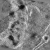
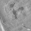
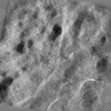
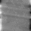
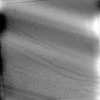
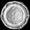
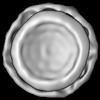
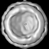
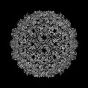
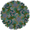
| Title | Tomographic reconstruction of the follicle cell nuclear periphery from high pressure frozen, freeze substituted and resin embedded D. melanogaster egg chamber | |||||||||
 Map data Map data | ||||||||||
 Sample Sample |
| |||||||||
 Keywords Keywords | Tomography / Drosophila / melanogaster / follicle cell / nuclear periphery / nucleus / freeze-substitution / VIRUS LIKE PARTICLE | |||||||||
| Biological species |  | |||||||||
| Method | electron tomography / cryo EM / negative staining | |||||||||
 Authors Authors | Klumpe S / Hampoelz B / Ronchi P / Beck M / Plitzko J | |||||||||
| Funding support |  Germany, 1 items Germany, 1 items
| |||||||||
 Citation Citation | Journal: Cell / Year: 2025 Title: In-cell structure and snapshots of copia retrotransposons in intact tissue by cryo-ET. Authors: Sven Klumpe / Kirsten A Senti / Florian Beck / Jenny Sachweh / Bernhard Hampoelz / Paolo Ronchi / Viola Oorschot / Marlene Brandstetter / Assa Yeroslaviz / John A G Briggs / Julius Brennecke ...Authors: Sven Klumpe / Kirsten A Senti / Florian Beck / Jenny Sachweh / Bernhard Hampoelz / Paolo Ronchi / Viola Oorschot / Marlene Brandstetter / Assa Yeroslaviz / John A G Briggs / Julius Brennecke / Martin Beck / Jürgen M Plitzko /  Abstract: Long terminal repeat (LTR) retrotransposons belong to the transposable elements (TEs), autonomously replicating genetic elements that integrate into the host's genome. Among animals, Drosophila ...Long terminal repeat (LTR) retrotransposons belong to the transposable elements (TEs), autonomously replicating genetic elements that integrate into the host's genome. Among animals, Drosophila melanogaster serves as an important model organism for TE research and contains several LTR retrotransposons, including the Ty1-copia family, which is evolutionarily related to retroviruses and forms virus-like particles (VLPs). In this study, we use cryo-focused ion beam (FIB) milling and lift-out approaches to visualize copia VLPs in ovarian cells and intact egg chambers, resolving the in situ copia capsid structure to 7.7 Å resolution by cryoelectron tomography (cryo-ET). Although cytoplasmic copia VLPs vary in size, nuclear VLPs are homogeneous and form densely packed clusters, supporting a model in which nuclear import acts as a size selector. Analyzing flies deficient in the TE-suppressing PIWI-interacting RNA (piRNA) pathway, we observe copia's translocation into the nucleus during spermatogenesis. Our findings provide insights into the replication cycle and cellular structural biology of an active LTR retrotransposon. | |||||||||
| History |
|
- Structure visualization
| Supplemental images |
|---|

- Downloads & links
-EMDB archive
| Map data | emd_19644.map.gz | 1.1 GB |  EMDB map data format EMDB map data format | |
|---|---|---|---|---|
| Header (meta data) | emd-19644-v30.xmlemd-19644.xml | 9.8 KB 9.8 KB | Display Display | EMDB header |
| Images |  emd_19644.png emd_19644.png | 230.3 KB | ||
| Filedesc metadata | emd-19644.cif.gz | 3.9 KB | ||
| Archive directory |  http://ftp.pdbj.org/pub/emdb/structures/EMD-19644ftp://ftp.pdbj.org/pub/emdb/structures/EMD-19644 http://ftp.pdbj.org/pub/emdb/structures/EMD-19644ftp://ftp.pdbj.org/pub/emdb/structures/EMD-19644 | HTTPS FTP |
-Validation report
| Summary document | emd_19644_validation.pdf.gz | 507.2 KB | Display | EMDB validaton report |
|---|---|---|---|---|
| Full document | emd_19644_full_validation.pdf.gz | 506.7 KB | Display | |
| Data in XML | emd_19644_validation.xml.gz | 4 KB | Display | |
| Data in CIF | emd_19644_validation.cif.gz | 4.7 KB | Display | |
| Arichive directory | https://ftp.pdbj.org/pub/emdb/validation_reports/EMD-19644ftp://ftp.pdbj.org/pub/emdb/validation_reports/EMD-19644 | HTTPS FTP |
-Related structure data
-Links
| EMDB pages | EMDB (EBI/PDBe) / EMDataResource |
|---|
-Map
| File | Download / File: emd_19644.map.gz / Format: CCP4 / Size: 1.5 GB / Type: IMAGE STORED AS SIGNED INTEGER (2 BYTES) | ||||||||||||||||||||||||||||||||
|---|---|---|---|---|---|---|---|---|---|---|---|---|---|---|---|---|---|---|---|---|---|---|---|---|---|---|---|---|---|---|---|---|---|
| Projections & slices | Image control
Images are generated by Spider. generated in cubic-lattice coordinate | ||||||||||||||||||||||||||||||||
| Voxel size | X=Y=Z: 12.2 Å | ||||||||||||||||||||||||||||||||
| Density |
| ||||||||||||||||||||||||||||||||
| Symmetry | Space group: 1 | ||||||||||||||||||||||||||||||||
| Details | EMDB XML:
|
 Z (Sec.)
 Y (Row.)
 X (Col.)


-Supplemental data
- Sample components
-Entire : D. melanogaster egg chamber from tj>Sec31::RFP flies
| Entire | Name: D. melanogaster egg chamber from tj>Sec31::RFP flies |
|---|---|
| Components |
|
-Supramolecule #1: D. melanogaster egg chamber from tj>Sec31::RFP flies
| Supramolecule | Name: D. melanogaster egg chamber from tj>Sec31::RFP flies / type: tissue / ID: 1 / Parent: 0 Details: Females from F1 scored against CyO from following genotypes: traffic jam-GAL4/CyO;; and w* P{UAS-Sec31.RFP}13.5; snaSco/CyO, P{ftz-lacB}E3 (Bloomington #86533); |
|---|---|
| Source (natural) | Organism: |
-Experimental details
-Structure determination
| Method | negative staining, cryo EM |
|---|---|
 Processing Processing | electron tomography |
| Aggregation state | tissue |
-Sample preparation
| Buffer | pH: 7.4 |
|---|---|
| Staining | Type: NEGATIVE / Material: Uranyl Acetate |
| Sugar embedding | Material: Lowicryl HM20 |
| Vitrification | Cryogen name: NITROGEN |
| Details | Dissected egg chambers |
| High pressure freezing | Instrument: OTHER Details: The value given for _em_high_pressure_freezing.instrument is HPM 010. This is not in a list of allowed values {'EMS-002 RAPID IMMERSION FREEZER', 'BAL-TEC HPM 010', 'OTHER', 'LEICA EM ...Details: The value given for _em_high_pressure_freezing.instrument is HPM 010. This is not in a list of allowed values {'EMS-002 RAPID IMMERSION FREEZER', 'BAL-TEC HPM 010', 'OTHER', 'LEICA EM HPM100', 'LEICA EM PACT2', 'LEICA EM PACT'} so OTHER is written into the XML file. |
| Cryo protectant | 20% Ficoll 70 kDa |
| Sectioning | Ultramicrotomy - Instrument: Leica EM UC7 / Ultramicrotomy - Temperature: 293 K / Ultramicrotomy - Final thickness: 300 |
- Electron microscopy
| Microscope | FEI TECNAI F30 |
|---|---|
| Image recording | Film or detector model: OTHER / Average electron dose: 10.0 e/Å2 / Details: Gatan OneView |
| Electron beam | Acceleration voltage: 300 kV / Electron source:  FIELD EMISSION GUN FIELD EMISSION GUN |
| Electron optics | Illumination mode: FLOOD BEAM / Imaging mode: BRIGHT FIELD / Nominal defocus max: 0.5 µm / Nominal defocus min: 0.5 µm |
| Experimental equipment |  Model: Tecnai F30 / Image courtesy: FEI Company |
-Image processing
| Final reconstruction | Software - Name: IMOD / Number images used: 121 |
|---|
