Journal: Nat Cell Biol / Year: 2025 Title: In situ architecture of the human prohibitin complex. Authors: Felix Lange / Michael Ratz / Jan-Niklas Dohrke / Maxence Le Vasseur / Dirk Wenzel / Peter Ilgen / Dietmar Riedel / Stefan Jakobs / Abstract: Prohibitins are a highly conserved family of proteins that have been implicated in a variety of functions including mitochondrial stress signalling and housekeeping, cell cycle progression, ...Prohibitins are a highly conserved family of proteins that have been implicated in a variety of functions including mitochondrial stress signalling and housekeeping, cell cycle progression, apoptosis, lifespan regulation and many others. The human prohibitins prohibitin 1 and prohibitin 2 have been proposed to act as scaffolds within the mitochondrial inner membrane, but their molecular organization has remained elusive. Here we determined the molecular organization of the human prohibitin complex within the mitochondrial inner membrane using an integrative structural biology approach combining quantitative western blotting, cryo-electron tomography, subtomogram averaging and molecular modelling. The proposed bell-shaped structure consists of 11 alternating prohibitin 1 and prohibitin 2 molecules. This study reveals an average of about 43 prohibitin complexes per crista, covering 1-3% of the crista membrane area. These findings provide a structural basis for understanding the functional contributions of prohibitins to the integrity and spatial organization of the mitochondrial inner membrane.
Entire : human prohibitin complex formed by PHB1 and PHB2
Entire
Name: human prohibitin complex formed by PHB1 and PHB2
Components
Cell: human prohibitin complex formed by PHB1 and PHB2
Protein or peptide: Prohibitin 1
Protein or peptide: Prohibitin-2
-
Supramolecule #1: human prohibitin complex formed by PHB1 and PHB2
Supramolecule
Name: human prohibitin complex formed by PHB1 and PHB2 / type: cell / ID: 1 / Parent: 0 / Macromolecule list: all Details: 11 molecules of PHB1 and PHB2 form the human prohibitin complex
Source (natural)
Organism: Homo sapiens (human)
-
Macromolecule #1: Prohibitin 1
Macromolecule
Name: Prohibitin 1 / type: protein_or_peptide / ID: 1 / Number of copies: 6 / Enantiomer: LEVO
Cryogen name: ETHANE / Chamber humidity: 95 % / Chamber temperature: 277.15 K / Instrument: FEI VITROBOT MARK IV
-
Electron microscopy
Microscope
FEI TITAN KRIOS
Temperature
Min: 80.0 K / Max: 93.0 K
Specialist optics
Energy filter - Name: GIF Bioquantum / Energy filter - Slit width: 20 eV
Image recording
Film or detector model: GATAN K3 BIOQUANTUM (6k x 4k) / Digitization - Dimensions - Width: 5760 pixel / Digitization - Dimensions - Height: 4092 pixel / Number real images: 4 / Average exposure time: 0.53 sec. / Average electron dose: 120.0 e/Å2
Electron beam
Acceleration voltage: 300 kV / Electron source: FIELD EMISSION GUN
In the structure databanks used in Yorodumi, some data are registered as the other names, "COVID-19 virus" and "2019-nCoV". Here are the details of the virus and the list of structure data.
Jan 31, 2019. EMDB accession codes are about to change! (news from PDBe EMDB page)
EMDB accession codes are about to change! (news from PDBe EMDB page)
The allocation of 4 digits for EMDB accession codes will soon come to an end. Whilst these codes will remain in use, new EMDB accession codes will include an additional digit and will expand incrementally as the available range of codes is exhausted. The current 4-digit format prefixed with “EMD-” (i.e. EMD-XXXX) will advance to a 5-digit format (i.e. EMD-XXXXX), and so on. It is currently estimated that the 4-digit codes will be depleted around Spring 2019, at which point the 5-digit format will come into force.
The EM Navigator/Yorodumi systems omit the EMD- prefix.
Related info.:Q: What is EMD? / ID/Accession-code notation in Yorodumi/EM Navigator
Yorodumi is a browser for structure data from EMDB, PDB, SASBDB, etc.
This page is also the successor to EM Navigator detail page, and also detail information page/front-end page for Omokage search.
The word "yorodu" (or yorozu) is an old Japanese word meaning "ten thousand". "mi" (miru) is to see.
Related info.:EMDB / PDB / SASBDB / Comparison of 3 databanks / Yorodumi Search / Aug 31, 2016. New EM Navigator & Yorodumi / Yorodumi Papers / Jmol/JSmol / Function and homology information / Changes in new EM Navigator and Yorodumi
 Movie
Movie Controller


 Open data
 Basic information
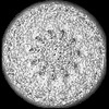
 Map data
 Sample
 Keywords
 Function and homology information
 Homo sapiens (human)
 Authors
 Germany, European Union, 5 items
  Citation

 Structure visualization
 Downloads & links
 emd_19459.png
 http://ftp.pdbj.org/pub/emdb/structures/EMD-19459
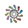

 Z (Sec.)
 Y (Row.)
 X (Col.)


 Sample components
 Processing
 Electron microscopy
 FIELD EMISSION GUN

