 Movie
Movie Controller

[English] 日本語
 Yorodumi
- EMDB-19340: SPNS2:sfGFP hetero dimer assembled by Di-Gluebody - sfGFP local r... -
+ Open data

- Basic information
| Entry |  | |||||||||
|---|---|---|---|---|---|---|---|---|---|---|
| Title | SPNS2:sfGFP hetero dimer assembled by Di-Gluebody - sfGFP local refinement | |||||||||
 Map data Map data | EMhancer: SPNS2:sfGFP hetero dimer assembled by Di-Gluebody - sfGFP local refinement | |||||||||
 Sample Sample |
| |||||||||
 Keywords Keywords | DNA helicase / Di-Gluebody / HYDROLASE | |||||||||
| Function / homology | Green fluorescent protein, GFP / Green fluorescent protein-related / Green fluorescent protein / Green fluorescent protein / bioluminescence / generation of precursor metabolites and energy / Green fluorescent protein Function and homology information Function and homology information | |||||||||
| Biological species |   Aequorea victoria (jellyfish) / Aequorea victoria (jellyfish) /  | |||||||||
| Method | single particle reconstruction / cryo EM / Resolution: 3.75 Å | |||||||||
 Authors Authors | Yi G / Ye M / Mamalis D / Sauer DB / von Delft F / Davis BG / Gilbert RJC | |||||||||
| Funding support |  United Kingdom, 1 items United Kingdom, 1 items
| |||||||||
 Citation Citation | Journal: Nat Chem Biol / Year: 2026 Title: Covalently constrained 'Di-Gembodies' enable parallel structure solutions by cryo-EM. Authors: Gangshun Yi / Dimitrios Mamalis / Mingda Ye / Loic Carrique / Michael Fairhead / Huanyu Li / Katharina L Duerr / Peijun Zhang / David B Sauer / Frank von Delft / Benjamin G Davis / Robert J C Gilbert /  Abstract: Whilst cryo-electron microscopy(cryo-EM) has become a routine methodology in structural biology, obtaining high-resolution cryo-EM structures of small proteins (<100 kDa) and increasing overall throughput remain challenging. One approach to augment protein size and improve particle alignment involves the use of binding proteins or protein-based scaffolds. However, a given imaging scaffold or linking module may prove inadequate for structure solution and availability of such scaffolds remains limited. Here, we describe a strategy that exploits covalent dimerization of nanobodies to trap an engineered, predisposed nanobody-to-nanobody interface, giving Di-Gembodies as modular constructs created in homomeric and heteromeric forms. By exploiting side-chain-to-side-chain assembly, they can simultaneously display two copies of the same or two distinct proteins through a subunit interface that provides sufficient constraint required for cryo-EM structure determination. We validate this method with multiple soluble and membrane structural targets, down to 14 kDa, demonstrating a flexible and scalable platform for expanded protein structure determination. | |||||||||
| History |
|
- Structure visualization
| Supplemental images |
|---|

- Downloads & links
-EMDB archive
| Map data | emd_19340.map.gz | 151.8 MB | EMDB map data format | |
|---|---|---|---|---|
| Header (meta data) | emd-19340-v30.xmlemd-19340.xml | 28.9 KB 28.9 KB | Display Display | EMDB header |
| FSC (resolution estimation) | emd_19340_fsc.xml | 11.6 KB | Display | FSC data file |
| Images |  emd_19340.png emd_19340.png | 93.2 KB | ||
| Masks | emd_19340_msk_1.map | 163.6 MB | Mask map | |
| Filedesc metadata | emd-19340.cif.gz | 7 KB | ||
| Others | emd_19340_additional_1.map.gzemd_19340_half_map_1.map.gzemd_19340_half_map_2.map.gz | 154.3 MB 152 MB 152 MB | ||
| Archive directory |  http://ftp.pdbj.org/pub/emdb/structures/EMD-19340ftp://ftp.pdbj.org/pub/emdb/structures/EMD-19340 http://ftp.pdbj.org/pub/emdb/structures/EMD-19340ftp://ftp.pdbj.org/pub/emdb/structures/EMD-19340 | HTTPS FTP |
-Related structure data
| Related structure data |  8rleMC  8rl5C  8rl6C  8rl7C  8rl8C  8rl9C  8rlaC  8rlbC  8rlcC  8rldC 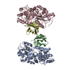 9fgvC  9fgxC  9fgyC  9fkqC M: atomic model generated by this map C: citing same article ( |
|---|---|
| Similar structure data |


-Links
| EMDB pages | EMDB (EBI/PDBe) / EMDataResource |
|---|---|
| Related items in Molecule of the Month |


-Map
| File | Download / File: emd_19340.map.gz / Format: CCP4 / Size: 163.6 MB / Type: IMAGE STORED AS FLOATING POINT NUMBER (4 BYTES) | ||||||||||||||||||||||||||||||||||||
|---|---|---|---|---|---|---|---|---|---|---|---|---|---|---|---|---|---|---|---|---|---|---|---|---|---|---|---|---|---|---|---|---|---|---|---|---|---|
| Annotation | EMhancer: SPNS2:sfGFP hetero dimer assembled by Di-Gluebody - sfGFP local refinement | ||||||||||||||||||||||||||||||||||||
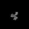
| Projections & slices | Image control
Images are generated by Spider. | ||||||||||||||||||||||||||||||||||||
| Voxel size | X=Y=Z: 0.932 Å | ||||||||||||||||||||||||||||||||||||
| Density |
| ||||||||||||||||||||||||||||||||||||
| Symmetry | Space group: 1 | ||||||||||||||||||||||||||||||||||||
| Details | EMDB XML:
|
 Z (Sec.)
 Y (Row.)
 X (Col.)


-Supplemental data
-Mask #1
| File | emd_19340_msk_1.map | ||||||||||||
|---|---|---|---|---|---|---|---|---|---|---|---|---|---|
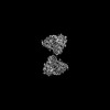
| Projections & Slices |
| ||||||||||||
| Density Histograms |


-Additional map: SPNS2:sfGFP hetero dimer assembled by Di-Gluebody - sfGFP...
| File | emd_19340_additional_1.map | ||||||||||||
|---|---|---|---|---|---|---|---|---|---|---|---|---|---|
| Annotation | SPNS2:sfGFP hetero dimer assembled by Di-Gluebody - sfGFP local refinement | ||||||||||||
| Projections & Slices |
| ||||||||||||
| Density Histograms |


-Half map: half map A
| File | emd_19340_half_map_1.map | ||||||||||||
|---|---|---|---|---|---|---|---|---|---|---|---|---|---|
| Annotation | half map A | ||||||||||||
| Projections & Slices |
| ||||||||||||
| Density Histograms |


-Half map: half map B
| File | emd_19340_half_map_2.map | ||||||||||||
|---|---|---|---|---|---|---|---|---|---|---|---|---|---|
| Annotation | half map B | ||||||||||||
| Projections & Slices |
| ||||||||||||
| Density Histograms |


- Sample components
-Entire : Local refinement of GFP part of the SPNS2:sfGFP heterodimer assem...
| Entire | Name: Local refinement of GFP part of the SPNS2:sfGFP heterodimer assembled by Di-Gluebody |
|---|---|
| Components |
|
-Supramolecule #1: Local refinement of GFP part of the SPNS2:sfGFP heterodimer assem...
| Supramolecule | Name: Local refinement of GFP part of the SPNS2:sfGFP heterodimer assembled by Di-Gluebody type: complex / ID: 1 / Parent: 0 / Macromolecule list: all Details: GbEnhancer and GbC4 were assembled via a disulfide to form a hetero Di-Gluebody |
|---|---|
| Molecular weight | Theoretical: 112 KDa |
-Supramolecule #2: Green fluorescent protein
| Supramolecule | Name: Green fluorescent protein / type: complex / ID: 2 / Parent: 1 / Details: Green fluorescent protein |
|---|---|
| Source (natural) | Organism: Aequorea victoria (jellyfish) |
-Supramolecule #3: Di-Gluebody
| Supramolecule | Name: Di-Gluebody / type: complex / ID: 3 / Parent: 1 / Details: Gluebody GbEnhancer and Gluebody GbC4 |
|---|---|
| Source (natural) | Organism: |
-Macromolecule #1: Green fluorescent protein
| Macromolecule | Name: Green fluorescent protein / type: protein_or_peptide / ID: 1 / Number of copies: 1 / Enantiomer: LEVO |
|---|---|
| Source (natural) | Organism: Aequorea victoria (jellyfish) |
| Molecular weight | Theoretical: 26.81923 KDa |
| Recombinant expression | Organism:  |
| Sequence | String: MSKGEELFTG VVPILVELDG DVNGHKFSVR GEGEGDATNG KLTLKFICTT GKLPVPWPTL VTTLTYGVQC FSRYPDHMKR HDFFKSAMP EGYVQERTIS FKDDGTYKTR AEVKFEGDTL VNRIELKGID FKEDGNILGH KLEYNFNSHN VYITADKQKN G IKANFKIR ...String: MSKGEELFTG VVPILVELDG DVNGHKFSVR GEGEGDATNG KLTLKFICTT GKLPVPWPTL VTTLTYGVQC FSRYPDHMKR HDFFKSAMP EGYVQERTIS FKDDGTYKTR AEVKFEGDTL VNRIELKGID FKEDGNILGH KLEYNFNSHN VYITADKQKN G IKANFKIR HNVEDGSVQL ADHYQQNTPI GDGPVLLPDN HYLSTQSVLS KDPNEKRDHM VLLEFVTAAG ITHGMDELYK UniProtKB: Green fluorescent protein |
-Macromolecule #2: Gluebody GbEnhancer
| Macromolecule | Name: Gluebody GbEnhancer / type: protein_or_peptide / ID: 2 / Number of copies: 1 / Enantiomer: LEVO |
|---|---|
| Source (natural) | Organism: |
| Molecular weight | Theoretical: 12.689139 KDa |
| Recombinant expression | Organism: |
| Sequence | String: QVQLVENGGA CVKPGGSLRL SCAASGFPVN RYSMRWYRQA PGKEREWVAG MSSAGDRSSY EDSVKGRFTI SRDDARNTVY LQMNSLKPE DTAVYYCNVN VGFEYWGQGT QVMVS |
-Macromolecule #3: Gluebody GbC4
| Macromolecule | Name: Gluebody GbC4 / type: protein_or_peptide / ID: 3 / Number of copies: 1 / Enantiomer: LEVO |
|---|---|
| Source (natural) | Organism: |
| Molecular weight | Theoretical: 12.982609 KDa |
| Recombinant expression | Organism: |
| Sequence | String: SQGQLVENGG GCVKAGGSLR LSCAASQGTL SNLVTGWFRR APGKEREFVA NIGRDGLTVY SNSVKGRFTI SRDRAKNTVY LQMDSLKPE DTAVYYCAGR LSRFPGEYDY WSKGTPVMVS |
-Experimental details
-Structure determination
| Method | cryo EM |
|---|---|
 Processing Processing | single particle reconstruction |
| Aggregation state | particle |
-Sample preparation
| Concentration | 4.2 mg/mL | ||||||||||||
|---|---|---|---|---|---|---|---|---|---|---|---|---|---|
| Buffer | pH: 7.5 Component:
| ||||||||||||
| Grid | Model: Quantifoil R1.2/1.3 / Material: COPPER / Mesh: 300 / Pretreatment - Type: GLOW DISCHARGE | ||||||||||||
| Vitrification | Cryogen name: ETHANE |
- Electron microscopy
| Microscope | FEI TITAN KRIOS |
|---|---|
| Image recording | Film or detector model: FEI FALCON IV (4k x 4k) / Number real images: 8674 / Average electron dose: 50.0 e/Å2 |
| Electron beam | Acceleration voltage: 300 kV / Electron source:  FIELD EMISSION GUN FIELD EMISSION GUN |
| Electron optics | Illumination mode: FLOOD BEAM / Imaging mode: BRIGHT FIELD / Nominal defocus max: 2.5 µm / Nominal defocus min: 1.5 µm |
| Experimental equipment |  Model: Titan Krios / Image courtesy: FEI Company |


