 Movie
Movie Controller

[English] 日本語
 Yorodumi
- EMDB-1860: Visualising an RNA genome poised for release from its receptor co... -
+ Open data

- Basic information
| Entry | Database: EMDB / ID: EMD-1860 | |||||||||
|---|---|---|---|---|---|---|---|---|---|---|
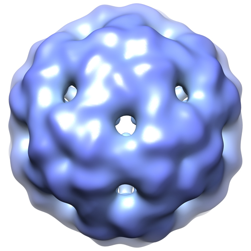
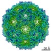
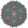
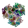
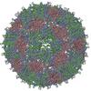
| Title | Visualising an RNA genome poised for release from its receptor complex. | |||||||||
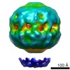
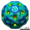
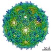
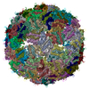
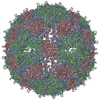
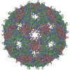
 Map data Map data | This is a cryo-EM icosahedrally-averaged reconstruction of bacteriophage MS2 bound to an F-pilus with a 5-fold axis. | |||||||||
 Sample Sample |
| |||||||||
 Keywords Keywords | MS2 / F-pili / virus-receptor complex | |||||||||
| Biological species |   Enterobacterio phage MS2 (virus) Enterobacterio phage MS2 (virus) | |||||||||
| Method | single particle reconstruction / cryo EM / Resolution: 19.0 Å | |||||||||
 Authors Authors | Toropova K / Stockley PG / Ranson NA | |||||||||
 Citation Citation | Journal: J Mol Biol / Year: 2011 Title: Visualising a viral RNA genome poised for release from its receptor complex. Authors: Katerina Toropova / Peter G Stockley / Neil A Ranson /  Abstract: We describe the cryo-electron microscopy structure of bacteriophage MS2 bound to its receptor, the bacterial F-pilus. The virus contacts the pilus at a capsid 5-fold vertex, thus locating the surface- ...We describe the cryo-electron microscopy structure of bacteriophage MS2 bound to its receptor, the bacterial F-pilus. The virus contacts the pilus at a capsid 5-fold vertex, thus locating the surface-accessible portion of the single copy of the pilin-binding maturation protein present in virions. This arrangement allows a 5-fold averaged map to be calculated, showing for the first time in any virus-receptor complex the nonuniform distribution of RNA within the capsid. Strikingly, at the vertex that contacts the pilus, a rod of density that may include contributions from both genome and maturation protein sits above a channel that goes through the capsid to the outside. This density is reminiscent of the DNA density observed in the exit channel of double-stranded DNA phages, suggesting that the RNA-maturation protein complex is poised to leave the capsid as the first step of the infection process. | |||||||||
| History |
|
- Structure visualization
| Movie |
 Movie viewer Movie viewer |
|---|---|
| Structure viewer | EM map: SurfViewMolmilJmol/JSmol |
| Supplemental images |
- Downloads & links
-EMDB archive
| Map data | emd_1860.map.gz | 25.1 MB | EMDB map data format | |
|---|---|---|---|---|
| Header (meta data) | emd-1860-v30.xmlemd-1860.xml | 9 KB 9 KB | Display Display | EMDB header |
| Images |  1860_1860_emdb.jpg 1860_1860_emdb.jpg | 116.6 KB | ||
| Archive directory |  http://ftp.pdbj.org/pub/emdb/structures/EMD-1860ftp://ftp.pdbj.org/pub/emdb/structures/EMD-1860 http://ftp.pdbj.org/pub/emdb/structures/EMD-1860ftp://ftp.pdbj.org/pub/emdb/structures/EMD-1860 | HTTPS FTP |
-Validation report
| Summary document | emd_1860_validation.pdf.gz | 211.8 KB | Display | EMDB validaton report |
|---|---|---|---|---|
| Full document | emd_1860_full_validation.pdf.gz | 210.9 KB | Display | |
| Data in XML | emd_1860_validation.xml.gz | 6.2 KB | Display | |
| Arichive directory | https://ftp.pdbj.org/pub/emdb/validation_reports/EMD-1860ftp://ftp.pdbj.org/pub/emdb/validation_reports/EMD-1860 | HTTPS FTP |
-Related structure data

-Links
| EMDB pages | EMDB (EBI/PDBe) / EMDataResource |
|---|
-Map
| File | Download / File: emd_1860.map.gz / Format: CCP4 / Size: 29.8 MB / Type: IMAGE STORED AS FLOATING POINT NUMBER (4 BYTES) | ||||||||||||||||||||||||||||||||||||||||||||||||||||||||||||||||||||
|---|---|---|---|---|---|---|---|---|---|---|---|---|---|---|---|---|---|---|---|---|---|---|---|---|---|---|---|---|---|---|---|---|---|---|---|---|---|---|---|---|---|---|---|---|---|---|---|---|---|---|---|---|---|---|---|---|---|---|---|---|---|---|---|---|---|---|---|---|---|
| Annotation | This is a cryo-EM icosahedrally-averaged reconstruction of bacteriophage MS2 bound to an F-pilus with a 5-fold axis. | ||||||||||||||||||||||||||||||||||||||||||||||||||||||||||||||||||||
| Projections & slices | Image control
Images are generated by Spider. | ||||||||||||||||||||||||||||||||||||||||||||||||||||||||||||||||||||
| Voxel size | X=Y=Z: 1.96 Å | ||||||||||||||||||||||||||||||||||||||||||||||||||||||||||||||||||||
| Density |
| ||||||||||||||||||||||||||||||||||||||||||||||||||||||||||||||||||||
| Symmetry | Space group: 1 | ||||||||||||||||||||||||||||||||||||||||||||||||||||||||||||||||||||
| Details | EMDB XML:
CCP4 map header:
| ||||||||||||||||||||||||||||||||||||||||||||||||||||||||||||||||||||
 Z (Sec.)
 Y (Row.)
 X (Col.)


-Supplemental data
- Sample components
-Entire : Bacteriophage MS2 in complex with F-pilus
| Entire | Name: Bacteriophage MS2 in complex with F-pilus |
|---|---|
| Components |
|
-Supramolecule #1000: Bacteriophage MS2 in complex with F-pilus
| Supramolecule | Name: Bacteriophage MS2 in complex with F-pilus / type: sample / ID: 1000 Oligomeric state: One to one, icosahedral virus bound to filamentous pilus Number unique components: 2 |
|---|
-Supramolecule #1: Enterobacterio phage MS2
| Supramolecule | Name: Enterobacterio phage MS2 / type: virus / ID: 1 / Name.synonym: MS2 Details: MS2 in complex with F-pilus, icosahedrally-averaged. NCBI-ID: 12022 / Sci species name: Enterobacterio phage MS2 / Virus type: VIRION / Virus isolate: STRAIN / Virus enveloped: No / Virus empty: No / Syn species name: MS2 |
|---|---|
| Host (natural) | Organism: |
| Virus shell | Shell ID: 1 / Diameter: 280 Å / T number (triangulation number): 3 |
-Supramolecule #2: F-PILUS
| Supramolecule | Name: F-PILUS / type: organelle_or_cellular_component / ID: 2 / Name.synonym: F-PILUS / Oligomeric state: helical multimer / Recombinant expression: No |
|---|---|
| Source (natural) | Organism: |
-Experimental details
-Structure determination
| Method | cryo EM |
|---|---|
 Processing Processing | single particle reconstruction |
| Aggregation state | particle |
-Sample preparation
| Buffer | pH: 7.8 / Details: 0.1 M Tris-HCl, 0.5 mM EDTA |
|---|---|
| Grid | Details: Lacey carbon copper mesh grid |
| Vitrification | Cryogen name: ETHANE / Chamber temperature: 22 K / Instrument: HOMEMADE PLUNGER Details: Vitrification instrument: Double-sided custom pneumatic blotter Method: 1.6s blot |
- Electron microscopy
| Microscope | FEI TECNAI 20 |
|---|---|
| Image recording | Category: FILM / Film or detector model: KODAK SO-163 FILM / Digitization - Scanner: OTHER / Digitization - Sampling interval: 9.88 µm / Number real images: 252 / Average electron dose: 18 e/Å2 / Od range: 1 / Bits/pixel: 16 |
| Electron beam | Acceleration voltage: 200 kV / Electron source:  FIELD EMISSION GUN FIELD EMISSION GUN |
| Electron optics | Calibrated magnification: 50400 / Illumination mode: FLOOD BEAM / Imaging mode: BRIGHT FIELD / Cs: 2.0 mm / Nominal magnification: 50000 |
| Sample stage | Specimen holder: Side entry / Specimen holder model: GATAN LIQUID NITROGEN |
-Image processing
| CTF correction | Details: Phase flipping each particle |
|---|---|
| Final reconstruction | Applied symmetry - Point group: I (icosahedral) / Algorithm: OTHER / Resolution.type: BY AUTHOR / Resolution: 19.0 Å / Resolution method: FSC 0.5 CUT-OFF / Software - Name: Spider / Number images used: 2739 |
| Final two d classification | Number classes: 78 |
