ジャーナル: Nat Microbiol / 年: 2024 タイトル: Molecular plasticity of herpesvirus nuclear egress analysed in situ. 著者: Vojtěch Pražák / Yuliia Mironova / Daven Vasishtan / Christoph Hagen / Ulrike Laugks / Yannick Jensen / Saskia Sanders / John M Heumann / Jens B Bosse / Barbara G Klupp / Thomas C ...著者: Vojtěch Pražák / Yuliia Mironova / Daven Vasishtan / Christoph Hagen / Ulrike Laugks / Yannick Jensen / Saskia Sanders / John M Heumann / Jens B Bosse / Barbara G Klupp / Thomas C Mettenleiter / Michael Grange / Kay Grünewald / 要旨: The viral nuclear egress complex (NEC) allows herpesvirus capsids to escape from the nucleus without compromising the nuclear envelope integrity. The NEC lattice assembles on the inner nuclear ...The viral nuclear egress complex (NEC) allows herpesvirus capsids to escape from the nucleus without compromising the nuclear envelope integrity. The NEC lattice assembles on the inner nuclear membrane and mediates the budding of nascent nucleocapsids into the perinuclear space and their subsequent release into the cytosol. Its essential role makes it a potent antiviral target, necessitating structural information in the context of a cellular infection. Here we determined structures of NEC-capsid interfaces in situ using electron cryo-tomography, showing a substantial structural heterogeneity. In addition, while the capsid is associated with budding initiation, it is not required for curvature formation. By determining the NEC structure in several conformations, we show that curvature arises from an asymmetric assembly of disordered and hexagonally ordered lattice domains independent of pUL25 or other viral capsid vertex components. Our results advance our understanding of the mechanism of nuclear egress in the context of a living cell.
 ムービー
ムービー コントローラー


 データを開く
 基本情報
基本情報
 マップデータ
 試料
試料 キーワード
キーワード
 Human alphaherpesvirus 1 (ヘルペスウイルス)
 データ登録者
 英国,
英国,  ドイツ, 5件
  引用
引用
 構造の表示
構造の表示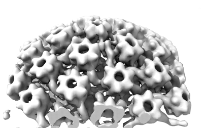
 ダウンロードとリンク
 EMDBマップデータ形式
 emd_18483.png
 http://ftp.pdbj.org/pub/emdb/structures/EMD-18483


 Z (Sec.)
 Y (Row.)
 X (Col.)


 試料の構成要素
 解析
解析 電子顕微鏡法
 FIELD EMISSION GUN

