 Movie
Movie Controller

[English] 日本語
 Yorodumi
- EMDB-18480: Pseudorabies virus nuclear C-capsid (WT) vertices determined in situ -
+ Open data

- Basic information
| Entry |  | ||||||||||||||||||
|---|---|---|---|---|---|---|---|---|---|---|---|---|---|---|---|---|---|---|---|
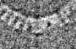
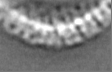
| Title | Pseudorabies virus nuclear C-capsid (WT) vertices determined in situ | ||||||||||||||||||
 Map data Map data | Unmasked | ||||||||||||||||||
 Sample Sample |
| ||||||||||||||||||
 Keywords Keywords | Capsid / capsid vertex specific component / in situ / virus | ||||||||||||||||||
| Biological species |  Suid herpesvirus 1 strain Kaplan Suid herpesvirus 1 strain Kaplan | ||||||||||||||||||
| Method | subtomogram averaging / cryo EM / Resolution: 29.0 Å | ||||||||||||||||||
 Authors Authors | Mironova Y / Prazak V / Vasishtan D | ||||||||||||||||||
| Funding support |  United Kingdom, United Kingdom,  Germany, 5 items Germany, 5 items
| ||||||||||||||||||
 Citation Citation | Journal: Nat Microbiol / Year: 2024 Title: Molecular plasticity of herpesvirus nuclear egress analysed in situ. Authors: Vojtěch Pražák / Yuliia Mironova / Daven Vasishtan / Christoph Hagen / Ulrike Laugks / Yannick Jensen / Saskia Sanders / John M Heumann / Jens B Bosse / Barbara G Klupp / Thomas C ...Authors: Vojtěch Pražák / Yuliia Mironova / Daven Vasishtan / Christoph Hagen / Ulrike Laugks / Yannick Jensen / Saskia Sanders / John M Heumann / Jens B Bosse / Barbara G Klupp / Thomas C Mettenleiter / Michael Grange / Kay Grünewald /  Abstract: The viral nuclear egress complex (NEC) allows herpesvirus capsids to escape from the nucleus without compromising the nuclear envelope integrity. The NEC lattice assembles on the inner nuclear ...The viral nuclear egress complex (NEC) allows herpesvirus capsids to escape from the nucleus without compromising the nuclear envelope integrity. The NEC lattice assembles on the inner nuclear membrane and mediates the budding of nascent nucleocapsids into the perinuclear space and their subsequent release into the cytosol. Its essential role makes it a potent antiviral target, necessitating structural information in the context of a cellular infection. Here we determined structures of NEC-capsid interfaces in situ using electron cryo-tomography, showing a substantial structural heterogeneity. In addition, while the capsid is associated with budding initiation, it is not required for curvature formation. By determining the NEC structure in several conformations, we show that curvature arises from an asymmetric assembly of disordered and hexagonally ordered lattice domains independent of pUL25 or other viral capsid vertex components. Our results advance our understanding of the mechanism of nuclear egress in the context of a living cell. | ||||||||||||||||||
| History |
|
- Structure visualization
| Supplemental images |
|---|

- Downloads & links
-EMDB archive
| Map data | emd_18480.map.gz | 3.2 MB |  EMDB map data format EMDB map data format | |
|---|---|---|---|---|
| Header (meta data) | emd-18480-v30.xmlemd-18480.xml | 19.3 KB 19.3 KB | Display Display | EMDB header |
| FSC (resolution estimation) | emd_18480_fsc.xml | 4.3 KB | Display | FSC data file |
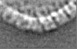
| Images | 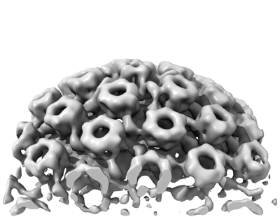 emd_18480.png emd_18480.png | 74.7 KB | ||
| Filedesc metadata | emd-18480.cif.gz | 4.7 KB | ||
| Others | emd_18480_additional_1.map.gzemd_18480_additional_2.map.gzemd_18480_half_map_1.map.gzemd_18480_half_map_2.map.gz | 3.2 MB 3.2 MB 3.2 MB 3.2 MB | ||
| Archive directory |  http://ftp.pdbj.org/pub/emdb/structures/EMD-18480ftp://ftp.pdbj.org/pub/emdb/structures/EMD-18480 http://ftp.pdbj.org/pub/emdb/structures/EMD-18480ftp://ftp.pdbj.org/pub/emdb/structures/EMD-18480 | HTTPS FTP |
-Related structure data
| Related structure data | C: citing same article ( |
|---|


-Links
| EMDB pages | EMDB (EBI/PDBe) / EMDataResource |
|---|
-Map
| File | Download / File: emd_18480.map.gz / Format: CCP4 / Size: 3.4 MB / Type: IMAGE STORED AS FLOATING POINT NUMBER (4 BYTES) | ||||||||||||||||||||||||||||||||||||
|---|---|---|---|---|---|---|---|---|---|---|---|---|---|---|---|---|---|---|---|---|---|---|---|---|---|---|---|---|---|---|---|---|---|---|---|---|---|
| Annotation | Unmasked | ||||||||||||||||||||||||||||||||||||
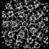
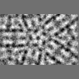
| Projections & slices | Image control
Images are generated by Spider. generated in cubic-lattice coordinate | ||||||||||||||||||||||||||||||||||||
| Voxel size | X=Y=Z: 7.1 Å | ||||||||||||||||||||||||||||||||||||
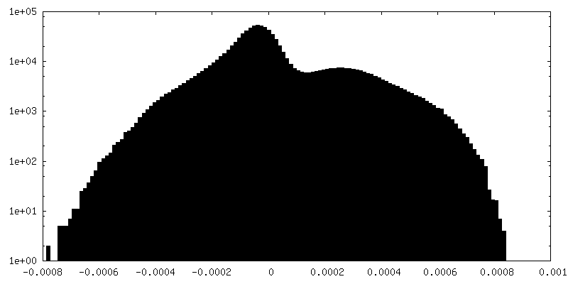
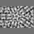
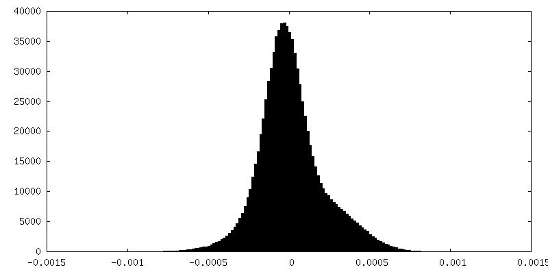
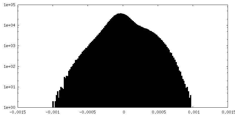
| Density |
| ||||||||||||||||||||||||||||||||||||
| Symmetry | Space group: 1 | ||||||||||||||||||||||||||||||||||||
| Details | EMDB XML:
|
 Z (Sec.)
 Y (Row.)
 X (Col.)


-Supplemental data
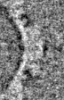
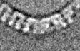
-Additional map: Sub-tomogram average of the nuclear A-capsids, unmasked
| File | emd_18480_additional_1.map | ||||||||||||
|---|---|---|---|---|---|---|---|---|---|---|---|---|---|
| Annotation | Sub-tomogram average of the nuclear A-capsids, unmasked | ||||||||||||
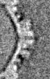
| Projections & Slices |
| ||||||||||||
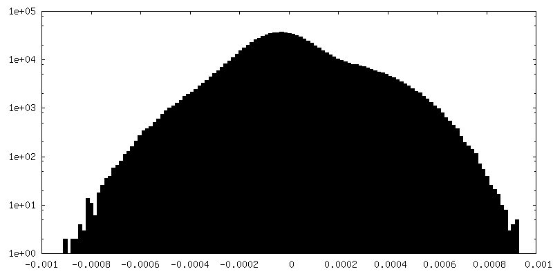
| Density Histograms |


-Additional map: Sub-tomogram average of the nuclear B-capsids, unmasked
| File | emd_18480_additional_2.map | ||||||||||||
|---|---|---|---|---|---|---|---|---|---|---|---|---|---|
| Annotation | Sub-tomogram average of the nuclear B-capsids, unmasked | ||||||||||||
| Projections & Slices |
| ||||||||||||
| Density Histograms |


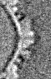
-Half map: Unfiltered
| File | emd_18480_half_map_1.map | ||||||||||||
|---|---|---|---|---|---|---|---|---|---|---|---|---|---|
| Annotation | Unfiltered | ||||||||||||
| Projections & Slices |
| ||||||||||||
| Density Histograms |


-Half map: Unfiltered
| File | emd_18480_half_map_2.map | ||||||||||||
|---|---|---|---|---|---|---|---|---|---|---|---|---|---|
| Annotation | Unfiltered | ||||||||||||
| Projections & Slices |
| ||||||||||||
| Density Histograms |


- Sample components
-Entire : PrV C-capsid in the nucleus of porcine epithelial-like embryonic ...
| Entire | Name: PrV C-capsid in the nucleus of porcine epithelial-like embryonic EFN-R kidney cells |
|---|---|
| Components |
|
-Supramolecule #1: PrV C-capsid in the nucleus of porcine epithelial-like embryonic ...
| Supramolecule | Name: PrV C-capsid in the nucleus of porcine epithelial-like embryonic EFN-R kidney cells type: cell / ID: 1 / Parent: 0 / Details: Cells were infected with wt PrV (Kaplan) for 10h |
|---|---|
| Source (natural) | Organism: Suid herpesvirus 1 strain Kaplan |
-Experimental details
-Structure determination
| Method | cryo EM |
|---|---|
 Processing Processing | subtomogram averaging |
| Aggregation state | cell |
-Sample preparation
| Buffer | pH: 7.5 |
|---|---|
| Vitrification | Cryogen name: ETHANE-PROPANE / Chamber humidity: 70 % / Chamber temperature: 310.15 K / Instrument: LEICA EM GP Details: Vitrified samples were milled using dual beam cryo-FIB-SEM (Aquilos). |
- Electron microscopy
| Microscope | TFS KRIOS |
|---|---|
| Specialist optics | Energy filter - Name: GIF Bioquantum / Energy filter - Slit width: 20 eV |
| Image recording | Film or detector model: GATAN K3 BIOQUANTUM (6k x 4k) / Average exposure time: 0.4 sec. / Average electron dose: 2.5 e/Å2 |
| Electron beam | Acceleration voltage: 300 kV / Electron source:  FIELD EMISSION GUN FIELD EMISSION GUN |
| Electron optics | C2 aperture diameter: 50.0 µm / Illumination mode: FLOOD BEAM / Imaging mode: BRIGHT FIELD / Cs: 2.7 mm / Nominal defocus max: 6.0 µm / Nominal defocus min: 6.0 µm / Nominal magnification: 26000 |
| Sample stage | Specimen holder model: FEI TITAN KRIOS AUTOGRID HOLDER / Cooling holder cryogen: NITROGEN |
| Experimental equipment |  Model: Titan Krios / Image courtesy: FEI Company |
-Image processing
| Final reconstruction | Applied symmetry - Point group: C5 (5 fold cyclic) / Algorithm: BACK PROJECTION / Resolution.type: BY AUTHOR / Resolution: 29.0 Å / Resolution method: FSC 0.143 CUT-OFF / Software - Name: PEET / Number subtomograms used: 1920 |
|---|---|
| Extraction | Number tomograms: 15 / Number images used: 1920 / Software - Name: IMOD |
| Final angle assignment | Type: OTHER / Software - Name: PEET |
| FSC plot (resolution estimation) |  |
