Movie
Movie Controller

[English] 日本語
 Yorodumi
- EMDB-1836: Conformational changes of Adeno-associated Virus type 1 induced b... -
+ Open data

- Basic information
| Entry | Database: EMDB / ID: EMD-1836 | |||||||||
|---|---|---|---|---|---|---|---|---|---|---|
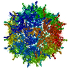
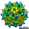
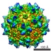
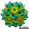
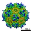
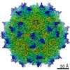
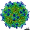
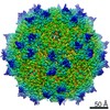
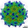
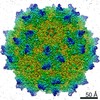
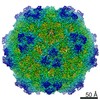
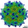
| Title | Conformational changes of Adeno-associated Virus type 1 induced by genome packaging | |||||||||
 Map data Map data | AAV1 group 1 (empty particles) | |||||||||
 Sample Sample |
| |||||||||
 Keywords Keywords | AAV-1 virus structure | |||||||||
| Biological species |  Adeno-associated virus - 1 Adeno-associated virus - 1 | |||||||||
| Method | single particle reconstruction / cryo EM / negative staining / Resolution: 9.6 Å | |||||||||
 Authors Authors | Gerlach B / Kleinschmidt JA / Bottcher B | |||||||||
 Citation Citation | Journal: J Mol Biol / Year: 2011 Title: Conformational changes in adeno-associated virus type 1 induced by genome packaging. Authors: Britta Gerlach / Jürgen A Kleinschmidt / Bettina Böttcher /  Abstract: Adeno-associated virus (AAV) is frequently used as a vector for gene therapy. The viral capsid consists of three structural proteins (VP1, VP2, and VP3) that have a common C-terminal core (VP3), with ...Adeno-associated virus (AAV) is frequently used as a vector for gene therapy. The viral capsid consists of three structural proteins (VP1, VP2, and VP3) that have a common C-terminal core (VP3), with N-terminal extensions of increasing length in VP2 and VP1. The capsid encloses a single-stranded genome of up to 4.7 kb, which is packaged into empty capsids. The N-terminal extension of VP1 carries a phospholipase domain that becomes accessible during infection in the endosomal pathway. We have used cryo-electron microscopy and image reconstruction to determine subnanometer-resolution structures of recombinant AAV1 that has packaged different amounts of a 3.6-kb recombinant genome. The maps show that the AAV1 capsid undergoes continuous conformational changes upon packaging of the genome. The rearrangements occur at the inner capsid surface and lead to constrictions of the pores at the 5-fold symmetry axes and to subtle movements of the β-sheet regions of the capsid proteins. In fully packaged particles, the genome forms stem-like features that contact the inner capsid surface at the 3-fold symmetry axes. We think that the reorganization of the inner surface has an impact on the viral life cycle during infection, preparing the externalization of phospholipase domains through the pores at the 5-fold symmetry axes and possibly genome release. | |||||||||
| History |
|
- Structure visualization
| Movie |
 Movie viewer Movie viewer |
|---|---|
| Structure viewer | EM map: SurfViewMolmilJmol/JSmol |
| Supplemental images |

- Downloads & links
-EMDB archive
| Map data | emd_1836.map.gz | 6.6 MB | EMDB map data format | |
|---|---|---|---|---|
| Header (meta data) | emd-1836-v30.xmlemd-1836.xml | 9.2 KB 9.2 KB | Display Display | EMDB header |
| Images | 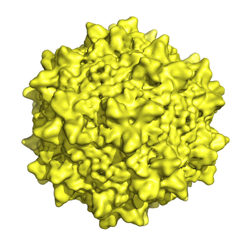 1836.png 1836.png | 576.1 KB | ||
| Archive directory |  http://ftp.pdbj.org/pub/emdb/structures/EMD-1836ftp://ftp.pdbj.org/pub/emdb/structures/EMD-1836 http://ftp.pdbj.org/pub/emdb/structures/EMD-1836ftp://ftp.pdbj.org/pub/emdb/structures/EMD-1836 | HTTPS FTP |
-Validation report
| Summary document | emd_1836_validation.pdf.gz | 265.3 KB | Display | EMDB validaton report |
|---|---|---|---|---|
| Full document | emd_1836_full_validation.pdf.gz | 264.4 KB | Display | |
| Data in XML | emd_1836_validation.xml.gz | 5.8 KB | Display | |
| Arichive directory | https://ftp.pdbj.org/pub/emdb/validation_reports/EMD-1836ftp://ftp.pdbj.org/pub/emdb/validation_reports/EMD-1836 | HTTPS FTP |
-Related structure data

-Links
| EMDB pages | EMDB (EBI/PDBe) / EMDataResource |
|---|
-Map
| File | Download / File: emd_1836.map.gz / Format: CCP4 / Size: 6.9 MB / Type: IMAGE STORED AS FLOATING POINT NUMBER (4 BYTES) | ||||||||||||||||||||||||||||||||||||||||||||||||||||||||||||||||||||
|---|---|---|---|---|---|---|---|---|---|---|---|---|---|---|---|---|---|---|---|---|---|---|---|---|---|---|---|---|---|---|---|---|---|---|---|---|---|---|---|---|---|---|---|---|---|---|---|---|---|---|---|---|---|---|---|---|---|---|---|---|---|---|---|---|---|---|---|---|---|
| Annotation | AAV1 group 1 (empty particles) | ||||||||||||||||||||||||||||||||||||||||||||||||||||||||||||||||||||
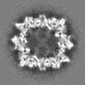
| Projections & slices | Image control
Images are generated by Spider. | ||||||||||||||||||||||||||||||||||||||||||||||||||||||||||||||||||||
| Voxel size | X=Y=Z: 2.8 Å | ||||||||||||||||||||||||||||||||||||||||||||||||||||||||||||||||||||
| Density |
| ||||||||||||||||||||||||||||||||||||||||||||||||||||||||||||||||||||
| Symmetry | Space group: 1 | ||||||||||||||||||||||||||||||||||||||||||||||||||||||||||||||||||||
| Details | EMDB XML:
CCP4 map header:
| ||||||||||||||||||||||||||||||||||||||||||||||||||||||||||||||||||||
 Z (Sec.)
 Y (Row.)
 X (Col.)


-Supplemental data
- Sample components
-Entire : Adeno-associated Virus Type 1
| Entire | Name: Adeno-associated Virus Type 1 |
|---|---|
| Components |
|
-Supramolecule #1000: Adeno-associated Virus Type 1
| Supramolecule | Name: Adeno-associated Virus Type 1 / type: sample / ID: 1000 Details: Recombinant AAV1 capsid, particles belong to group 1 (empty) Oligomeric state: VP1, VP2 and VP3 in T1 arrangement / Number unique components: 1 |
|---|
-Supramolecule #1: Adeno-associated virus - 1
| Supramolecule | Name: Adeno-associated virus - 1 / type: virus / ID: 1 / Name.synonym: AAV 1 / NCBI-ID: 85106 / Sci species name: Adeno-associated virus - 1 / Virus type: VIRUS-LIKE PARTICLE / Virus isolate: SEROTYPE / Virus enveloped: No / Virus empty: Yes / Syn species name: AAV 1 |
|---|---|
| Host (natural) | Organism:  Homo sapiens (human) / synonym: VERTEBRATES Homo sapiens (human) / synonym: VERTEBRATES |
| Virus shell | Shell ID: 1 / Diameter: 240 Å / T number (triangulation number): 1 |
-Experimental details
-Structure determination
| Method | negative staining, cryo EM |
|---|---|
 Processing Processing | single particle reconstruction |
| Aggregation state | particle |
-Sample preparation
| Staining | Type: NEGATIVE / Details: Vitrified |
|---|---|
| Grid | Details: 400 mesh perforated carbon |
| Vitrification | Cryogen name: ETHANE / Chamber humidity: 100 % / Chamber temperature: 96 K / Instrument: HOMEMADE PLUNGER / Details: Vitrification instrument: Controlled environment / Method: Blot for 15 s before plunging |
- Electron microscopy
| Microscope | FEI/PHILIPS CM200FEG |
|---|---|
| Temperature | Average: 95 K |
| Alignment procedure | Legacy - Astigmatism: At 200,000 times magnification on graininess of carbon |
| Image recording | Category: FILM / Film or detector model: KODAK SO-163 FILM / Digitization - Scanner: ZEISS SCAI / Digitization - Sampling interval: 7 µm / Number real images: 105 / Details: Data binning 2x2 Px, before processing / Bits/pixel: 8 |
| Electron beam | Acceleration voltage: 200 kV / Electron source:  FIELD EMISSION GUN FIELD EMISSION GUN |
| Electron optics | Illumination mode: FLOOD BEAM / Imaging mode: BRIGHT FIELD / Nominal defocus max: 3.5 µm / Nominal defocus min: 1.5 µm / Nominal magnification: 50000 |
| Sample stage | Specimen holder: Eucentric / Specimen holder model: GATAN LIQUID NITROGEN |
-Image processing
| Details | Particles were sorted according to their density profiles. This reconstruction represents group 1, which consists of mainly empty particles. |
|---|---|
| CTF correction | Details: Maps during combination |
| Final reconstruction | Applied symmetry - Point group: I (icosahedral) / Algorithm: OTHER / Resolution.type: BY AUTHOR / Resolution: 9.6 Å / Resolution method: FSC 0.5 CUT-OFF / Software - Name: MRC / Number images used: 3427 |
-Atomic model buiding 1
| Initial model | PDB ID: |
|---|---|
| Software | Name: Chimera |
| Refinement | Space: REAL |