- EMDB-18156: Combined map of Candida albicans 80S ribosome in complex with cep... -
+
Open data
ID or keywords:
Loading...
-
Basic information
Entry
Database: EMDB / ID: EMD-18156
Title
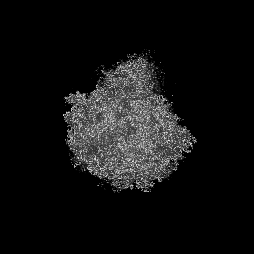
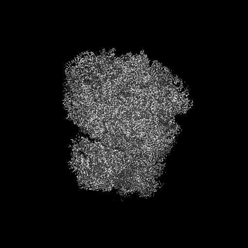
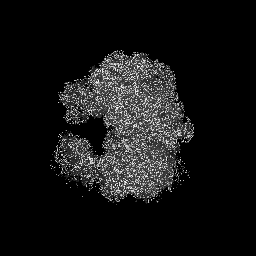
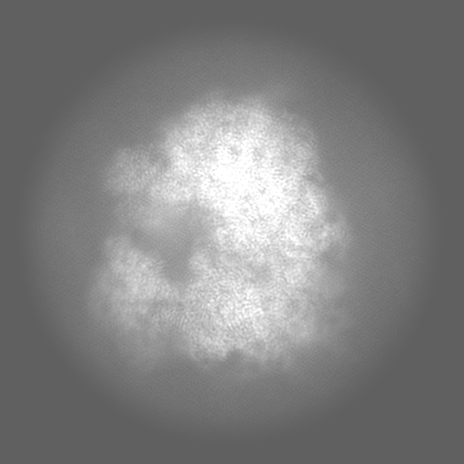
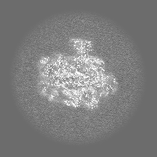
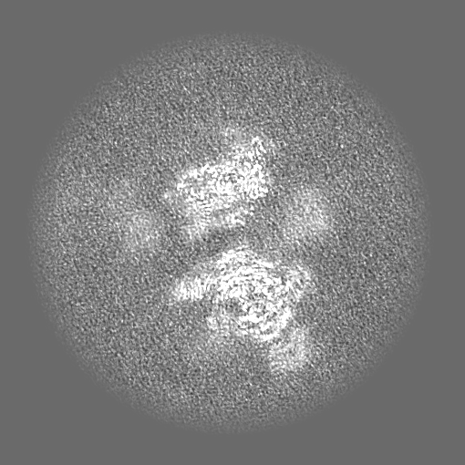
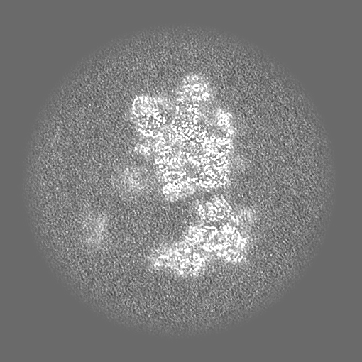
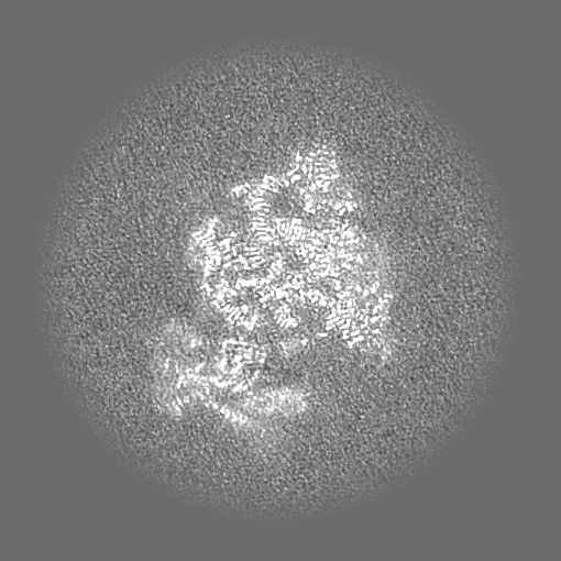
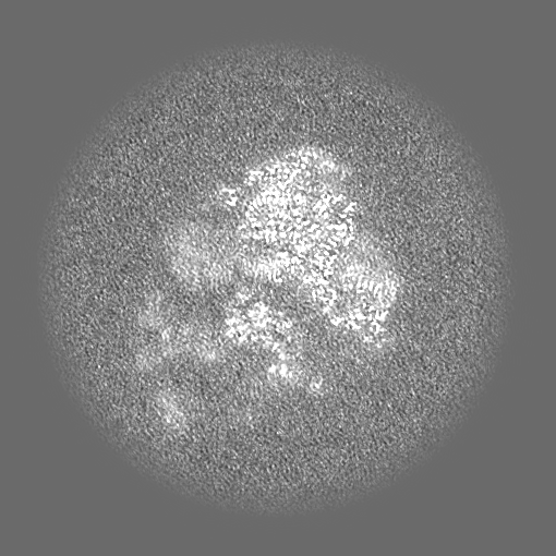
Combined map of Candida albicans 80S ribosome in complex with cephaeline
Map data
Composite map
Sample
Complex: The Candida albicans ribosome in complew with cephaeline
Keywords
Ribosome / Candida albicans / cephaeline
Function / homology
Function and homology information
yeast-form cell wall / hyphal cell wall / preribosome / positive regulation of translational fidelity / pre-mRNA 5'-splice site binding / nonfunctional rRNA decay / cleavage in ITS2 between 5.8S rRNA and LSU-rRNA of tricistronic rRNA transcript (SSU-rRNA, 5.8S rRNA, LSU-rRNA) / preribosome, small subunit precursor / negative regulation of mRNA splicing, via spliceosome / preribosome, large subunit precursor ...yeast-form cell wall / hyphal cell wall / preribosome / positive regulation of translational fidelity / pre-mRNA 5'-splice site binding / nonfunctional rRNA decay / cleavage in ITS2 between 5.8S rRNA and LSU-rRNA of tricistronic rRNA transcript (SSU-rRNA, 5.8S rRNA, LSU-rRNA) / preribosome, small subunit precursor / negative regulation of mRNA splicing, via spliceosome / preribosome, large subunit precursor / translational elongation / 90S preribosome / endonucleolytic cleavage to generate mature 3'-end of SSU-rRNA from (SSU-rRNA, 5.8S rRNA, LSU-rRNA) / ribosomal subunit export from nucleus / translational termination / protein-RNA complex assembly / endonucleolytic cleavage in ITS1 to separate SSU-rRNA from 5.8S rRNA and LSU-rRNA from tricistronic rRNA transcript (SSU-rRNA, 5.8S rRNA, LSU-rRNA) / ribosomal small subunit export from nucleus / DNA-(apurinic or apyrimidinic site) endonuclease activity / cytosolic ribosome / ribosomal large subunit biogenesis / maturation of LSU-rRNA from tricistronic rRNA transcript (SSU-rRNA, 5.8S rRNA, LSU-rRNA) / maturation of SSU-rRNA from tricistronic rRNA transcript (SSU-rRNA, 5.8S rRNA, LSU-rRNA) / maturation of SSU-rRNA / small-subunit processome / maintenance of translational fidelity / rRNA processing / large ribosomal subunit / ribosomal small subunit assembly / ribosome biogenesis / ribosomal small subunit biogenesis / 5S rRNA binding / ribosomal large subunit assembly / small ribosomal subunit / small ribosomal subunit rRNA binding / large ribosomal subunit rRNA binding / cytosolic small ribosomal subunit / cytosolic large ribosomal subunit / cytoplasmic translation / negative regulation of translation / rRNA binding / structural constituent of ribosome / ribosome / translation / ribonucleoprotein complex / mRNA binding / nucleolus / cell surface / RNA binding / zinc ion binding / metal ion binding / nucleus / cytoplasm / cytosol Similarity search - Function
: / : / Ribosomal protein S26e signature. / Ribosomal protein L41 / Ribosomal protein L41 / Ribosomal protein L13e, conserved site / Ribosomal protein L13e signature. / Ribosomal protein S21e, conserved site / Ribosomal protein S21e signature. / Ribosomal protein S26e ...: / : / Ribosomal protein S26e signature. / Ribosomal protein L41 / Ribosomal protein L41 / Ribosomal protein L13e, conserved site / Ribosomal protein L13e signature. / Ribosomal protein S21e, conserved site / Ribosomal protein S21e signature. / Ribosomal protein S26e / Ribosomal protein L29e / Ribosomal protein S26e superfamily / Ribosomal protein S26e / Ribosomal L29e protein family / Ribosomal protein L22e / Ribosomal protein L22e superfamily / Ribosomal L22e protein family / Ribosomal protein L27e, conserved site / Ribosomal protein L27e signature. / Ribosomal protein L13e / Ribosomal protein L13e / Ribosomal protein L38e / Ribosomal protein S5, eukaryotic/archaeal / Ribosomal protein L38e superfamily / Ribosomal L38e protein family / : / Ribosomal protein L19, eukaryotic / Ribosomal protein S21e / Ribosomal protein S21e superfamily / Ribosomal protein S21e / Ribosomal protein L6e signature. / 60S ribosomal protein L18a/ L20, eukaryotes / Ribosomal protein S2, eukaryotic / Ribosomal protein L10e, conserved site / Ribosomal protein L10e signature. / Ribosomal protein L19/L19e conserved site / Ribosomal protein L19e signature. / Ribosomal protein L44e signature. / Ribosomal protein L24e, conserved site / Ribosomal protein L24e signature. / Ribosomal protein L18/L18-A/B/e, conserved site / Ribosomal protein L18e signature. / 40S Ribosomal protein S10 / Ribosomal protein L10e / Ribosomal protein L34e, conserved site / Ribosomal protein L34e signature. / Plectin/S10, N-terminal / Plectin/S10 domain / Ribosomal protein L5 eukaryotic, C-terminal / Ribosomal L18 C-terminal region / Ribosomal protein L23/L25, N-terminal / Ribosomal protein L23, N-terminal domain / 50S ribosomal protein L18Ae/60S ribosomal protein L20 and L18a / : / Ribosomal protein L36e signature. / Ribosomal L40e family / Ribosomal protein L30e signature 1. / Ribosomal protein 50S-L18Ae/60S-L20/60S-L18A / Ribosomal proteins 50S-L18Ae/60S-L20/60S-L18A / Ribosomal protein S10, eukaryotic/archaeal / Ribosomal protein S30 / Ribosomal protein S30 / Ribosomal protein L44e / Ribosomal protein L44 / Ribosomal protein 60S L18 and 50S L18e / Ribosomal protein L35Ae, conserved site / Ribosomal protein L35Ae signature. / Eukaryotic Ribosomal Protein L27, KOW domain / Ribosomal_L40e / Ribosomal protein L40e / Ribosomal protein L40e superfamily / : / Ribosomal protein L27e / Ribosomal protein L27e superfamily / Ribosomal L27e protein family / Ribosomal Protein L6, KOW domain / Ribosomal protein S8e subdomain, eukaryotes / : / Ribosomal protein S17e, conserved site / Ribosomal protein S17e signature. / Ribosomal protein L39e, conserved site / Ribosomal protein L39e signature. / 60S ribosomal protein L35 / Ribosomal protein S7e signature. / Ribosomal protein L30e signature 2. / Ribosomal protein L7A/L8 / Ribosomal protein L30e, conserved site / Ribosomal protein S2, eukaryotic/archaeal / : / Ribosomal protein L6e / Ribosomal protein L34Ae / Ribosomal protein L34e / 60S ribosomal protein L19 / 40S ribosomal protein S29/30S ribosomal protein S14 type Z / Ribosomal protein L13, eukaryotic/archaeal / 60S ribosomal protein L6E / Ribosomal protein S3, eukaryotic/archaeal / Ribosomal protein L7, eukaryotic / Ribosomal protein L30, N-terminal / Ribosomal L30 N-terminal domain Similarity search - Domain/homology
Small ribosomal subunit protein eS4 / Large ribosomal subunit protein eL37 / Large ribosomal subunit protein eL38 / Small ribosomal subunit protein uS19 / Large ribosomal subunit protein eL19 / Large ribosomal subunit protein eL40 / Large ribosomal subunit protein eL22 / Large ribosomal subunit protein eL30 / Small ribosomal subunit protein uS17 / Large ribosomal subunit protein eL43 ...Small ribosomal subunit protein eS4 / Large ribosomal subunit protein eL37 / Large ribosomal subunit protein eL38 / Small ribosomal subunit protein uS19 / Large ribosomal subunit protein eL19 / Large ribosomal subunit protein eL40 / Large ribosomal subunit protein eL22 / Large ribosomal subunit protein eL30 / Small ribosomal subunit protein uS17 / Large ribosomal subunit protein eL43 / Small ribosomal subunit protein eS30 / Small ribosomal subunit protein uS14 / 60S ribosomal protein L42-B / Ribosomal protein S7 / 40S ribosomal protein S17-B / 40S ribosomal protein S26 / Ribosomal protein L22 / 60S ribosomal protein L7-A / 60S ribosomal protein L28 / 60S ribosomal protein L33-A / 40S ribosomal protein S27 / 40S ribosomal protein S10-A / Small ribosomal subunit protein uS10 / Small ribosomal subunit protein uS3 / 40S ribosomal protein S6 / Ribosomal protein L13 / Small ribosomal subunit protein uS5 / 60S ribosomal protein L20 / 40S ribosomal protein S28-B / 60S ribosomal protein L8 / Small ribosomal subunit protein eS1 / Small ribosomal subunit protein uS2 / 40S ribosomal protein S24 / 60S ribosomal protein L9-B / 60S ribosomal protein L23-A / 60S ribosomal protein L24-A / 60S ribosomal protein L18-A / 40S ribosomal protein S19-A / 40S ribosomal protein S7 / 60S ribosomal protein L29 / 40S ribosomal protein S22-A / 40S ribosomal protein S8 / Ribosomal protein S23 (S12) / Small ribosomal subunit protein uS11 / 60S ribosomal protein L34-B / 60S ribosomal protein L21-A / 60S ribosomal protein L31-B / 60S ribosomal protein L11-B / Ribosomal protein S4 / 60S ribosomal protein L2-B / 60S ribosomal protein L14-B / Ribosomal protein L24 / 40S ribosomal protein S18-B / 40S ribosomal protein S13 / 60S ribosomal protein L4-B / 60S ribosomal protein L13 / Large ribosomal subunit protein uL23 / Ribosomal protein L29 / Large ribosomal subunit protein eL32 / Small ribosomal subunit protein uS9 / Large ribosomal subunit protein eL36 / Small ribosomal subunit protein eS32 / Large ribosomal subunit protein uL3 / Large ribosomal subunit protein eL15 / Large ribosomal subunit protein uL18 / Large ribosomal subunit protein uL16 / Large ribosomal subunit protein eL39 / 60S ribosomal protein L6 / Large ribosomal subunit protein eL27 / Small ribosomal subunit protein eS21 Similarity search - Component
Biological species
Candida albicans (yeast)
Method
single particle reconstruction / cryo EM / Resolution: 2.45 Å
In the structure databanks used in Yorodumi, some data are registered as the other names, "COVID-19 virus" and "2019-nCoV". Here are the details of the virus and the list of structure data.
Jan 31, 2019. EMDB accession codes are about to change! (news from PDBe EMDB page)
EMDB accession codes are about to change! (news from PDBe EMDB page)
The allocation of 4 digits for EMDB accession codes will soon come to an end. Whilst these codes will remain in use, new EMDB accession codes will include an additional digit and will expand incrementally as the available range of codes is exhausted. The current 4-digit format prefixed with “EMD-” (i.e. EMD-XXXX) will advance to a 5-digit format (i.e. EMD-XXXXX), and so on. It is currently estimated that the 4-digit codes will be depleted around Spring 2019, at which point the 5-digit format will come into force.
The EM Navigator/Yorodumi systems omit the EMD- prefix.
Related info.:Q: What is EMD? / ID/Accession-code notation in Yorodumi/EM Navigator
Yorodumi is a browser for structure data from EMDB, PDB, SASBDB, etc.
This page is also the successor to EM Navigator detail page, and also detail information page/front-end page for Omokage search.
The word "yorodu" (or yorozu) is an old Japanese word meaning "ten thousand". "mi" (miru) is to see.
Related info.:EMDB / PDB / SASBDB / Comparison of 3 databanks / Yorodumi Search / Aug 31, 2016. New EM Navigator & Yorodumi / Yorodumi Papers / Jmol/JSmol / Function and homology information / Changes in new EM Navigator and Yorodumi
 Movie
Movie Controller

 Yorodumi
 Open data
 Basic information
 Map data
 Sample
 Keywords
 Function and homology information
 Candida albicans (yeast)
 Authors
 France, 1 items
  Citation
 Structure visualization
 Downloads & links
 emd_18156.png
 http://ftp.pdbj.org/pub/emdb/structures/EMD-18156


 Z (Sec.)
 Y (Row.)
 X (Col.)


 Sample components
 Processing
 Electron microscopy
 FIELD EMISSION GUN

