Movie
Movie Controller

[English] 日本語
 Yorodumi
- EMDB-18151: Focused map on the body of the SSU of the Candida albicans 80S ri... -
+ Open data

- Basic information
| Entry |  | |||||||||
|---|---|---|---|---|---|---|---|---|---|---|
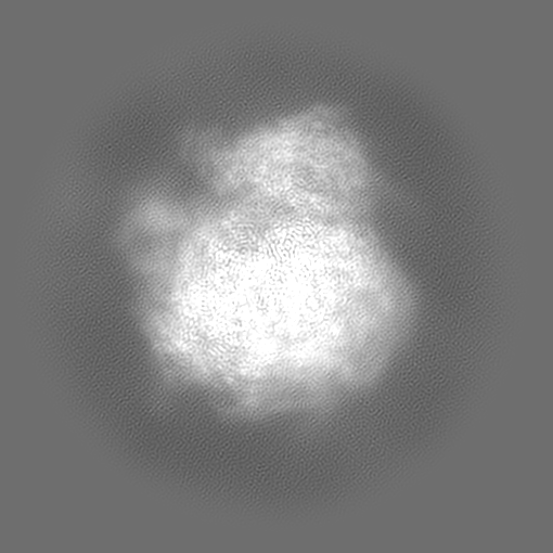
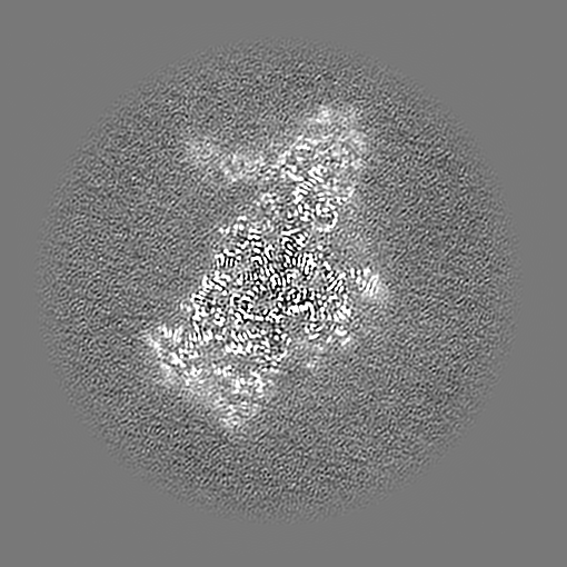
| Title | Focused map on the body of the SSU of the Candida albicans 80S ribosome in complex with cephaeline | |||||||||
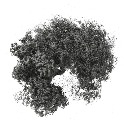
 Map data Map data | Cryo-EM map of the C. albicans ribosome focused on the body of the SSU | |||||||||
 Sample Sample |
| |||||||||
 Keywords Keywords | Ribosome / Candida albicans / cephaeline | |||||||||
| Biological species |  Candida albicans (yeast) Candida albicans (yeast) | |||||||||
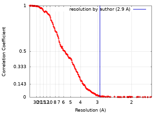
| Method | single particle reconstruction / cryo EM / Resolution: 2.9 Å | |||||||||
 Authors Authors | Kolosova O / Zgadzay Y / Stetsenko A / Atamas A / Jenner L / Guskov A / Yusupov M | |||||||||
| Funding support |  France, 1 items France, 1 items
| |||||||||
 Citation Citation | Journal: Biopolym Cell / Year: 2023 Title: Structural characterization of cephaeline binding to the eukaryotic ribosome using Cryo-Electron Microscopy Authors: Kolosova O / Zgadzay Y / Stetsenko A / Atamas A / Wu C / Jenner L / Sachs SM / Guskov A / Yusupov M | |||||||||
| History |
|
- Structure visualization
| Supplemental images |
|---|
- Downloads & links
-EMDB archive
| Map data | emd_18151.map.gz | 394.2 MB |  EMDB map data format EMDB map data format | |
|---|---|---|---|---|
| Header (meta data) | emd-18151-v30.xmlemd-18151.xml | 11.8 KB 11.8 KB | Display Display | EMDB header |
| FSC (resolution estimation) | emd_18151_fsc.xml | 22 KB | Display | FSC data file |
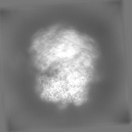
| Images |  emd_18151.png emd_18151.png | 206.8 KB | ||
| Filedesc metadata | emd-18151.cif.gz | 3.8 KB | ||
| Others | emd_18151_half_map_1.map.gzemd_18151_half_map_2.map.gz | 387.3 MB 387.3 MB | ||
| Archive directory |  http://ftp.pdbj.org/pub/emdb/structures/EMD-18151ftp://ftp.pdbj.org/pub/emdb/structures/EMD-18151 http://ftp.pdbj.org/pub/emdb/structures/EMD-18151ftp://ftp.pdbj.org/pub/emdb/structures/EMD-18151 | HTTPS FTP |
-Related structure data


-Links
| EMDB pages | EMDB (EBI/PDBe) / EMDataResource |
|---|
-Map
| File | Download / File: emd_18151.map.gz / Format: CCP4 / Size: 506 MB / Type: IMAGE STORED AS FLOATING POINT NUMBER (4 BYTES) | ||||||||||||||||||||||||||||||||||||
|---|---|---|---|---|---|---|---|---|---|---|---|---|---|---|---|---|---|---|---|---|---|---|---|---|---|---|---|---|---|---|---|---|---|---|---|---|---|
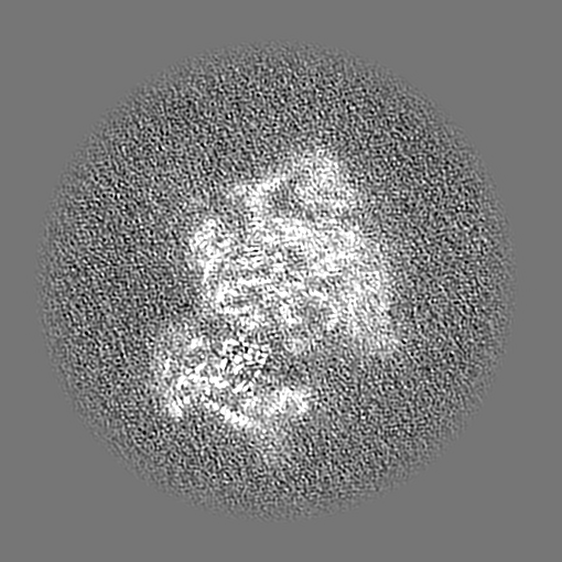
| Annotation | Cryo-EM map of the C. albicans ribosome focused on the body of the SSU | ||||||||||||||||||||||||||||||||||||
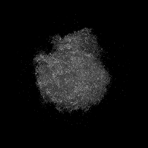
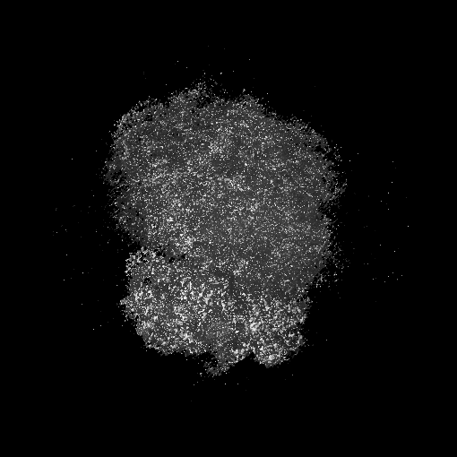
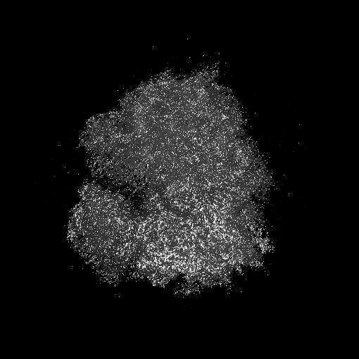
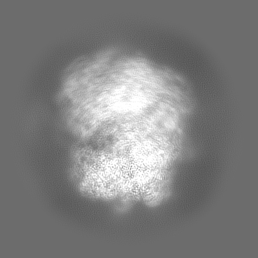
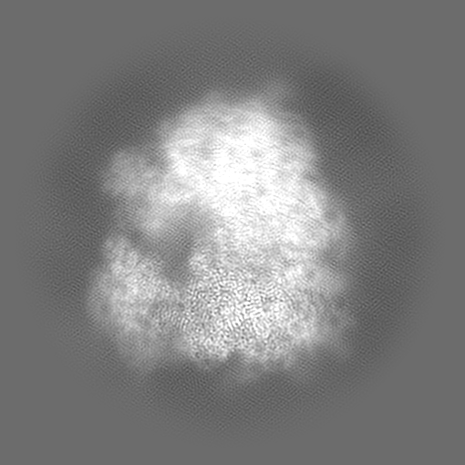
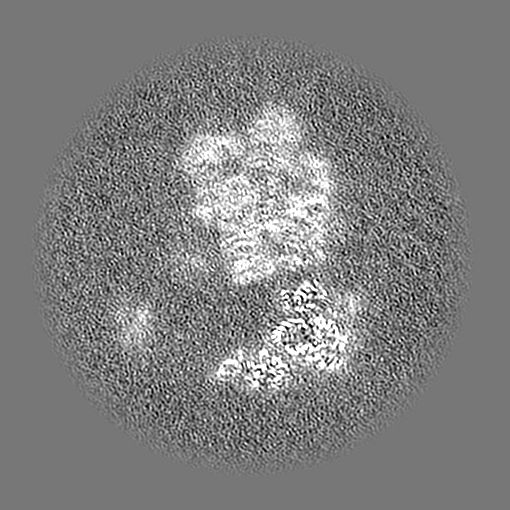
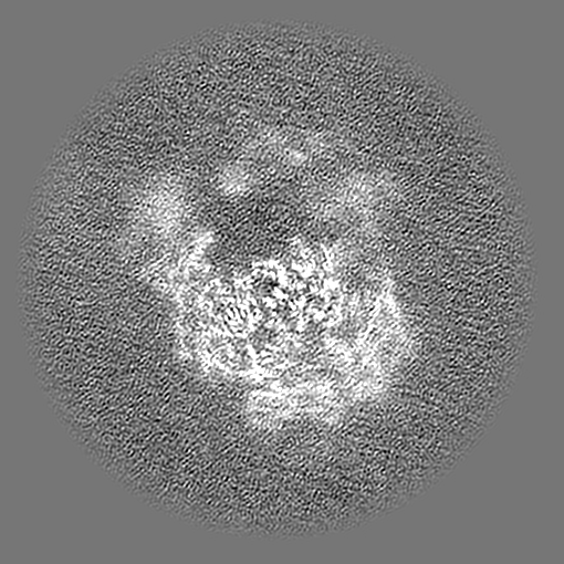
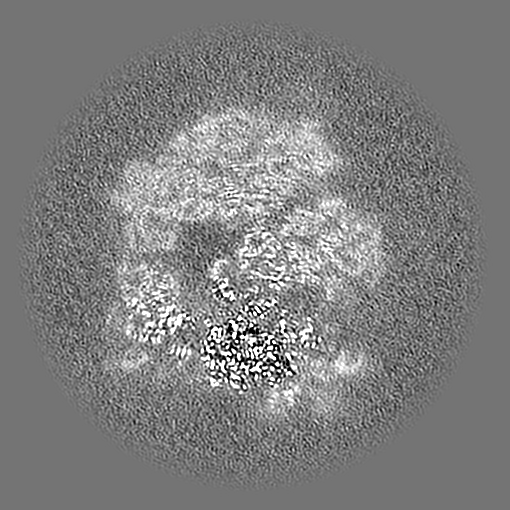
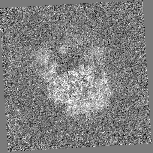
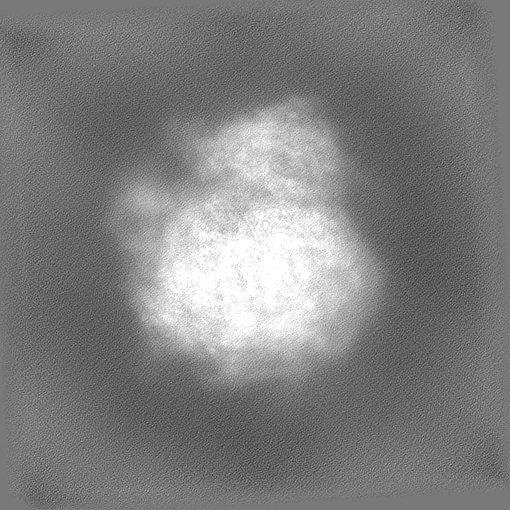
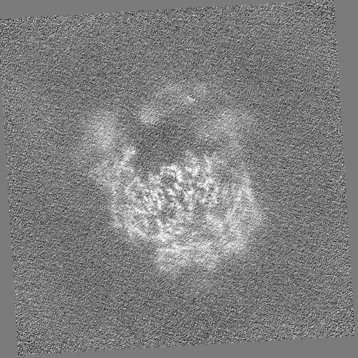
| Projections & slices | Image control
Images are generated by Spider. | ||||||||||||||||||||||||||||||||||||
| Voxel size | X=Y=Z: 0.836 Å | ||||||||||||||||||||||||||||||||||||
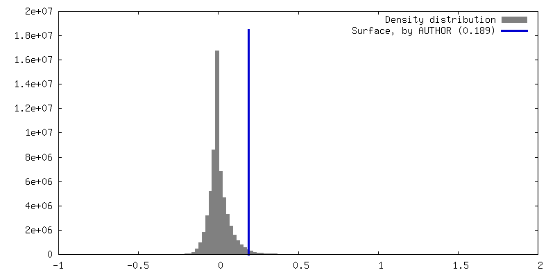
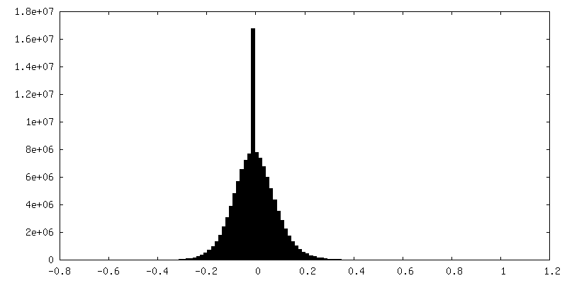
| Density |
| ||||||||||||||||||||||||||||||||||||
| Symmetry | Space group: 1 | ||||||||||||||||||||||||||||||||||||
| Details | EMDB XML:
|
 Z (Sec.)
 Y (Row.)
 X (Col.)


-Supplemental data
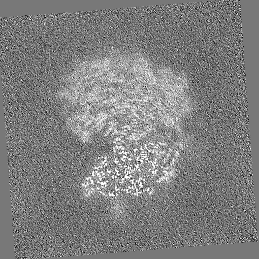
-Half map: Half map A
| File | emd_18151_half_map_1.map | ||||||||||||
|---|---|---|---|---|---|---|---|---|---|---|---|---|---|
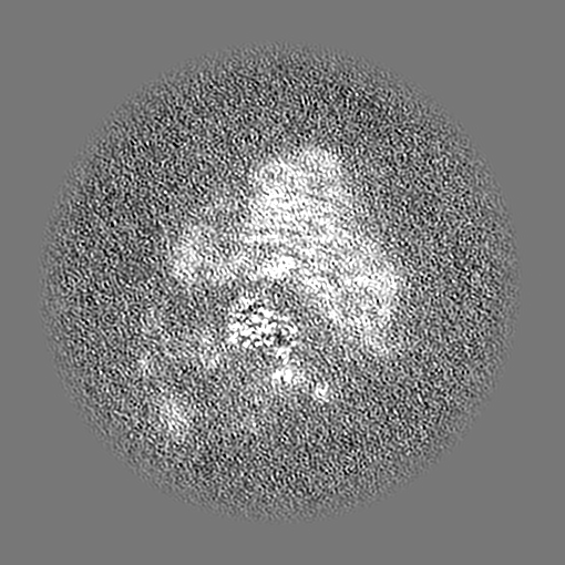
| Annotation | Half map A | ||||||||||||
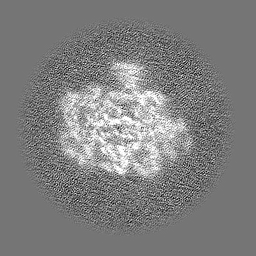
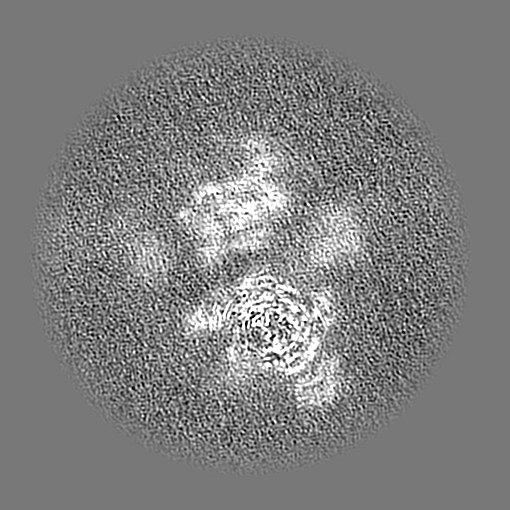
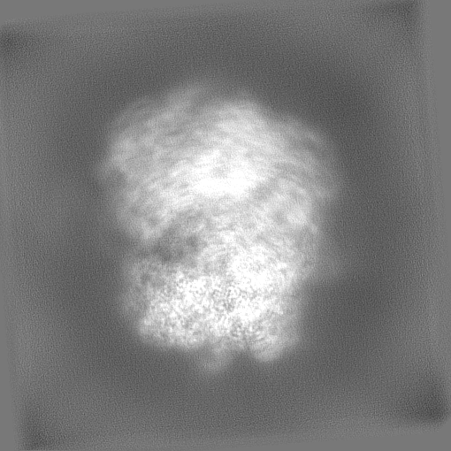
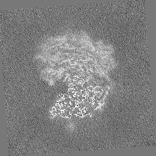
| Projections & Slices |
| ||||||||||||
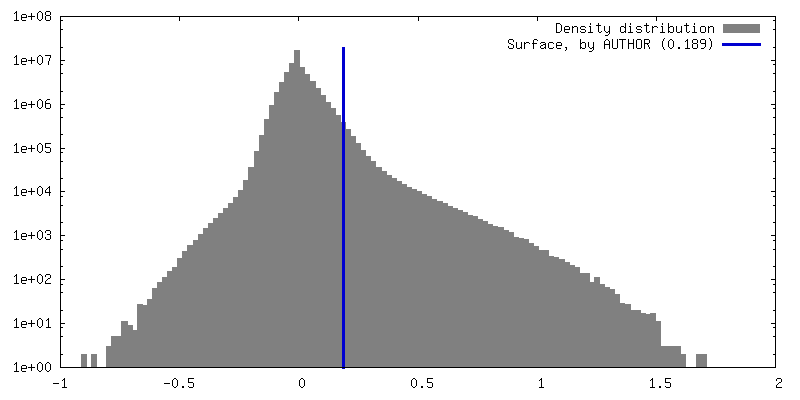
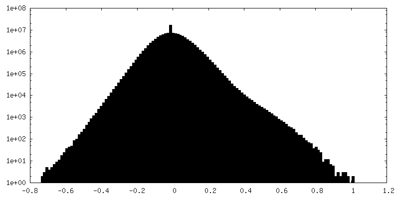
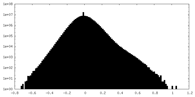
| Density Histograms |


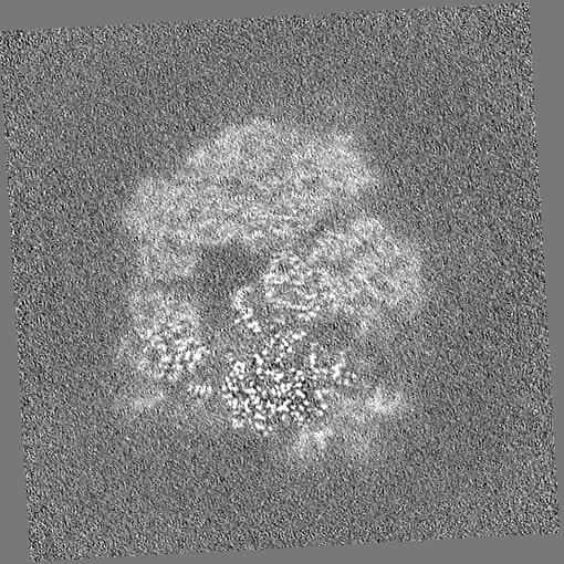

-Half map: Half map B
| File | emd_18151_half_map_2.map | ||||||||||||
|---|---|---|---|---|---|---|---|---|---|---|---|---|---|
| Annotation | Half map B | ||||||||||||
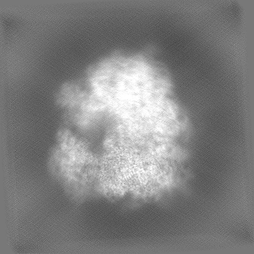
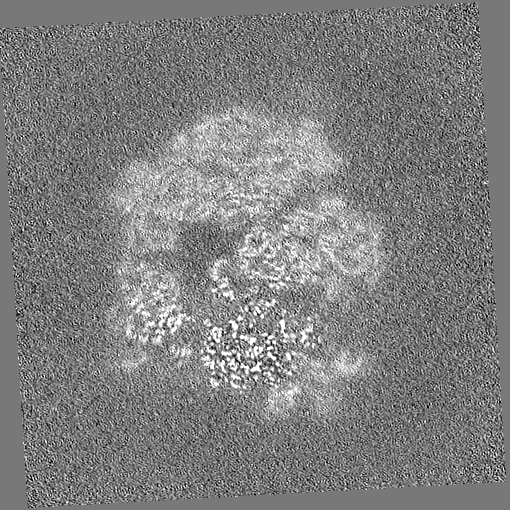
| Projections & Slices |
| ||||||||||||
| Density Histograms |
- Sample components
-Entire : The Candida albicans ribosome in complew with cephaeline
| Entire | Name: The Candida albicans ribosome in complew with cephaeline |
|---|---|
| Components |
|
-Supramolecule #1: The Candida albicans ribosome in complew with cephaeline
| Supramolecule | Name: The Candida albicans ribosome in complew with cephaeline type: complex / ID: 1 / Parent: 0 |
|---|---|
| Source (natural) | Organism: Candida albicans (yeast) |
-Experimental details
-Structure determination
| Method | cryo EM |
|---|---|
 Processing Processing | single particle reconstruction |
| Aggregation state | particle |
-Sample preparation
| Buffer | pH: 7 |
|---|---|
| Vitrification | Cryogen name: ETHANE |
- Electron microscopy
| Microscope | FEI TITAN KRIOS |
|---|---|
| Image recording | Film or detector model: GATAN K3 (6k x 4k) / Average electron dose: 50.0 e/Å2 |
| Electron beam | Acceleration voltage: 300 kV / Electron source:  FIELD EMISSION GUN FIELD EMISSION GUN |
| Electron optics | Illumination mode: FLOOD BEAM / Imaging mode: BRIGHT FIELD / Nominal defocus max: 2.4 µm / Nominal defocus min: 0.9 µm |
| Experimental equipment |  Model: Titan Krios / Image courtesy: FEI Company |