 Movie
Movie Controller

[English] 日本語
 Yorodumi
- EMDB-1805: Structure of human complement C8, a precursor to membrane attack. -
+ Open data

- Basic information
| Entry | Database: EMDB / ID: EMD-1805 | |||||||||
|---|---|---|---|---|---|---|---|---|---|---|
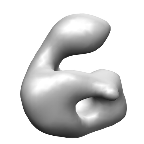
| Title | Structure of human complement C8, a precursor to membrane attack. | |||||||||
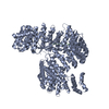
 Map data Map data | This is the EM reconstruction of human C8 | |||||||||
 Sample Sample |
| |||||||||
 Keywords Keywords | C8 / complement / membrane attack complex / MAC / terminal pathway | |||||||||
| Function / homology | Membrane attack complex component/perforin/complement C9 / Thrombospondin type-1 (TSP1) repeat / complement activation, alternative pathway / complement activation, classical pathway / Lipocalin/cytosolic fatty-acid binding domain Function and homology information Function and homology information | |||||||||
| Biological species |  Homo sapiens (human) Homo sapiens (human) | |||||||||
| Method | single particle reconstruction / negative staining / Resolution: 25.0 Å | |||||||||
 Authors Authors | Bubeck D / Roversi P / Donev R / Morgan BP / Llorca O / Lea SM | |||||||||
 Citation Citation | Journal: J Mol Biol / Year: 2011 Title: Structure of human complement C8, a precursor to membrane attack. Authors: Doryen Bubeck / Pietro Roversi / Rossen Donev / B Paul Morgan / Oscar Llorca / Susan M Lea /  Abstract: Complement component C8 plays a pivotal role in the formation of the membrane attack complex (MAC), an important antibacterial immune effector. C8 initiates membrane penetration and coordinates MAC ...Complement component C8 plays a pivotal role in the formation of the membrane attack complex (MAC), an important antibacterial immune effector. C8 initiates membrane penetration and coordinates MAC pore formation. High-resolution structures of C8 subunits have provided some insight into the function of the C8 heterotrimer; however, there is no structural information describing how the intersubunit organization facilitates MAC assembly. We have determined the structure of C8 by electron microscopy and fitted the C8α-MACPF (membrane attack complex/perforin)-C8γ co-crystal structure and a homology model for C8β-MACPF into the density. Here, we demonstrate that both the C8γ protrusion and the C8α-MACPF region that inserts into the membrane upon activation are accessible. | |||||||||
| History |
|
- Structure visualization
| Movie |
Movie viewer |
|---|---|
| Structure viewer | EM map: SurfViewMolmilJmol/JSmol |
| Supplemental images |
- Downloads & links
-EMDB archive
| Map data | emd_1805.map.gz | 126.8 KB | EMDB map data format | |
|---|---|---|---|---|
| Header (meta data) | emd-1805-v30.xmlemd-1805.xml | 11.3 KB 11.3 KB | Display Display | EMDB header |
| Images |  1805.jpg 1805.jpg | 68.1 KB | ||
| Archive directory |  http://ftp.pdbj.org/pub/emdb/structures/EMD-1805ftp://ftp.pdbj.org/pub/emdb/structures/EMD-1805 http://ftp.pdbj.org/pub/emdb/structures/EMD-1805ftp://ftp.pdbj.org/pub/emdb/structures/EMD-1805 | HTTPS FTP |
-Validation report
| Summary document | emd_1805_validation.pdf.gz | 200.6 KB | Display | EMDB validaton report |
|---|---|---|---|---|
| Full document | emd_1805_full_validation.pdf.gz | 199.7 KB | Display | |
| Data in XML | emd_1805_validation.xml.gz | 4.9 KB | Display | |
| Arichive directory | https://ftp.pdbj.org/pub/emdb/validation_reports/EMD-1805ftp://ftp.pdbj.org/pub/emdb/validation_reports/EMD-1805 | HTTPS FTP |
-Related structure data
| Similar structure data |
|---|


-Links
| EMDB pages | EMDB (EBI/PDBe) / EMDataResource |
|---|
-Map
| File | Download / File: emd_1805.map.gz / Format: CCP4 / Size: 422.9 KB / Type: IMAGE STORED AS FLOATING POINT NUMBER (4 BYTES) | ||||||||||||||||||||||||||||||||||||||||||||||||||||||||||||||||||||
|---|---|---|---|---|---|---|---|---|---|---|---|---|---|---|---|---|---|---|---|---|---|---|---|---|---|---|---|---|---|---|---|---|---|---|---|---|---|---|---|---|---|---|---|---|---|---|---|---|---|---|---|---|---|---|---|---|---|---|---|---|---|---|---|---|---|---|---|---|---|
| Annotation | This is the EM reconstruction of human C8 | ||||||||||||||||||||||||||||||||||||||||||||||||||||||||||||||||||||
| Projections & slices | Image control
Images are generated by Spider. | ||||||||||||||||||||||||||||||||||||||||||||||||||||||||||||||||||||
| Voxel size | X=Y=Z: 4.7 Å | ||||||||||||||||||||||||||||||||||||||||||||||||||||||||||||||||||||
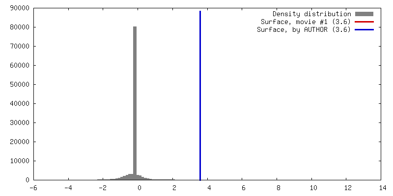
| Density |
| ||||||||||||||||||||||||||||||||||||||||||||||||||||||||||||||||||||
| Symmetry | Space group: 1 | ||||||||||||||||||||||||||||||||||||||||||||||||||||||||||||||||||||
| Details | EMDB XML:
CCP4 map header:
| ||||||||||||||||||||||||||||||||||||||||||||||||||||||||||||||||||||
 Z (Sec.)
 Y (Row.)
 X (Col.)


-Supplemental data
- Sample components
-Entire : Human complement C8
| Entire | Name: Human complement C8 |
|---|---|
| Components |
|
-Supramolecule #1000: Human complement C8
| Supramolecule | Name: Human complement C8 / type: sample / ID: 1000 Details: The sample was purified by size exclusion chromatography and the purity checked by SDS-PAGE Oligomeric state: Heterotrimer of C8alpha C8beta and C8gamma Number unique components: 3 |
|---|---|
| Molecular weight | Theoretical: 150 KDa |
-Macromolecule #1: Human complement C8 alpha subunit
| Macromolecule | Name: Human complement C8 alpha subunit / type: protein_or_peptide / ID: 1 / Name.synonym: C8 alpha / Number of copies: 1 / Oligomeric state: Monomer / Recombinant expression: No |
|---|---|
| Source (natural) | Organism: Homo sapiens (human) / synonym: Human / Tissue: Serum / Location in cell: Extracellular |
| Molecular weight | Theoretical: 65.163 KDa |
| Sequence | GO: complement activation, alternative pathway / InterPro: Thrombospondin type-1 (TSP1) repeat |
-Macromolecule #2: Human complement C8 beta subunit
| Macromolecule | Name: Human complement C8 beta subunit / type: protein_or_peptide / ID: 2 / Name.synonym: C8 beta / Number of copies: 1 / Oligomeric state: Monomer / Recombinant expression: No |
|---|---|
| Source (natural) | Organism: Homo sapiens (human) / synonym: Human / Tissue: Serum / Location in cell: Extracellular |
| Sequence | GO: complement activation, classical pathway InterPro: Membrane attack complex component/perforin/complement C9 |
-Macromolecule #3: Human complement C8 gamma subunit
| Macromolecule | Name: Human complement C8 gamma subunit / type: protein_or_peptide / ID: 3 / Name.synonym: C8 gamma / Number of copies: 1 / Oligomeric state: Monomer / Recombinant expression: No |
|---|---|
| Source (natural) | Organism: Homo sapiens (human) / synonym: Human / Tissue: Serum / Location in cell: Extracellular |
| Sequence | GO: complement activation, alternative pathway / InterPro: Lipocalin/cytosolic fatty-acid binding domain |
-Experimental details
-Structure determination
| Method | negative staining |
|---|---|
 Processing Processing | single particle reconstruction |
| Aggregation state | particle |
-Sample preparation
| Concentration | 0.03 mg/mL |
|---|---|
| Buffer | pH: 7.4 Details: 100 mM NaCl, 0.15 mM CaCl2, 0.5 mM MgCl2, 5 mM Imidazole pH 7.4. |
| Staining | Type: NEGATIVE / Details: 0.75% uranyl formate using the two-drop method |
| Grid | Details: Carbon-coated copper-palladium grid, which was glow-discharged for 10 seconds at 20 mA |
| Vitrification | Cryogen name: NONE / Instrument: OTHER |
- Electron microscopy
| Microscope | FEI TECNAI F30 |
|---|---|
| Image recording | Digitization - Scanner: ZEISS SCAI / Digitization - Sampling interval: 7 µm / Average electron dose: 10 e/Å2 / Bits/pixel: 4 |
| Electron beam | Acceleration voltage: 300 kV / Electron source: OTHER |
| Electron optics | Illumination mode: OTHER / Imaging mode: OTHER / Nominal magnification: 59000 |
| Sample stage | Specimen holder: OTHER / Specimen holder model: OTHER |
| Experimental equipment |  Model: Tecnai F30 / Image courtesy: FEI Company |
-Image processing
| Details | Micrographs were digitized using a SCAI scanner at a step size of 7 um and binned by a factor of 4 resulting in a pixel size of 4.74 A per pixel |
|---|---|
| Final reconstruction | Applied symmetry - Point group: C1 (asymmetric) / Resolution.type: BY AUTHOR / Resolution: 25.0 Å / Resolution method: FSC 0.5 CUT-OFF / Software - Name: EMAN / Number images used: 5167 |
| Final two d classification | Number classes: 362 |
-Atomic model buiding 1
| Initial model | PDB ID: Chain - #0 - Chain ID: A / Chain - #1 - Chain ID: C |
|---|---|
| Software | Name:  UCSF Chimera UCSF Chimera |
| Details | PDBEntryID_givenInChain. Protocol: Real space rigid body. Fitted as a rigid body in real space |
| Refinement | Space: REAL / Protocol: RIGID BODY FIT / Target criteria: R-factor |

