 Movie
Movie Controller

[English] 日本語
 Yorodumi
- EMDB-1779: EM structure of bacteriophage SPP1 distal tail protein (GP 19.1):... -
+ Open data

- Basic information
| Entry | Database: EMDB / ID: EMD-1779 | |||||||||
|---|---|---|---|---|---|---|---|---|---|---|
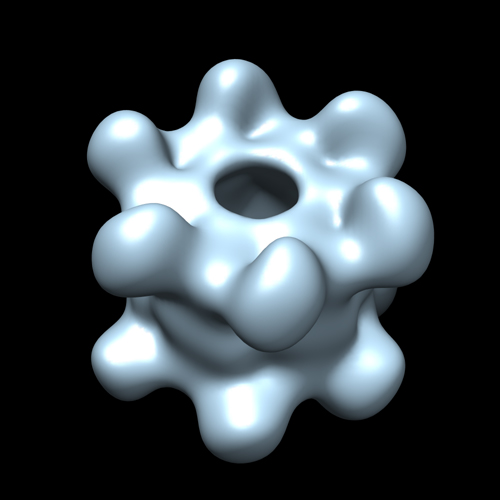
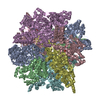
| Title | EM structure of bacteriophage SPP1 distal tail protein (GP 19.1): a baseplate hub paradigm in gram positive infecting phages | |||||||||
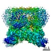
 Map data Map data | EM map of Dit (GP19.1) from SPP1 Bacteriophage | |||||||||
 Sample Sample |
| |||||||||
 Keywords Keywords | Dit / GP 19.1 / SPP1 / Bacteriophage | |||||||||
| Biological species |  Bacillus phage SPP1 (virus) Bacillus phage SPP1 (virus) | |||||||||
| Method | single particle reconstruction / negative staining / Resolution: 21.5 Å | |||||||||
 Authors Authors | Veesler D / Robin G / Lichiere J / Auzat I / Tavares P / Bron P / Campanacci V / Cambillau C | |||||||||
 Citation Citation | Journal: J Biol Chem / Year: 2010 Title: Crystal structure of bacteriophage SPP1 distal tail protein (gp19.1): a baseplate hub paradigm in gram-positive infecting phages. Authors: David Veesler / Gautier Robin / Julie Lichière / Isabelle Auzat / Paulo Tavares / Patrick Bron / Valérie Campanacci / Christian Cambillau /  Abstract: Siphophage SPP1 infects the gram-positive bacterium Bacillus subtilis using its long non-contractile tail and tail-tip. Electron microscopy (EM) previously allowed a low resolution assignment of most ...Siphophage SPP1 infects the gram-positive bacterium Bacillus subtilis using its long non-contractile tail and tail-tip. Electron microscopy (EM) previously allowed a low resolution assignment of most orf products belonging to these regions. We report here the structure of the SPP1 distal tail protein (Dit, gp19.1). The combination of x-ray crystallography, EM, and light scattering established that Dit is a back-to-back dimer of hexamers. However, Dit fitting in the virion EM maps was only possible with a hexamer located between the tail-tube and the tail-tip. Structure comparison revealed high similarity between Dit and a central component of lactophage baseplates. Sequence similarity search expanded its relatedness to several phage proteins, suggesting that Dit is a docking platform for the tail adsorption apparatus in Siphoviridae infecting gram-positive bacteria and that its architecture is a paradigm for these hub proteins. Dit structural similarity extends also to non-contractile and contractile phage tail proteins (gpV(N) and XkdM) as well as to components of the bacterial type 6 secretion system, supporting an evolutionary connection between all these devices. | |||||||||
| History |
|
- Structure visualization
| Movie |
 Movie viewer Movie viewer |
|---|---|
| Structure viewer | EM map: SurfViewMolmilJmol/JSmol |
| Supplemental images |
- Downloads & links
-EMDB archive
| Map data | emd_1779.map.gz | 7.4 MB | EMDB map data format | |
|---|---|---|---|---|
| Header (meta data) | emd-1779-v30.xmlemd-1779.xml | 9.4 KB 9.4 KB | Display Display | EMDB header |
| Images |  emd1779.jpg emd1779.jpg | 95.9 KB | ||
| Archive directory |  http://ftp.pdbj.org/pub/emdb/structures/EMD-1779ftp://ftp.pdbj.org/pub/emdb/structures/EMD-1779 http://ftp.pdbj.org/pub/emdb/structures/EMD-1779ftp://ftp.pdbj.org/pub/emdb/structures/EMD-1779 | HTTPS FTP |
-Validation report
| Summary document | emd_1779_validation.pdf.gz | 211.5 KB | Display | EMDB validaton report |
|---|---|---|---|---|
| Full document | emd_1779_full_validation.pdf.gz | 210.6 KB | Display | |
| Data in XML | emd_1779_validation.xml.gz | 5.8 KB | Display | |
| Arichive directory | https://ftp.pdbj.org/pub/emdb/validation_reports/EMD-1779ftp://ftp.pdbj.org/pub/emdb/validation_reports/EMD-1779 | HTTPS FTP |
-Related structure data

-Links
| EMDB pages | EMDB (EBI/PDBe) / EMDataResource |
|---|
-Map
| File | Download / File: emd_1779.map.gz / Format: CCP4 / Size: 7.8 MB / Type: IMAGE STORED AS FLOATING POINT NUMBER (4 BYTES) | ||||||||||||||||||||||||||||||||||||||||||||||||||||||||||||||||||||
|---|---|---|---|---|---|---|---|---|---|---|---|---|---|---|---|---|---|---|---|---|---|---|---|---|---|---|---|---|---|---|---|---|---|---|---|---|---|---|---|---|---|---|---|---|---|---|---|---|---|---|---|---|---|---|---|---|---|---|---|---|---|---|---|---|---|---|---|---|---|
| Annotation | EM map of Dit (GP19.1) from SPP1 Bacteriophage | ||||||||||||||||||||||||||||||||||||||||||||||||||||||||||||||||||||
| Projections & slices | Image control
Images are generated by Spider. | ||||||||||||||||||||||||||||||||||||||||||||||||||||||||||||||||||||
| Voxel size | X=Y=Z: 2.19 Å | ||||||||||||||||||||||||||||||||||||||||||||||||||||||||||||||||||||
| Density |
| ||||||||||||||||||||||||||||||||||||||||||||||||||||||||||||||||||||
| Symmetry | Space group: 1 | ||||||||||||||||||||||||||||||||||||||||||||||||||||||||||||||||||||
| Details | EMDB XML:
CCP4 map header:
| ||||||||||||||||||||||||||||||||||||||||||||||||||||||||||||||||||||
 Z (Sec.)
 Y (Row.)
 X (Col.)


-Supplemental data
- Sample components
-Entire : GP 19.1 protein from B. subtilis phage SPP1 Bacteriophage
| Entire | Name: GP 19.1 protein from B. subtilis phage SPP1 Bacteriophage |
|---|---|
| Components |
|
-Supramolecule #1000: GP 19.1 protein from B. subtilis phage SPP1 Bacteriophage
| Supramolecule | Name: GP 19.1 protein from B. subtilis phage SPP1 Bacteriophage type: sample / ID: 1000 / Details: The sample was monodisperse / Oligomeric state: Dodecameric / Number unique components: 1 |
|---|---|
| Molecular weight | Experimental: 325.629 KDa / Theoretical: 28.489 KDa Method: Molar mass and hydrodynamic radius determination by SEC, MALS,UV,QELS and RI. |
-Macromolecule #1: GP19.1
| Macromolecule | Name: GP19.1 / type: protein_or_peptide / ID: 1 / Name.synonym: Dit / Number of copies: 12 / Oligomeric state: Dodecameric / Recombinant expression: Yes |
|---|---|
| Source (natural) | Organism: Bacillus phage SPP1 (virus) |
-Experimental details
-Structure determination
| Method | negative staining |
|---|---|
 Processing Processing | single particle reconstruction |
| Aggregation state | particle |
-Sample preparation
| Concentration | 0.010 mg/mL |
|---|---|
| Buffer | pH: 7.5 / Details: 10 mM HEPES, 150 mM NaCl |
| Staining | Type: NEGATIVE Details: 3 microLitres of freshly prepared protein were applied on a glow-discharged carbon-coated grid. The excess of Dit solution was blotted and 4 microliters of 1% Uranyl- Acetate was applied ...Details: 3 microLitres of freshly prepared protein were applied on a glow-discharged carbon-coated grid. The excess of Dit solution was blotted and 4 microliters of 1% Uranyl- Acetate was applied twice on the grid and incubated for 1 min. Grids were then dried and kept in a desiccator cabinet until use |
| Grid | Details: 400-Mesh Carbon-Coated Copper Grid |
| Vitrification | Cryogen name: NONE / Instrument: OTHER |
- Electron microscopy
| Microscope | JEOL 2200FS |
|---|---|
| Specialist optics | Energy filter - Lower energy threshold: 0.0 eV / Energy filter - Upper energy threshold: 20.0 eV |
| Details | Low-dose imaging |
| Image recording | Category: FILM / Film or detector model: KODAK SO-163 FILM / Digitization - Scanner: NIKON SUPER COOLSCAN 9000 / Digitization - Sampling interval: 10 µm / Number real images: 8 / Average electron dose: 18 e/Å2 / Bits/pixel: 8 |
| Electron beam | Acceleration voltage: 200 kV / Electron source:  FIELD EMISSION GUN FIELD EMISSION GUN |
| Electron optics | Calibrated magnification: 45710 / Illumination mode: FLOOD BEAM / Imaging mode: BRIGHT FIELD / Nominal defocus max: 1.2 µm / Nominal defocus min: 0.3 µm / Nominal magnification: 50000 |
| Sample stage | Specimen holder: Eucentric / Specimen holder model: JEOL |
-Image processing
| CTF correction | Details: Each particle |
|---|---|
| Final reconstruction | Applied symmetry - Point group: D6 (2x6 fold dihedral) / Algorithm: OTHER / Resolution.type: BY AUTHOR / Resolution: 21.5 Å / Resolution method: FSC 0.5 CUT-OFF / Software - Name: IMAGIC V / Number images used: 9027 |
