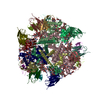
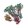
Journal: J Biol Chem / Year: 2010 Title: Crystal structure of bacteriophage SPP1 distal tail protein (gp19.1): a baseplate hub paradigm in gram-positive infecting phages. Authors: David Veesler / Gautier Robin / Julie Lichière / Isabelle Auzat / Paulo Tavares / Patrick Bron / Valérie Campanacci / Christian Cambillau / Abstract: Siphophage SPP1 infects the gram-positive bacterium Bacillus subtilis using its long non-contractile tail and tail-tip. Electron microscopy (EM) previously allowed a low resolution assignment of most ...Siphophage SPP1 infects the gram-positive bacterium Bacillus subtilis using its long non-contractile tail and tail-tip. Electron microscopy (EM) previously allowed a low resolution assignment of most orf products belonging to these regions. We report here the structure of the SPP1 distal tail protein (Dit, gp19.1). The combination of x-ray crystallography, EM, and light scattering established that Dit is a back-to-back dimer of hexamers. However, Dit fitting in the virion EM maps was only possible with a hexamer located between the tail-tube and the tail-tip. Structure comparison revealed high similarity between Dit and a central component of lactophage baseplates. Sequence similarity search expanded its relatedness to several phage proteins, suggesting that Dit is a docking platform for the tail adsorption apparatus in Siphoviridae infecting gram-positive bacteria and that its architecture is a paradigm for these hub proteins. Dit structural similarity extends also to non-contractile and contractile phage tail proteins (gpV(N) and XkdM) as well as to components of the bacterial type 6 secretion system, supporting an evolutionary connection between all these devices.
In the structure databanks used in Yorodumi, some data are registered as the other names, "COVID-19 virus" and "2019-nCoV". Here are the details of the virus and the list of structure data.
Jan 31, 2019. EMDB accession codes are about to change! (news from PDBe EMDB page)
EMDB accession codes are about to change! (news from PDBe EMDB page)
The allocation of 4 digits for EMDB accession codes will soon come to an end. Whilst these codes will remain in use, new EMDB accession codes will include an additional digit and will expand incrementally as the available range of codes is exhausted. The current 4-digit format prefixed with “EMD-” (i.e. EMD-XXXX) will advance to a 5-digit format (i.e. EMD-XXXXX), and so on. It is currently estimated that the 4-digit codes will be depleted around Spring 2019, at which point the 5-digit format will come into force.
The EM Navigator/Yorodumi systems omit the EMD- prefix.
Related info.:Q: What is EMD? / ID/Accession-code notation in Yorodumi/EM Navigator
Yorodumi is a browser for structure data from EMDB, PDB, SASBDB, etc.
This page is also the successor to EM Navigator detail page, and also detail information page/front-end page for Omokage search.
The word "yorodu" (or yorozu) is an old Japanese word meaning "ten thousand". "mi" (miru) is to see.
Related info.:EMDB / PDB / SASBDB / Comparison of 3 databanks / Yorodumi Search / Aug 31, 2016. New EM Navigator & Yorodumi / Yorodumi Papers / Jmol/JSmol / Function and homology information / Changes in new EM Navigator and Yorodumi
 Movie
Movie Controller

 Yorodumi
 Open data
 Basic information
 Components
 Keywords
 Function and homology information
 BACILLUS PHAGE SPP1 (virus)
 X-RAY DIFFRACTION /
  Authors
 Citation
 Structure visualization
 Downloads & links
 Other downloads
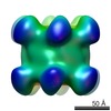
 PDBj
PDBj Assembly
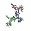


 Sample preparation
 Processing