 Movie
Movie Controller


+ Open data

- Basic information
| Entry | Database: EMDB / ID: EMD-1778 | |||||||||
|---|---|---|---|---|---|---|---|---|---|---|
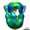
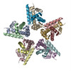
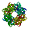
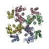
| Title | Structure of the Nuclear Chaperone Nucleoplamsin. | |||||||||
 Map data Map data | This a map of the nuclear chaperone nucleoplasmin. | |||||||||
 Sample Sample |
| |||||||||
 Keywords Keywords | Nucleoplasmin / Histone H2A / Histone H2B / Chaperone / Chromatin / Nuclear-chaperone / Histone-chaperone | |||||||||
| Function / homology | Nucleoplasmin family Function and homology information Function and homology information | |||||||||
| Biological species | ||||||||||
| Method | single particle reconstruction / negative staining / Resolution: 21.0 Å | |||||||||
 Authors Authors | Ramos I / Martin-Benito J / Finn R / Bretana L / Aloria K / Arizmendi JM / Ausio J / Muga A / Valpuesta JM / Prado A | |||||||||
 Citation Citation | Journal: J Biol Chem / Year: 2010 Title: Nucleoplasmin binds histone H2A-H2B dimers through its distal face. Authors: Isbaal Ramos / Jaime Martín-Benito / Ron Finn / Laura Bretaña / Kerman Aloria / Jesús M Arizmendi / Juan Ausió / Arturo Muga / José M Valpuesta / Adelina Prado /  Abstract: Nucleoplasmin (NP) is a pentameric chaperone that regulates the condensation state of chromatin extracting specific basic proteins from sperm chromatin and depositing H2A-H2B histone dimers. It has ...Nucleoplasmin (NP) is a pentameric chaperone that regulates the condensation state of chromatin extracting specific basic proteins from sperm chromatin and depositing H2A-H2B histone dimers. It has been proposed that histones could bind to either the lateral or distal face of the pentameric structure. Here, we combine different biochemical and biophysical techniques to show that natural, hyperphosphorylated NP can bind five H2A-H2B dimers and that the amount of bound ligand depends on the overall charge (phosphorylation level) of the chaperone. Three-dimensional reconstruction of NP/H2A-H2B complex carried out by electron microscopy reveals that histones interact with the chaperone distal face. Limited proteolysis and mass spectrometry indicate that the interaction results in protection of the histone fold and most of the H2A and H2B C-terminal tails. This structural information can help to understand the function of NP as a histone chaperone. | |||||||||
| History |
|
- Structure visualization
| Movie |
Movie viewer |
|---|---|
| Structure viewer | EM map: SurfViewMolmilJmol/JSmol |
| Supplemental images |
 UCSF Chimera
- Downloads & links
-EMDB archive
| Map data | emd_1778.map.gz | 1.8 MB | EMDB map data format | |
|---|---|---|---|---|
| Header (meta data) | emd-1778-v30.xmlemd-1778.xml | 9.2 KB 9.2 KB | Display Display | EMDB header |
| Images | emd_1778.tif | 756.1 KB | ||
| Archive directory |  http://ftp.pdbj.org/pub/emdb/structures/EMD-1778ftp://ftp.pdbj.org/pub/emdb/structures/EMD-1778 http://ftp.pdbj.org/pub/emdb/structures/EMD-1778ftp://ftp.pdbj.org/pub/emdb/structures/EMD-1778 | HTTPS FTP |
-Validation report
| Summary document | emd_1778_validation.pdf.gz | 201.1 KB | Display | EMDB validaton report |
|---|---|---|---|---|
| Full document | emd_1778_full_validation.pdf.gz | 200.2 KB | Display | |
| Data in XML | emd_1778_validation.xml.gz | 4.7 KB | Display | |
| Arichive directory | https://ftp.pdbj.org/pub/emdb/validation_reports/EMD-1778ftp://ftp.pdbj.org/pub/emdb/validation_reports/EMD-1778 | HTTPS FTP |
-Related structure data


-Links
| EMDB pages | EMDB (EBI/PDBe) / EMDataResource |
|---|
-Map
| File | Download / File: emd_1778.map.gz / Format: CCP4 / Size: 1.9 MB / Type: IMAGE STORED AS FLOATING POINT NUMBER (4 BYTES) | ||||||||||||||||||||||||||||||||||||||||||||||||||||||||||||||||||||
|---|---|---|---|---|---|---|---|---|---|---|---|---|---|---|---|---|---|---|---|---|---|---|---|---|---|---|---|---|---|---|---|---|---|---|---|---|---|---|---|---|---|---|---|---|---|---|---|---|---|---|---|---|---|---|---|---|---|---|---|---|---|---|---|---|---|---|---|---|---|
| Annotation | This a map of the nuclear chaperone nucleoplasmin. | ||||||||||||||||||||||||||||||||||||||||||||||||||||||||||||||||||||
| Projections & slices | Image control
Images are generated by Spider. | ||||||||||||||||||||||||||||||||||||||||||||||||||||||||||||||||||||
| Voxel size | X=Y=Z: 2.3 Å | ||||||||||||||||||||||||||||||||||||||||||||||||||||||||||||||||||||
| Density |
| ||||||||||||||||||||||||||||||||||||||||||||||||||||||||||||||||||||
| Symmetry | Space group: 1 | ||||||||||||||||||||||||||||||||||||||||||||||||||||||||||||||||||||
| Details | EMDB XML:
CCP4 map header:
| ||||||||||||||||||||||||||||||||||||||||||||||||||||||||||||||||||||
 Z (Sec.)
 Y (Row.)
 X (Col.)


-Supplemental data
- Sample components
-Entire : Nucleoplasmin chaperone
| Entire | Name: Nucleoplasmin chaperone |
|---|---|
| Components |
|
-Supramolecule #1000: Nucleoplasmin chaperone
| Supramolecule | Name: Nucleoplasmin chaperone / type: sample / ID: 1000 / Oligomeric state: Pentameric / Number unique components: 1 |
|---|---|
| Molecular weight | Experimental: 110 KDa / Theoretical: 110 KDa |
-Macromolecule #1: Nucleoplasmin
| Macromolecule | Name: Nucleoplasmin / type: protein_or_peptide / ID: 1 / Name.synonym: Nucleoplasmin / Number of copies: 5 / Oligomeric state: Pentamer / Recombinant expression: No |
|---|---|
| Source (natural) | Organism: |
| Molecular weight | Experimental: 110 KDa / Theoretical: 110 KDa |
| Sequence | InterPro: Nucleoplasmin family |
-Experimental details
-Structure determination
| Method | negative staining |
|---|---|
 Processing Processing | single particle reconstruction |
| Aggregation state | particle |
-Sample preparation
| Buffer | pH: 7.5 / Details: 2mM MgCl2, 240mM NaCl, 25 mM Tris-HCl |
|---|---|
| Staining | Type: NEGATIVE Details: Grids were stained with 2% w/v Uranyl Acetate solution for 1 minute. |
| Grid | Details: 200 mesh CuRh grid |
| Vitrification | Cryogen name: NONE / Instrument: OTHER |
- Electron microscopy
| Microscope | JEOL 1200EXII |
|---|---|
| Alignment procedure | Legacy - Astigmatism: Objective lens astigmatism was corrected at 200,000 times magnification |
| Image recording | Category: FILM / Film or detector model: KODAK SO-163 FILM / Digitization - Scanner: ZEISS SCAI / Digitization - Sampling interval: 2.3 µm / Number real images: 10 / Details: downsampling factor of 2 / Bits/pixel: 8 |
| Electron beam | Acceleration voltage: 100 kV / Electron source: TUNGSTEN HAIRPIN |
| Electron optics | Illumination mode: FLOOD BEAM / Imaging mode: BRIGHT FIELD / Cs: 5.6 mm / Nominal defocus max: 2.5 µm / Nominal defocus min: 1.0 µm / Nominal magnification: 60000 |
| Sample stage | Specimen holder: Eucentric / Specimen holder model: JEOL |
-Image processing
| CTF correction | Details: Each plate |
|---|---|
| Final reconstruction | Applied symmetry - Point group: C5 (5 fold cyclic) / Algorithm: OTHER / Resolution.type: BY AUTHOR / Resolution: 21.0 Å / Resolution method: FSC 0.5 CUT-OFF / Software - Name: EMAN, XMIPP, SPIDER / Number images used: 9862 |
