 ムービー
ムービー コントローラー


+ データを開く

- 基本情報
基本情報
| 登録情報 | データベース: EMDB / ID: EMD-1775 | |||||||||
|---|---|---|---|---|---|---|---|---|---|---|
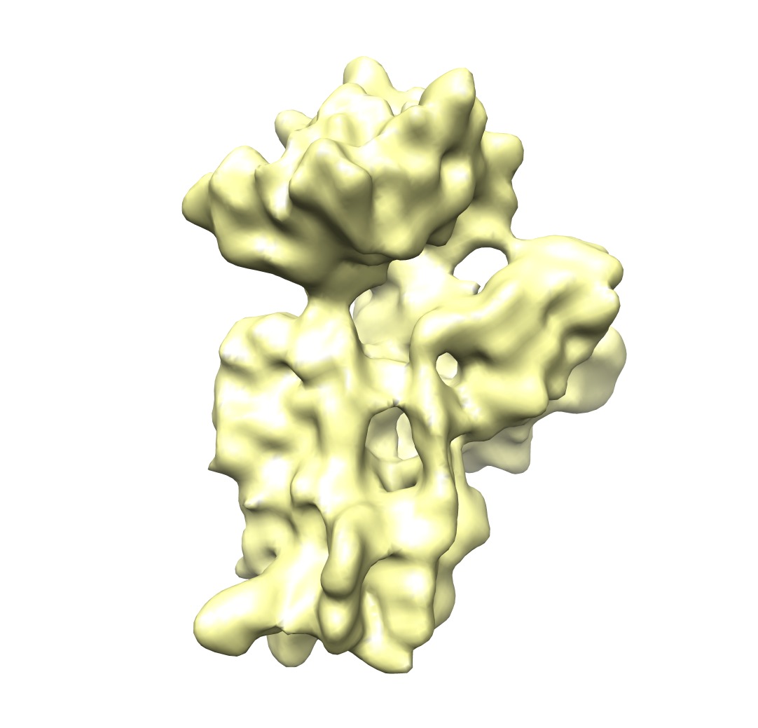
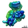
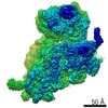
| タイトル | Understanding Ribosome Assembly: the Structure of in vivo Assembled Immature 30S Subunits Revealed by Cryo-electron Microscopy | |||||||||
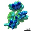
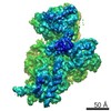
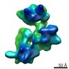
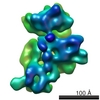
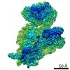
 マップデータ マップデータ | Surface rendering of 30S ribosomal subunit from wild type E. coli cells. Map was used as a control. | |||||||||
 試料 試料 |
| |||||||||
| 生物種 |  | |||||||||
| 手法 | 単粒子再構成法 / クライオ電子顕微鏡法 / 解像度: 14.1 Å | |||||||||
 データ登録者 データ登録者 | Jomaa A / Stewart G / Benito JM / Zielke R / Campbell T / Maddock J / Brown E / Ortega J | |||||||||
 引用 引用 | ジャーナル: RNA / 年: 2011 タイトル: Understanding ribosome assembly: the structure of in vivo assembled immature 30S subunits revealed by cryo-electron microscopy. 著者: Ahmad Jomaa / Geordie Stewart / Jaime Martín-Benito / Ryszard Zielke / Tracey L Campbell / Janine R Maddock / Eric D Brown / Joaquin Ortega /  要旨: Four decades after early in vitro assembly studies demonstrated that ribosome assembly is a controlled process, our understanding of ribosome assembly is still incomplete. Just as structure ...Four decades after early in vitro assembly studies demonstrated that ribosome assembly is a controlled process, our understanding of ribosome assembly is still incomplete. Just as structure determination has been so important to understanding ribosome function, so too will it be critical to sorting out the assembly process. Here, we used a viable deletion in the yjeQ gene, a recognized ribosome assembly factor, to isolate and structurally characterize immature 30S subunits assembled in vivo. These small ribosome subunits contained unprocessed 17S rRNA and lacked some late ribosomal proteins. Cryo-electron microscopy reconstructions revealed that the presence of precursor sequences in the rRNA induces a severe distortion in the 3' minor domain of the subunit involved in the decoding of mRNA and interaction with the large ribosome subunit. These findings suggest that rRNA processing events induce key local conformational changes directing the structure toward the mature assembly. We concluded that rRNA processing, folding, and the entry of tertiary r-proteins are interdependent events in the late stages of 30S subunit assembly. In addition, we demonstrate how studies of emerging assembly factors in ribosome biogenesis can help to elucidate the path of subunit assembly in vivo. | |||||||||
| 履歴 |
|
- 構造の表示
構造の表示
| ムービー |
 ムービービューア ムービービューア |
|---|---|
| 構造ビューア | EMマップ: SurfViewMolmilJmol/JSmol |
| 添付画像 |
- ダウンロードとリンク
-EMDBアーカイブ
| マップデータ | emd_1775.map.gz | 7.4 MB | EMDBマップデータ形式 | |
|---|---|---|---|---|
| ヘッダ (付随情報) | emd-1775-v30.xmlemd-1775.xml | 9.8 KB 9.8 KB | 表示 表示 | EMDBヘッダ |
| 画像 | 1775.tif | 501.1 KB | ||
| アーカイブディレクトリ |  http://ftp.pdbj.org/pub/emdb/structures/EMD-1775ftp://ftp.pdbj.org/pub/emdb/structures/EMD-1775 http://ftp.pdbj.org/pub/emdb/structures/EMD-1775ftp://ftp.pdbj.org/pub/emdb/structures/EMD-1775 | HTTPS FTP |
-検証レポート
| 文書・要旨 | emd_1775_validation.pdf.gz | 231.8 KB | 表示 | EMDB検証レポート |
|---|---|---|---|---|
| 文書・詳細版 | emd_1775_full_validation.pdf.gz | 230.9 KB | 表示 | |
| XML形式データ | emd_1775_validation.xml.gz | 5.2 KB | 表示 | |
| アーカイブディレクトリ | https://ftp.pdbj.org/pub/emdb/validation_reports/EMD-1775ftp://ftp.pdbj.org/pub/emdb/validation_reports/EMD-1775 | HTTPS FTP |
-関連構造データ
-リンク
| EMDBのページ | EMDB (EBI/PDBe) / EMDataResource |
|---|---|
| 「今月の分子」の関連する項目 |

-マップ
| ファイル | ダウンロード / ファイル: emd_1775.map.gz / 形式: CCP4 / 大きさ: 7.8 MB / タイプ: IMAGE STORED AS FLOATING POINT NUMBER (4 BYTES) | ||||||||||||||||||||||||||||||||||||||||||||||||||||||||||||||||||||
|---|---|---|---|---|---|---|---|---|---|---|---|---|---|---|---|---|---|---|---|---|---|---|---|---|---|---|---|---|---|---|---|---|---|---|---|---|---|---|---|---|---|---|---|---|---|---|---|---|---|---|---|---|---|---|---|---|---|---|---|---|---|---|---|---|---|---|---|---|---|
| 注釈 | Surface rendering of 30S ribosomal subunit from wild type E. coli cells. Map was used as a control. | ||||||||||||||||||||||||||||||||||||||||||||||||||||||||||||||||||||
| 投影像・断面図 | 画像のコントロール
画像は Spider により作成 | ||||||||||||||||||||||||||||||||||||||||||||||||||||||||||||||||||||
| ボクセルのサイズ | X=Y=Z: 2.54 Å | ||||||||||||||||||||||||||||||||||||||||||||||||||||||||||||||||||||
| 密度 |
| ||||||||||||||||||||||||||||||||||||||||||||||||||||||||||||||||||||
| 対称性 | 空間群: 1 | ||||||||||||||||||||||||||||||||||||||||||||||||||||||||||||||||||||
| 詳細 | EMDB XML:
CCP4マップ ヘッダ情報:
| ||||||||||||||||||||||||||||||||||||||||||||||||||||||||||||||||||||
 Z (Sec.)
 Y (Row.)
 X (Col.)


-添付データ
- 試料の構成要素
-全体 : Reconstruction of the mature 30S subunit from wild type E.coli cells
| 全体 | 名称: Reconstruction of the mature 30S subunit from wild type E.coli cells |
|---|---|
| 要素 |
|
-超分子 #1000: Reconstruction of the mature 30S subunit from wild type E.coli cells
| 超分子 | 名称: Reconstruction of the mature 30S subunit from wild type E.coli cells タイプ: sample / ID: 1000 詳細: The sample was thawed from storage at -80 degrees Celcius before being loaded onto the grid. Number unique components: 1 |
|---|---|
| 分子量 | 理論値: 800 KDa |
-超分子 #1: Small Ribosomal Subunit
| 超分子 | 名称: Small Ribosomal Subunit / タイプ: complex / ID: 1 / Name.synonym: 30S / 組換発現: No / Ribosome-details: ribosome-prokaryote: SSU 30S |
|---|---|
| 由来(天然) | 生物種: |
| 分子量 | 理論値: 850 KDa |
-実験情報
-構造解析
| 手法 | クライオ電子顕微鏡法 |
|---|---|
 解析 解析 | 単粒子再構成法 |
| 試料の集合状態 | particle |
-試料調製
| 濃度 | 5.8 mg/mL |
|---|---|
| 緩衝液 | pH: 7.5 詳細: 10 mM Tris-HCl pH 7.5, 10 mM magnesium acetate, 60 mM NH4Cl, 3 mM 2-mercaptoethanol |
| グリッド | 詳細: 400 Mesh Copper Grid |
| 凍結 | 凍結剤: ETHANE / チャンバー内湿度: 100 % / チャンバー内温度: 93 K / 装置: OTHER / 詳細: Vitrification instrument: FEI vitrobot / 手法: Blot 7 Seconds Twice |
- 電子顕微鏡法
| 顕微鏡 | JEOL 2010F |
|---|---|
| 温度 | 最低: 93 K / 最高: 93 K / 平均: 93 K |
| 日付 | 2008年12月10日 |
| 撮影 | カテゴリ: FILM / フィルム・検出器のモデル: KODAK SO-163 FILM デジタル化 - スキャナー: NIKON SUPER COOLSCAN 9000 デジタル化 - サンプリング間隔: 12.7 µm / 実像数: 29 / 平均電子線量: 10 e/Å2 / ビット/ピクセル: 16 |
| 電子線 | 加速電圧: 200 kV / 電子線源:  FIELD EMISSION GUN FIELD EMISSION GUN |
| 電子光学系 | 倍率(補正後): 50000 / 照射モード: FLOOD BEAM / 撮影モード: BRIGHT FIELD / Cs: 1.0 mm / 最大 デフォーカス(公称値): 3.9 µm / 最小 デフォーカス(公称値): 0.65 µm / 倍率(公称値): 50000 |
| 試料ステージ | 試料ホルダー: Side entry liquid nitrogen-cooled cryo specimen holder. 試料ホルダーモデル: GATAN LIQUID NITROGEN |
-画像解析
| 詳細 | Particles were picked with BOXER |
|---|---|
| CTF補正 | 詳細: Each Micrograph |
| 最終 再構成 | 想定した対称性 - 点群: C1 (非対称) / アルゴリズム: OTHER / 解像度のタイプ: BY AUTHOR / 解像度: 14.1 Å / 解像度の算出法: FSC 0.5 CUT-OFF / ソフトウェア - 名称: Xmipp / 使用した粒子像数: 25803 |
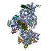
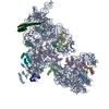
-原子モデル構築 1
| 初期モデル | PDB ID:  2z4k |
|---|---|
| ソフトウェア | 名称: Situs |
| 詳細 | Protocol: Rigid Body |
| 精密化 | 空間: REAL / プロトコル: RIGID BODY FIT |
