 Movie
Movie Controller


+ Open data

- Basic information
| Entry |  | |||||||||
|---|---|---|---|---|---|---|---|---|---|---|
| Title | MutSbeta bound to (CAG)2 DNA (open form) | |||||||||
 Map data Map data | MutSbeta bound to (CAG)2 DNA (open form) | |||||||||
 Sample Sample |
| |||||||||
 Keywords Keywords | Protein-DNA complex / DNA BINDING PROTEIN | |||||||||
| Function / homology |  Function and homology information Function and homology informationsomatic recombination of immunoglobulin genes involved in immune response / MutSbeta complex / Defective Mismatch Repair Associated With MSH3 / MutSalpha complex / Defective Mismatch Repair Associated With MSH6 / somatic recombination of immunoglobulin gene segments / Defective Mismatch Repair Associated With MSH2 / guanine/thymine mispair binding / B cell mediated immunity / maintenance of DNA repeat elements ...somatic recombination of immunoglobulin genes involved in immune response / MutSbeta complex / Defective Mismatch Repair Associated With MSH3 / MutSalpha complex / Defective Mismatch Repair Associated With MSH6 / somatic recombination of immunoglobulin gene segments / Defective Mismatch Repair Associated With MSH2 / guanine/thymine mispair binding / B cell mediated immunity / maintenance of DNA repeat elements / positive regulation of isotype switching to IgG isotypes / positive regulation of isotype switching to IgA isotypes / centromeric DNA binding / mismatched DNA binding / isotype switching / negative regulation of DNA recombination / mitotic recombination / response to UV-B / Mismatch repair (MMR) directed by MSH2:MSH3 (MutSbeta) / Mismatch repair (MMR) directed by MSH2:MSH6 (MutSalpha) / DNA damage tolerance / response to X-ray / ATP-dependent DNA damage sensor activity / ATP-dependent activity, acting on DNA / mismatch repair / B cell differentiation / male gonad development / TP53 Regulates Transcription of DNA Repair Genes / enzyme activator activity / negative regulation of neuron apoptotic process / double-stranded DNA binding / damaged DNA binding / chromosome, telomeric region / DNA repair / chromatin binding / enzyme binding / protein homodimerization activity / ATP hydrolysis activity / DNA binding / nucleoplasm / ATP binding / membrane / nucleus Similarity search - Function | |||||||||
| Biological species |  Homo sapiens (human) Homo sapiens (human) | |||||||||
| Method | single particle reconstruction / cryo EM / Resolution: 3.32 Å | |||||||||
 Authors Authors | Lee J-H / Thomsen M / Daub H / Steinbacher S / Sztyler A / Thieulin-Pardo G / Neudegger T / Plotnikov N / Iyer RR / Wilkinson H ...Lee J-H / Thomsen M / Daub H / Steinbacher S / Sztyler A / Thieulin-Pardo G / Neudegger T / Plotnikov N / Iyer RR / Wilkinson H / Monteagudo E / Felsenfeld DP / Haque T / Finley M / Dominguez C / Vogt TF / Prasad BC | |||||||||
| Funding support |  United States, 1 items United States, 1 items
| |||||||||
 Citation Citation | Journal: Nucleic Acids Res / Year: 2025 Title: Elucidation of multiple high-resolution states of human MutSβ by cryo-EM reveals interplay between ATP/ADP binding and heteroduplex DNA recognition. Authors: Jung-Hoon Lee / Maren Thomsen / Herwin Daub / Gabriel Thieulin-Pardo / Stefan Steinbacher / Agnieszka Sztyler / Vinay Dahiya / Tobias Neudegger / Celia Dominguez / Ravi R Iyer / Hilary A ...Authors: Jung-Hoon Lee / Maren Thomsen / Herwin Daub / Gabriel Thieulin-Pardo / Stefan Steinbacher / Agnieszka Sztyler / Vinay Dahiya / Tobias Neudegger / Celia Dominguez / Ravi R Iyer / Hilary A Wilkinson / Edith Monteagudo / Nikolay V Plotnikov / Dan P Felsenfeld / Tasir S Haque / Michael Finley / Julien Boudet / Thomas F Vogt / Brinda C Prasad /  Abstract: Human and mouse genetic studies have demonstrated a role for DNA mismatch repair (MMR) molecular machines in modulating the rate of somatic expansion of the huntingtin (HTT) CAG repeats, and onset ...Human and mouse genetic studies have demonstrated a role for DNA mismatch repair (MMR) molecular machines in modulating the rate of somatic expansion of the huntingtin (HTT) CAG repeats, and onset and progression of Huntington's Disease (HD). MutSβ, a key component of the MMR pathway, is a heterodimeric protein of MSH2 and MSH3 that recognizes and initiates the repair of extrahelical DNA extrusions. Loss-of-function of mouse Msh3 and reduced-expression alleles of human MSH3 lead to slower rates of somatic expansion and delayed disease onset in humans, signifying MSH3 as a promising therapeutic target for HD. Here we report biochemical and cryo-electron microscopy analyses of human MutSβ, demonstrating MutSβ undergoes conformational changes induced by nucleotide and DNA binding. We present multiple conformations of MutSβ including the DNA-free MutSβ compatible with precisely complementary base-paired homoduplex DNA binding, two distinct structures of MutSβ bound to (CAG)2 DNA, a sliding clamp form and a DNA-unbound, ATP-bound conformation. Along with evidence for novel conformational states adopted by MutSβ to initiate the MMR cascade, these structures provide a foundation for structure-guided drug discovery. | |||||||||
| History |
|
- Structure visualization
| Supplemental images |
|---|

- Downloads & links
-EMDB archive
| Map data | emd_16971.map.gz | 110.9 MB | EMDB map data format | |
|---|---|---|---|---|
| Header (meta data) | emd-16971-v30.xmlemd-16971.xml | 24.1 KB 24.1 KB | Display Display | EMDB header |
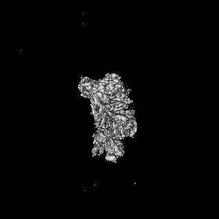
| Images |  emd_16971.png emd_16971.png | 23 KB | ||
| Filedesc metadata | emd-16971.cif.gz | 7.9 KB | ||
| Others | emd_16971_half_map_1.map.gzemd_16971_half_map_2.map.gz | 98.4 MB 98.3 MB | ||
| Archive directory |  https://data.pdbj.org/pub/emdb/structures/EMD-16971ftp://data.pdbj.org/pub/emdb/structures/EMD-16971 https://data.pdbj.org/pub/emdb/structures/EMD-16971ftp://data.pdbj.org/pub/emdb/structures/EMD-16971 | HTTPS FTP |
-Related structure data
| Related structure data |  8om9MC  8olxC  8om5C  8omaC  8omoC  8omqC  8rz7C  8rz8C  8rz9C M: atomic model generated by this map C: citing same article ( |
|---|---|
| Similar structure data |


-Links
| EMDB pages | EMDB (EBI/PDBe) / EMDataResource |
|---|---|
| Related items in Molecule of the Month |







-Map
| File | Download / File: emd_16971.map.gz / Format: CCP4 / Size: 125 MB / Type: IMAGE STORED AS FLOATING POINT NUMBER (4 BYTES) | ||||||||||||||||||||||||||||||||||||
|---|---|---|---|---|---|---|---|---|---|---|---|---|---|---|---|---|---|---|---|---|---|---|---|---|---|---|---|---|---|---|---|---|---|---|---|---|---|
| Annotation | MutSbeta bound to (CAG)2 DNA (open form) | ||||||||||||||||||||||||||||||||||||
| Projections & slices | Image control
Images are generated by Spider. | ||||||||||||||||||||||||||||||||||||
| Voxel size | X=Y=Z: 0.9142 Å | ||||||||||||||||||||||||||||||||||||
| Density |
| ||||||||||||||||||||||||||||||||||||
| Symmetry | Space group: 1 | ||||||||||||||||||||||||||||||||||||
| Details | EMDB XML:
|
 Z (Sec.)
 Y (Row.)
 X (Col.)



-Supplemental data
-Half map: MutSbeta bound to (CAG)2 DNA (open form)
| File | emd_16971_half_map_1.map | ||||||||||||
|---|---|---|---|---|---|---|---|---|---|---|---|---|---|
| Annotation | MutSbeta bound to (CAG)2 DNA (open form) | ||||||||||||
| Projections & Slices |
| ||||||||||||
| Density Histograms |




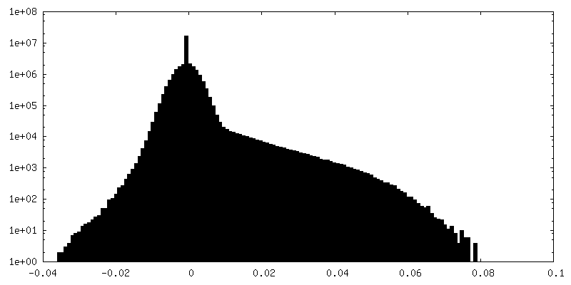
-Half map: MutSbeta bound to (CAG)2 DNA (open form)
| File | emd_16971_half_map_2.map | ||||||||||||
|---|---|---|---|---|---|---|---|---|---|---|---|---|---|
| Annotation | MutSbeta bound to (CAG)2 DNA (open form) | ||||||||||||
| Projections & Slices |
| ||||||||||||
| Density Histograms |



- Sample components
-Entire : MutSbeta bound to (CAG)2 DNA (open form)
| Entire | Name: MutSbeta bound to (CAG)2 DNA (open form) |
|---|---|
| Components |
|
-Supramolecule #1: MutSbeta bound to (CAG)2 DNA (open form)
| Supramolecule | Name: MutSbeta bound to (CAG)2 DNA (open form) / type: complex / ID: 1 / Parent: 0 / Macromolecule list: #1-#4 |
|---|---|
| Source (natural) | Organism: Homo sapiens (human) |
-Macromolecule #1: DNA mismatch repair protein Msh2
| Macromolecule | Name: DNA mismatch repair protein Msh2 / type: protein_or_peptide / ID: 1 / Number of copies: 1 / Enantiomer: LEVO |
|---|---|
| Source (natural) | Organism: Homo sapiens (human) |
| Molecular weight | Theoretical: 104.861875 KDa |
| Recombinant expression | Organism:   Spodoptera frugiperda (fall armyworm) Spodoptera frugiperda (fall armyworm) |
| Sequence | String: MAVQPKETLQ LESAAEVGFV RFFQGMPEKP TTTVRLFDRG DFYTAHGEDA LLAAREVFKT QGVIKYMGPA GAKNLQSVVL SKMNFESFV KDLLLVRQYR VEVYKNRAGN KASKENDWYL AYKASPGNLS QFEDILFGNN DMSASIGVVG VKMSAVDGQR Q VGVGYVDS ...String: MAVQPKETLQ LESAAEVGFV RFFQGMPEKP TTTVRLFDRG DFYTAHGEDA LLAAREVFKT QGVIKYMGPA GAKNLQSVVL SKMNFESFV KDLLLVRQYR VEVYKNRAGN KASKENDWYL AYKASPGNLS QFEDILFGNN DMSASIGVVG VKMSAVDGQR Q VGVGYVDS IQRKLGLCEF PDNDQFSNLE ALLIQIGPKE CVLPGGETAG DMGKLRQIIQ RGGILITERK KADFSTKDIY QD LNRLLKG KKGEQMNSAV LPEMENQVAV SSLSAVIKFL ELLSDDSNFG QFELTTFDFS QYMKLDIAAV RALNLFQGSV EDT TGSQSL AALLNKCKTP QGQRLVNQWI KQPLMDKNRI EERLNLVEAF VEDAELRQTL QEDLLRRFPD LNRLAKKFQR QAAN LQDCY RLYQGINQLP NVIQALEKHE GKHQKLLLAV FVTPLTDLRS DFSKFQEMIE TTLDMDQVEN HEFLVKPSFD PNLSE LREI MNDLEKKMQS TLISAARDLG LDPGKQIKLD SSAQFGYYFR VTCKEEKVLR NNKNFSTVDI QKNGVKFTNS KLTSLN EEY TKNKTEYEEA QDAIVKEIVN ISSGYVEPMQ TLNDVLAQLD AVVSFAHVSN GAPVPYVRPA ILEKGQGRII LKASRHA CV EVQDEIAFIP NDVYFEKDKQ MFHIITGPNM GGKSTYIRQT GVIVLMAQIG CFVPCESAEV SIVDCILARV GAGDSQLK G VSTFMAEMLE TASILRSATK DSLIIIDELG RGTSTYDGFG LAWAISEYIA TKIGAFCMFA THFHELTALA NQIPTVNNL HVTALTTEET LTMLYQVKKG VCDQSFGIHV AELANFPKHV IECAKQKALE LEEFQYIGES QGYDIMEPAA KKCYLEREQG EKIIQEFLS KVKQMPFTEM SEENITIKLK QLKAEVIAKN NSFVNEIISR IKVTT UniProtKB: DNA mismatch repair protein Msh2 |
-Macromolecule #2: DNA mismatch repair protein Msh3
| Macromolecule | Name: DNA mismatch repair protein Msh3 / type: protein_or_peptide / ID: 2 / Number of copies: 1 / Enantiomer: LEVO |
|---|---|
| Source (natural) | Organism: Homo sapiens (human) |
| Molecular weight | Theoretical: 127.618703 KDa |
| Recombinant expression | Organism: Spodoptera frugiperda (fall armyworm) |
| Sequence | String: MSRRKPASGG LAASSSAPAR QAVLSRFFQS TGSLKSTSSS TGAADQVDPG AAAAAAAAAA AAPPAPPAPA FPPQLPPHIA TEIDRRKKR PLENDGPVKK KVKKVQQKEG GSDLGMSGNS EPKKCLRTRN VSKSLEKLKE FCCDSALPQS RVQTESLQER F AVLPKCTD ...String: MSRRKPASGG LAASSSAPAR QAVLSRFFQS TGSLKSTSSS TGAADQVDPG AAAAAAAAAA AAPPAPPAPA FPPQLPPHIA TEIDRRKKR PLENDGPVKK KVKKVQQKEG GSDLGMSGNS EPKKCLRTRN VSKSLEKLKE FCCDSALPQS RVQTESLQER F AVLPKCTD FDDISLLHAK NAVSSEDSKR QINQKDTTLF DLSQFGSSNT SHENLQKTAS KSANKRSKSI YTPLELQYIE MK QQHKDAV LCVECGYKYR FFGEDAEIAA RELNIYCHLD HNFMTASIPT HRLFVHVRRL VAKGYKVGVV KQTETAALKA IGD NRSSLF SRKLTALYTK STLIGEDVNP LIKLDDAVNV DEIMTDTSTS YLLCISENKE NVRDKKKGNI FIGIVGVQPA TGEV VFDSF QDSASRSELE TRMSSLQPVE LLLPSALSEQ TEALIHRATS VSVQDDRIRV ERMDNIYFEY SHAFQAVTEF YAKDT VDIK GSQIISGIVN LEKPVICSLA AIIKYLKEFN LEKMLSKPEN FKQLSSKMEF MTINGTTLRN LEILQNQTDM KTKGSL LWV LDHTKTSFGR RKLKKWVTQP LLKLREINAR LDAVSEVLHS ESSVFGQIEN HLRKLPDIER GLCSIYHKKC STQEFFL IV KTLYHLKSEF QAIIPAVNSH IQSDLLRTVI LEIPELLSPV EHYLKILNEQ AAKVGDKTEL FKDLSDFPLI KKRKDEIQ G VIDEIRMHLQ EIRKILKNPS AQYVTVSGQE FMIEIKNSAV SCIPTDWVKV GSTKAVSRFH SPFIVENYRH LNQLREQLV LDCSAEWLDF LEKFSEHYHS LCKAVHHLAT VDCIFSLAKV AKQGDYCRPT VQEERKIVIK NGRHPVIDVL LGEQDQYVPN NTDLSEDSE RVMIITGPNM GGKSSYIKQV ALITIMAQIG SYVPAEEATI GIVDGIFTRM GAADNIYKGR STFMEELTDT A EIIRKATS QSLVILDELG RGTSTHDGIA IAYATLEYFI RDVKSLTLFV THYPPVCELE KNYSHQVGNY HMGFLVSEDE SK LDPGAAE QVPDFVTFLY QITRGIAARS YGLNVAKLAD VPGEILKKAA HKSKELEGLI NTKRKRLKYF AKLWTMHNAQ DLQ KWTEEF NMEETQTSLL H UniProtKB: DNA mismatch repair protein Msh3 |
-Macromolecule #3: DNA1
| Macromolecule | Name: DNA1 / type: dna / ID: 3 / Number of copies: 1 / Classification: DNA |
|---|---|
| Source (natural) | Organism: Homo sapiens (human) |
| Molecular weight | Theoretical: 8.968785 KDa |
| Sequence | String: (DA)(DG)(DG)(DA)(DT)(DC)(DC)(DA)(DG)(DC) (DA)(DG)(DA)(DG)(DC)(DT)(DC)(DG)(DG)(DT) (DG)(DC)(DA)(DA)(DT)(DT)(DC)(DA)(DG) |
-Macromolecule #4: DNA2
| Macromolecule | Name: DNA2 / type: dna / ID: 4 / Number of copies: 1 / Classification: DNA |
|---|---|
| Source (natural) | Organism: Homo sapiens (human) |
| Molecular weight | Theoretical: 6.133979 KDa |
| Sequence | String: (DC)(DT)(DG)(DA)(DA)(DT)(DT)(DG)(DC)(DA) (DC)(DC)(DG)(DA)(DG)(DC)(DT)(DG)(DA)(DT) |
-Macromolecule #5: ADENOSINE-5'-DIPHOSPHATE
| Macromolecule | Name: ADENOSINE-5'-DIPHOSPHATE / type: ligand / ID: 5 / Number of copies: 2 / Formula: ADP |
|---|---|
| Molecular weight | Theoretical: 427.201 Da |
| Chemical component information |  ChemComp-ADP: |
-Macromolecule #6: MAGNESIUM ION
| Macromolecule | Name: MAGNESIUM ION / type: ligand / ID: 6 / Number of copies: 2 / Formula: MG |
|---|---|
| Molecular weight | Theoretical: 24.305 Da |
-Experimental details
-Structure determination
| Method | cryo EM |
|---|---|
 Processing Processing | single particle reconstruction |
| Aggregation state | particle |
-Sample preparation
| Buffer | pH: 7.5 |
|---|---|
| Vitrification | Cryogen name: ETHANE-PROPANE |
- Electron microscopy
| Microscope | TFS GLACIOS |
|---|---|
| Image recording | Film or detector model: FEI FALCON IV (4k x 4k) / Average electron dose: 55.95 e/Å2 |
| Electron beam | Acceleration voltage: 200 kV / Electron source:  FIELD EMISSION GUN FIELD EMISSION GUN |
| Electron optics | Illumination mode: OTHER / Imaging mode: BRIGHT FIELD / Nominal defocus max: 2.5 µm / Nominal defocus min: 0.7000000000000001 µm |

