European Union, Germany, United Kingdom, United States, 5 items
Organization
Grant number
Country
European Research Council (ERC)
European Union
German Research Foundation (DFG)
Germany
The Lister Institute of Preventive Medicine
United Kingdom
National Institutes of Health/National Institute of General Medical Sciences (NIH/NIGMS)
United States
National Institutes of Health/National Institute of Neurological Disorders and Stroke (NIH/NINDS)
United States
Citation
Journal: Nature / Year: 2024 Title: UFM1 E3 ligase promotes recycling of 60S ribosomal subunits from the ER. Authors: Paul A DaRosa / Ivan Penchev / Samantha C Gumbin / Francesco Scavone / Magda Wąchalska / Joao A Paulo / Alban Ordureau / Joshua J Peter / Yogesh Kulathu / J Wade Harper / Thomas Becker / ...Authors: Paul A DaRosa / Ivan Penchev / Samantha C Gumbin / Francesco Scavone / Magda Wąchalska / Joao A Paulo / Alban Ordureau / Joshua J Peter / Yogesh Kulathu / J Wade Harper / Thomas Becker / Roland Beckmann / Ron R Kopito / Abstract: Reversible modification of target proteins by ubiquitin and ubiquitin-like proteins (UBLs) is widely used by eukaryotic cells to control protein fate and cell behaviour. UFM1 is a UBL that ...Reversible modification of target proteins by ubiquitin and ubiquitin-like proteins (UBLs) is widely used by eukaryotic cells to control protein fate and cell behaviour. UFM1 is a UBL that predominantly modifies a single lysine residue on a single ribosomal protein, uL24 (also called RPL26), on ribosomes at the cytoplasmic surface of the endoplasmic reticulum (ER). UFM1 conjugation (UFMylation) facilitates the rescue of 60S ribosomal subunits (60S) that are released after ribosome-associated quality-control-mediated splitting of ribosomes that stall during co-translational translocation of secretory proteins into the ER. Neither the molecular mechanism by which the UFMylation machinery achieves such precise target selection nor how this ribosomal modification promotes 60S rescue is known. Here we show that ribosome UFMylation in vivo occurs on free 60S and we present sequential cryo-electron microscopy snapshots of the heterotrimeric UFM1 E3 ligase (E3(UFM1)) engaging its substrate uL24. E3(UFM1) binds the L1 stalk, empty transfer RNA-binding sites and the peptidyl transferase centre through carboxy-terminal domains of UFL1, which results in uL24 modification more than 150 Å away. After catalysing UFM1 transfer, E3(UFM1) remains stably bound to its product, UFMylated 60S, forming a C-shaped clamp that extends all the way around the 60S from the transfer RNA-binding sites to the polypeptide tunnel exit. Our structural and biochemical analyses suggest a role for E3(UFM1) in post-termination release and recycling of the large ribosomal subunit from the ER membrane.
In the structure databanks used in Yorodumi, some data are registered as the other names, "COVID-19 virus" and "2019-nCoV". Here are the details of the virus and the list of structure data.
Jan 31, 2019. EMDB accession codes are about to change! (news from PDBe EMDB page)
EMDB accession codes are about to change! (news from PDBe EMDB page)
The allocation of 4 digits for EMDB accession codes will soon come to an end. Whilst these codes will remain in use, new EMDB accession codes will include an additional digit and will expand incrementally as the available range of codes is exhausted. The current 4-digit format prefixed with “EMD-” (i.e. EMD-XXXX) will advance to a 5-digit format (i.e. EMD-XXXXX), and so on. It is currently estimated that the 4-digit codes will be depleted around Spring 2019, at which point the 5-digit format will come into force.
The EM Navigator/Yorodumi systems omit the EMD- prefix.
Related info.:Q: What is EMD? / ID/Accession-code notation in Yorodumi/EM Navigator
Yorodumi is a browser for structure data from EMDB, PDB, SASBDB, etc.
This page is also the successor to EM Navigator detail page, and also detail information page/front-end page for Omokage search.
The word "yorodu" (or yorozu) is an old Japanese word meaning "ten thousand". "mi" (miru) is to see.
Related info.:EMDB / PDB / SASBDB / Comparison of 3 databanks / Yorodumi Search / Aug 31, 2016. New EM Navigator & Yorodumi / Yorodumi Papers / Jmol/JSmol / Function and homology information / Changes in new EM Navigator and Yorodumi
 Movie
Movie Controller

 Yorodumi
 Open data
 Basic information
 Map data
 Sample
 Keywords
 Homo sapiens (human)
 Authors
 Germany,
  United Kingdom,
  United States, 5 items
  Citation
 Structure visualization
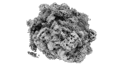
 Downloads & links
 EMDB map data format
 emd_16903.png
 http://ftp.pdbj.org/pub/emdb/structures/EMD-16903


 Z (Sec.)
 Y (Row.)
 X (Col.)


 Sample components
 Processing
 Electron microscopy
 FIELD EMISSION GUN
