 ムービー
ムービー コントローラー


+ データを開く

- 基本情報
基本情報
| 登録情報 |  | |||||||||||||||||||||||||||
|---|---|---|---|---|---|---|---|---|---|---|---|---|---|---|---|---|---|---|---|---|---|---|---|---|---|---|---|---|
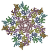
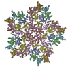
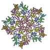
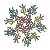
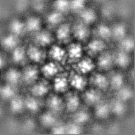
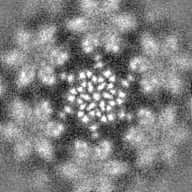
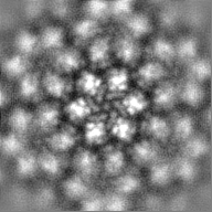
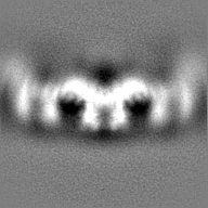
| タイトル | Subtomogram average of HIV-1 CA hexamer from capsid-like particles assembled with inositol hexakisphosphate | |||||||||||||||||||||||||||
 マップデータ マップデータ | Subtomogram average of HIV-1 CA protein hexamer from capsid-like particles assembled with inositol hexakisphosphate | |||||||||||||||||||||||||||
 試料 試料 |
| |||||||||||||||||||||||||||
| 生物種 |   Human immunodeficiency virus 1 (ヒト免疫不全ウイルス) Human immunodeficiency virus 1 (ヒト免疫不全ウイルス) | |||||||||||||||||||||||||||
| 手法 | サブトモグラム平均法 / クライオ電子顕微鏡法 / 解像度: 3.9 Å | |||||||||||||||||||||||||||
 データ登録者 データ登録者 | Tan A / Briggs JAG / Dick RA | |||||||||||||||||||||||||||
| 資金援助 |  米国, 米国,  英国, 英国,  ドイツ, 8件 ドイツ, 8件
| |||||||||||||||||||||||||||
 引用 引用 | ジャーナル: Proc Natl Acad Sci U S A / 年: 2023 タイトル: Structural insights into HIV-1 polyanion-dependent capsid lattice formation revealed by single particle cryo-EM. 著者: Carolyn M Highland / Aaron Tan / Clifton L Ricaña / John A G Briggs / Robert A Dick / 要旨: The HIV-1 capsid houses the viral genome and interacts extensively with host cell proteins throughout the viral life cycle. It is composed of capsid protein (CA), which assembles into a conical ...The HIV-1 capsid houses the viral genome and interacts extensively with host cell proteins throughout the viral life cycle. It is composed of capsid protein (CA), which assembles into a conical fullerene lattice composed of roughly 200 CA hexamers and 12 CA pentamers. Previous structural analyses of individual CA hexamers and pentamers have provided valuable insight into capsid structure and function, but detailed structural information about these assemblies in the broader context of the capsid lattice is lacking. In this study, we combined cryoelectron tomography and single particle analysis (SPA) cryoelectron microscopy to determine structures of continuous regions of the capsid lattice containing both hexamers and pentamers. We also developed a method of liposome scaffold-based in vitro lattice assembly ("lattice templating") that enabled us to directly study the lattice under a wider range of conditions than has previously been possible. Using this approach, we identified a critical role for inositol hexakisphosphate in pentamer formation and determined the structure of the CA lattice bound to the capsid-targeting antiretroviral drug GS-6207 (lenacapavir). Our work reveals key structural details of the mature HIV-1 CA lattice and establishes the combination of lattice templating and SPA as a robust strategy for studying retroviral capsid structure and capsid interactions with host proteins and antiviral compounds. | |||||||||||||||||||||||||||
| 履歴 |
|
- 構造の表示
構造の表示
| 添付画像 |
|---|

- ダウンロードとリンク
-EMDBアーカイブ
| マップデータ | emd_16699.map.gz | 25.1 MB |  EMDBマップデータ形式 EMDBマップデータ形式 | |
|---|---|---|---|---|
| ヘッダ (付随情報) | emd-16699-v30.xmlemd-16699.xml | 17.4 KB 17.4 KB | 表示 表示 | EMDBヘッダ |
| 画像 | 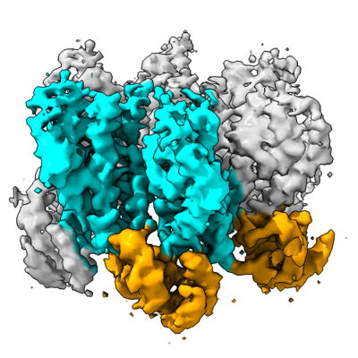 emd_16699.png emd_16699.png | 144.8 KB | ||
| その他 | emd_16699_half_map_1.map.gzemd_16699_half_map_2.map.gz | 25.1 MB 25.1 MB | ||
| アーカイブディレクトリ |  http://ftp.pdbj.org/pub/emdb/structures/EMD-16699ftp://ftp.pdbj.org/pub/emdb/structures/EMD-16699 http://ftp.pdbj.org/pub/emdb/structures/EMD-16699ftp://ftp.pdbj.org/pub/emdb/structures/EMD-16699 | HTTPS FTP |
-検証レポート
| 文書・要旨 | emd_16699_validation.pdf.gz | 1 MB | 表示 | EMDB検証レポート |
|---|---|---|---|---|
| 文書・詳細版 | emd_16699_full_validation.pdf.gz | 1 MB | 表示 | |
| XML形式データ | emd_16699_validation.xml.gz | 10.7 KB | 表示 | |
| CIF形式データ | emd_16699_validation.cif.gz | 12.5 KB | 表示 | |
| アーカイブディレクトリ | https://ftp.pdbj.org/pub/emdb/validation_reports/EMD-16699ftp://ftp.pdbj.org/pub/emdb/validation_reports/EMD-16699 | HTTPS FTP |
-関連構造データ


-リンク
| EMDBのページ | EMDB (EBI/PDBe) / EMDataResource |
|---|
-マップ
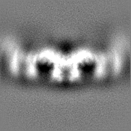
| ファイル | ダウンロード / ファイル: emd_16699.map.gz / 形式: CCP4 / 大きさ: 27 MB / タイプ: IMAGE STORED AS FLOATING POINT NUMBER (4 BYTES) | ||||||||||||||||||||||||||||||||||||
|---|---|---|---|---|---|---|---|---|---|---|---|---|---|---|---|---|---|---|---|---|---|---|---|---|---|---|---|---|---|---|---|---|---|---|---|---|---|
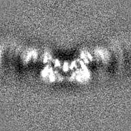
| 注釈 | Subtomogram average of HIV-1 CA protein hexamer from capsid-like particles assembled with inositol hexakisphosphate | ||||||||||||||||||||||||||||||||||||
| 投影像・断面図 | 画像のコントロール
画像は Spider により作成 | ||||||||||||||||||||||||||||||||||||
| ボクセルのサイズ | X=Y=Z: 1.379 Å | ||||||||||||||||||||||||||||||||||||
| 密度 |
| ||||||||||||||||||||||||||||||||||||
| 対称性 | 空間群: 1 | ||||||||||||||||||||||||||||||||||||
| 詳細 | EMDB XML:
|
 Z (Sec.)
 Y (Row.)
 X (Col.)


-添付データ
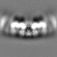
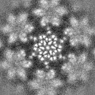
-ハーフマップ: Subtomogram average of HIV-1 CA protein hexamer from...
| ファイル | emd_16699_half_map_1.map | ||||||||||||
|---|---|---|---|---|---|---|---|---|---|---|---|---|---|
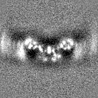
| 注釈 | Subtomogram average of HIV-1 CA protein hexamer from capsid-like particles assembled with inositol hexakisphosphate, half map A | ||||||||||||
| 投影像・断面図 |
| ||||||||||||
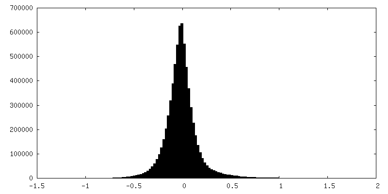
| 密度ヒストグラム |
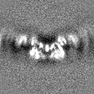
-ハーフマップ: Subtomogram average of HIV-1 CA protein hexamer from...
| ファイル | emd_16699_half_map_2.map | ||||||||||||
|---|---|---|---|---|---|---|---|---|---|---|---|---|---|
| 注釈 | Subtomogram average of HIV-1 CA protein hexamer from capsid-like particles assembled with inositol hexakisphosphate, half map B | ||||||||||||
| 投影像・断面図 |
| ||||||||||||
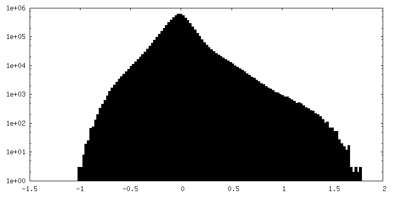
| 密度ヒストグラム |


- 試料の構成要素
-全体 : HIV-1 CA protein capsid-like particles assembled in the presence ...
| 全体 | 名称: HIV-1 CA protein capsid-like particles assembled in the presence of inositol hexakisphosphate |
|---|---|
| 要素 |
|
-超分子 #1: HIV-1 CA protein capsid-like particles assembled in the presence ...
| 超分子 | 名称: HIV-1 CA protein capsid-like particles assembled in the presence of inositol hexakisphosphate タイプ: complex / ID: 1 / キメラ: Yes / 親要素: 0 |
|---|---|
| 由来(天然) | 生物種: Human immunodeficiency virus 1 (ヒト免疫不全ウイルス) |
-実験情報
-構造解析
| 手法 | クライオ電子顕微鏡法 |
|---|---|
 解析 解析 | サブトモグラム平均法 |
| 試料の集合状態 | particle |
-試料調製
| 緩衝液 | pH: 6.2 構成要素:
| ||||||||||||
|---|---|---|---|---|---|---|---|---|---|---|---|---|---|
| グリッド | モデル: C-flat-2/2 / 材質: COPPER / メッシュ: 300 / 前処理 - タイプ: GLOW DISCHARGE / 前処理 - 時間: 30 sec. / 前処理 - 雰囲気: AIR 詳細: The grid was glow discharged for 30 seconds at a current of 20 mA. | ||||||||||||
| 凍結 | 凍結剤: ETHANE / チャンバー内湿度: 100 % / チャンバー内温度: 277.15 K / 装置: FEI VITROBOT MARK IV |
- 電子顕微鏡法
| 顕微鏡 | FEI TITAN KRIOS |
|---|---|
| 特殊光学系 | エネルギーフィルター - 名称: GIF Bioquantum / エネルギーフィルター - スリット幅: 20 eV |
| 撮影 | フィルム・検出器のモデル: GATAN K2 SUMMIT (4k x 4k) 検出モード: COUNTING / 平均電子線量: 3.0 e/Å2 |
| 電子線 | 加速電圧: 300 kV / 電子線源:  FIELD EMISSION GUN FIELD EMISSION GUN |
| 電子光学系 | 照射モード: FLOOD BEAM / 撮影モード: BRIGHT FIELD / Cs: 2.7 mm / 最大 デフォーカス(公称値): 4.5 µm / 最小 デフォーカス(公称値): 1.5 µm / 倍率(公称値): 105000 |
| 試料ステージ | 試料ホルダーモデル: FEI TITAN KRIOS AUTOGRID HOLDER ホルダー冷却材: NITROGEN |
| 実験機器 |  モデル: Titan Krios / 画像提供: FEI Company |
-画像解析
| 最終 再構成 | 想定した対称性 - 点群: C6 (6回回転対称) / アルゴリズム: BACK PROJECTION / 解像度のタイプ: BY AUTHOR / 解像度: 3.9 Å / 解像度の算出法: FSC 0.143 CUT-OFF / ソフトウェア - 名称: subTOM / 使用したサブトモグラム数: 89951 |
|---|---|
| 抽出 | トモグラム数: 66 / 使用した粒子像数: 570742 / ソフトウェア - 名称: MATLAB 詳細: HIV-1 CA protein capsid-like particles (CLPs) were segmented using the Ilastik software package. The vertex coordinates of the segmented volumes were used to define an oversampled grid of ...詳細: HIV-1 CA protein capsid-like particles (CLPs) were segmented using the Ilastik software package. The vertex coordinates of the segmented volumes were used to define an oversampled grid of points normal to the CLP surface in MATLAB. |
| 最終 角度割当 | タイプ: OTHER / ソフトウェア - 名称: subTOM |
