 Movie
Movie Controller


+ Open data

- Basic information
| Entry |  | |||||||||
|---|---|---|---|---|---|---|---|---|---|---|
| Title | Type Ib beta-amyloid 42 Filaments from Human Brain | |||||||||
 Map data Map data | ||||||||||
 Sample Sample |
| |||||||||
| Biological species |  Homo sapiens (human) Homo sapiens (human) | |||||||||
| Method | helical reconstruction / cryo EM / Resolution: 3.5 Å | |||||||||
 Authors Authors | Yang Y / Arseni D / Zhang W / Huang M / Lovestam SKA / Schweighauser M / Kotecha A / Murzin AG / Peak-Chew SY / Macdonald J ...Yang Y / Arseni D / Zhang W / Huang M / Lovestam SKA / Schweighauser M / Kotecha A / Murzin AG / Peak-Chew SY / Macdonald J / Lavenir I / Garringer HJ / Gelpi E / Newell KL / Kovacs GG / Vidal R / Ghetti B / Falcon B / Scheres HW / Goedert M | |||||||||
| Funding support |  United Kingdom, United Kingdom,  United States, 2 items United States, 2 items
| |||||||||
 Citation Citation | Journal: Science / Year: 2022 Title: Cryo-EM structures of amyloid-β 42 filaments from human brains. Authors: Yang Yang / Diana Arseni / Wenjuan Zhang / Melissa Huang / Sofia Lövestam / Manuel Schweighauser / Abhay Kotecha / Alexey G Murzin / Sew Y Peak-Chew / Jennifer Macdonald / Isabelle Lavenir ...Authors: Yang Yang / Diana Arseni / Wenjuan Zhang / Melissa Huang / Sofia Lövestam / Manuel Schweighauser / Abhay Kotecha / Alexey G Murzin / Sew Y Peak-Chew / Jennifer Macdonald / Isabelle Lavenir / Holly J Garringer / Ellen Gelpi / Kathy L Newell / Gabor G Kovacs / Ruben Vidal / Bernardino Ghetti / Benjamin Ryskeldi-Falcon / Sjors H W Scheres / Michel Goedert /    Abstract: Filament assembly of amyloid-β peptides ending at residue 42 (Aβ42) is a central event in Alzheimer’s disease. Here, we report the cryo–electron microscopy (cryo-EM) structures of Aβ42 ...Filament assembly of amyloid-β peptides ending at residue 42 (Aβ42) is a central event in Alzheimer’s disease. Here, we report the cryo–electron microscopy (cryo-EM) structures of Aβ42 filaments from human brains. Two structurally related S-shaped protofilament folds give rise to two types of filaments. Type I filaments were found mostly in the brains of individuals with sporadic Alzheimer’s disease, and type II filaments were found in individuals with familial Alzheimer’s disease and other conditions. The structures of Aβ42 filaments from the brain differ from those of filaments assembled in vitro. By contrast, in knock-in mice, Aβ42 deposits were made of type II filaments. Knowledge of Aβ42 filament structures from human brains may lead to the development of inhibitors of assembly and improved imaging agents. | |||||||||
| History |
|
- Structure visualization
| Supplemental images |
|---|
- Downloads & links
-EMDB archive
| Map data | emd_16434.map.gz | 8.9 MB |  EMDB map data format EMDB map data format | |
|---|---|---|---|---|
| Header (meta data) | emd-16434-v30.xmlemd-16434.xml | 14.4 KB 14.4 KB | Display Display | EMDB header |
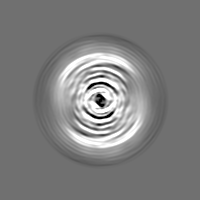
| FSC (resolution estimation) | emd_16434_fsc.xml | 7.2 KB | Display | FSC data file |
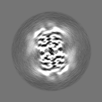
| Images |  emd_16434.png emd_16434.png | 61.1 KB | ||
| Others | emd_16434_half_map_1.map.gzemd_16434_half_map_2.map.gz | 10.3 MB 10.2 MB | ||
| Archive directory |  http://ftp.pdbj.org/pub/emdb/structures/EMD-16434ftp://ftp.pdbj.org/pub/emdb/structures/EMD-16434 http://ftp.pdbj.org/pub/emdb/structures/EMD-16434ftp://ftp.pdbj.org/pub/emdb/structures/EMD-16434 | HTTPS FTP |
-Related structure data

-Links
| EMDB pages | EMDB (EBI/PDBe) / EMDataResource |
|---|
-Map
| File | Download / File: emd_16434.map.gz / Format: CCP4 / Size: 30.5 MB / Type: IMAGE STORED AS FLOATING POINT NUMBER (4 BYTES) | ||||||||||||||||||||||||||||||||||||
|---|---|---|---|---|---|---|---|---|---|---|---|---|---|---|---|---|---|---|---|---|---|---|---|---|---|---|---|---|---|---|---|---|---|---|---|---|---|
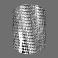
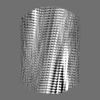
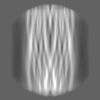
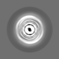
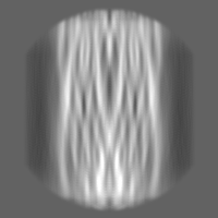
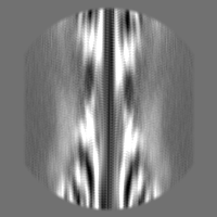
| Projections & slices | Image control
Images are generated by Spider. | ||||||||||||||||||||||||||||||||||||
| Voxel size | X=Y=Z: 0.93 Å | ||||||||||||||||||||||||||||||||||||
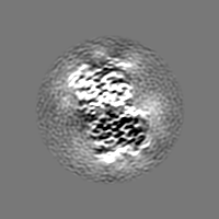
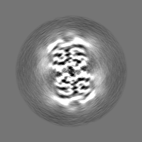
| Density |
| ||||||||||||||||||||||||||||||||||||
| Symmetry | Space group: 1 | ||||||||||||||||||||||||||||||||||||
| Details | EMDB XML:
|
 Z (Sec.)
 Y (Row.)
 X (Col.)


-Supplemental data
-Half map: #1
| File | emd_16434_half_map_1.map | ||||||||||||
|---|---|---|---|---|---|---|---|---|---|---|---|---|---|
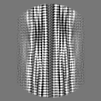
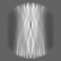
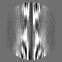
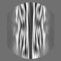
| Projections & Slices |
| ||||||||||||
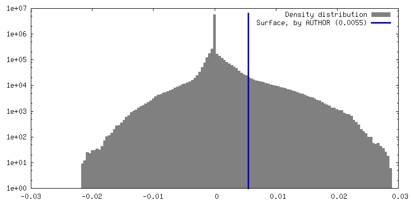
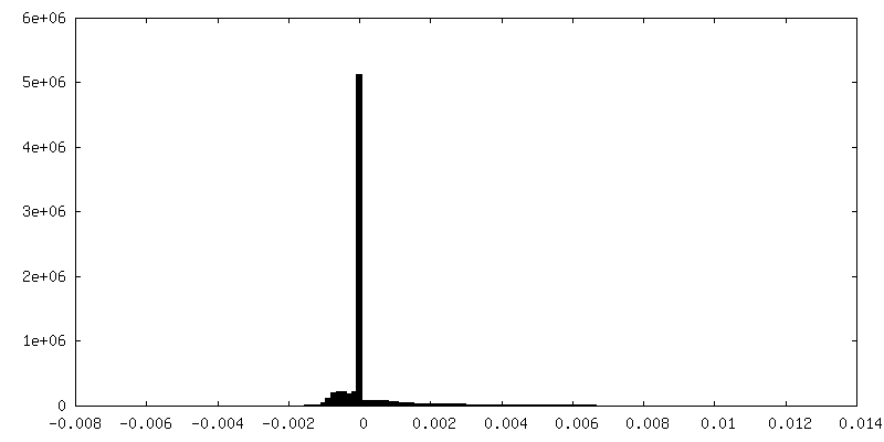
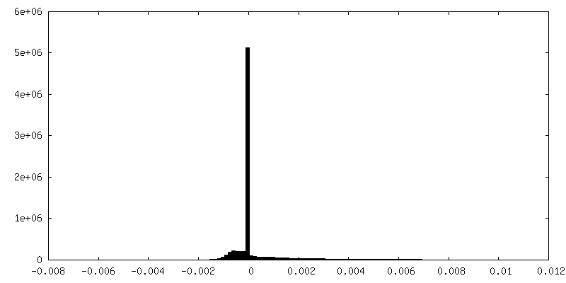
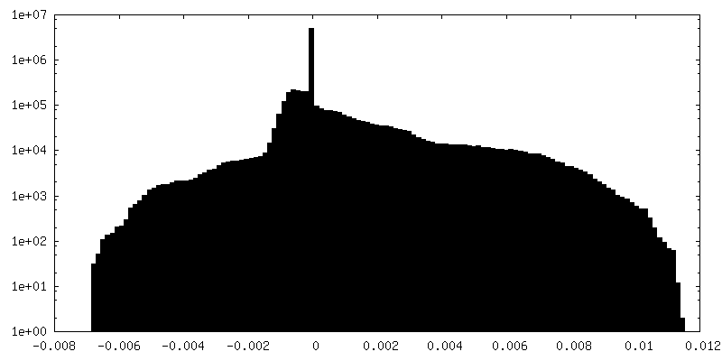
| Density Histograms |

-Half map: #2
| File | emd_16434_half_map_2.map | ||||||||||||
|---|---|---|---|---|---|---|---|---|---|---|---|---|---|
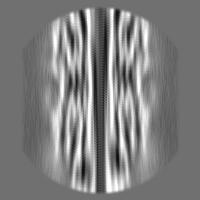
| Projections & Slices |
| ||||||||||||
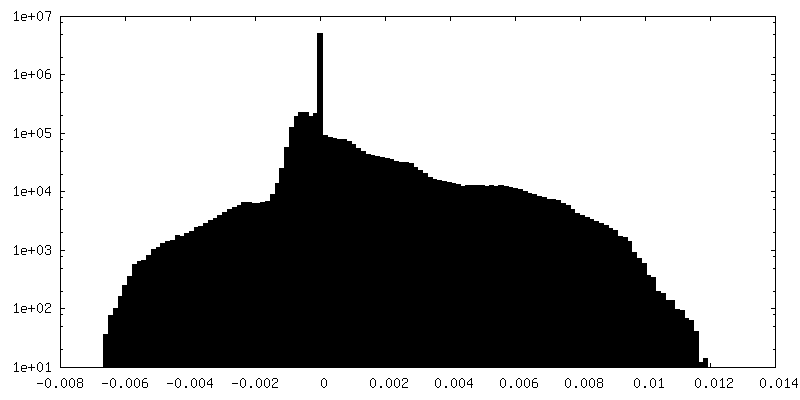
| Density Histograms |

- Sample components
-Entire : beta-amyloid 42 filaments extracted from the human brain with Alz...
| Entire | Name: beta-amyloid 42 filaments extracted from the human brain with Alzheimer's disease |
|---|---|
| Components |
|
-Supramolecule #1: beta-amyloid 42 filaments extracted from the human brain with Alz...
| Supramolecule | Name: beta-amyloid 42 filaments extracted from the human brain with Alzheimer's disease type: tissue / ID: 1 / Parent: 0 / Macromolecule list: all |
|---|---|
| Source (natural) | Organism: Homo sapiens (human) |
-Macromolecule #1: beta-amyloid 42
| Macromolecule | Name: beta-amyloid 42 / type: protein_or_peptide / ID: 1 / Enantiomer: LEVO |
|---|---|
| Source (natural) | Organism: Homo sapiens (human) |
| Sequence | String: DAEFRHDSGY EVHHQKLVFF AEDVGSNKGA IIGLMVGGVV IA |
-Experimental details
-Structure determination
| Method | cryo EM |
|---|---|
 Processing Processing | helical reconstruction |
| Aggregation state | filament |
-Sample preparation
| Buffer | pH: 7.5 |
|---|---|
| Vitrification | Cryogen name: ETHANE |
- Electron microscopy
| Microscope | FEI TITAN KRIOS |
|---|---|
| Image recording | Film or detector model: GATAN K3 (6k x 4k) / Average electron dose: 40.0 e/Å2 |
| Electron beam | Acceleration voltage: 300 kV / Electron source:  FIELD EMISSION GUN FIELD EMISSION GUN |
| Electron optics | Illumination mode: FLOOD BEAM / Imaging mode: BRIGHT FIELD / Nominal defocus max: 2.8000000000000003 µm / Nominal defocus min: 1.0 µm |
| Experimental equipment |  Model: Titan Krios / Image courtesy: FEI Company |
-Image processing
| Final reconstruction | Applied symmetry - Helical parameters - Δz: 2.4 Å Applied symmetry - Helical parameters - Δ&Phi: 179.2 ° Applied symmetry - Helical parameters - Axial symmetry: C1 (asymmetric) Resolution.type: BY AUTHOR / Resolution: 3.5 Å / Resolution method: FSC 0.143 CUT-OFF / Number images used: 50990 |
|---|---|
| Final angle assignment | Type: NOT APPLICABLE |
| FSC plot (resolution estimation) | 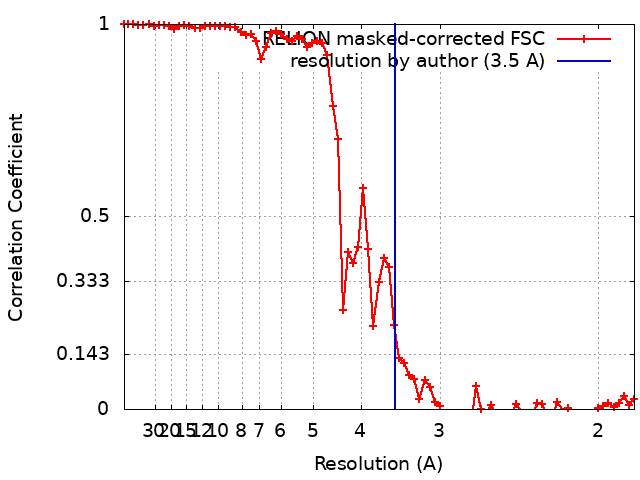 |
