 Movie
Movie Controller

[English] 日本語
 Yorodumi
- EMDB-1624: Three-dimensional structure of the recombinant, virus-like partic... -
+ Open data

- Basic information
| Entry | Database: EMDB / ID: EMD-1624 | |||||||||
|---|---|---|---|---|---|---|---|---|---|---|
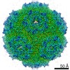
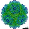
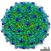
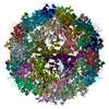
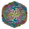
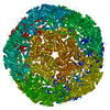
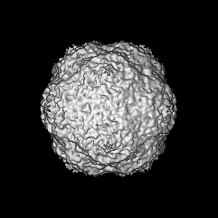
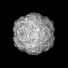
| Title | Three-dimensional structure of the recombinant, virus-like particle of Penaeus stylirostris densovirus (PstDNV) or infectious hypodermal and hematopoietic necrosis virus (IHHNV) | |||||||||
 Map data Map data | Penaeus stylirostris densovirus (PstDNV) or infectious hypodermal and hematopoietic necrosis virus (IHHNV) | |||||||||
 Sample Sample |
| |||||||||
 Keywords Keywords | virus-like particle / VLP / parvovirus / Densovirinae / shrimp pathogen / protein capsid | |||||||||
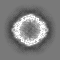
| Biological species |  Infectious hypodermal and hematopoietic necrosis virus Infectious hypodermal and hematopoietic necrosis virus | |||||||||
| Method | single particle reconstruction / cryo EM / Resolution: 6.17 Å | |||||||||
 Authors Authors | Kaufmann B / Bowman VD / Li Y / Szelei J / Tijssen P / Rossmann MG | |||||||||
 Citation Citation | Journal: J Virol / Year: 2010 Title: Structure of Penaeus stylirostris densovirus, a shrimp pathogen. Authors: Bärbel Kaufmann / Valorie D Bowman / Yi Li / Jozsef Szelei / Peter J Waddell / Peter Tijssen / Michael G Rossmann /  Abstract: Penaeus stylirostris densovirus (PstDNV), a pathogen of penaeid shrimp, causes significant damage to farmed and wild shrimp populations. In contrast to other parvoviruses, PstDNV probably has only ...Penaeus stylirostris densovirus (PstDNV), a pathogen of penaeid shrimp, causes significant damage to farmed and wild shrimp populations. In contrast to other parvoviruses, PstDNV probably has only one type of capsid protein that lacks the phospholipase A2 activity that has been implicated as a requirement during parvoviral host cell infection. The structure of recombinant virus-like particles, composed of 60 copies of the 37.5-kDa coat protein, the smallest parvoviral capsid protein reported thus far, was determined to 2.5-Å resolution by X-ray crystallography. The structure represents the first near-atomic resolution structure within the genus Brevidensovirus. The capsid protein has a β-barrel "jelly roll" motif similar to that found in many icosahedral viruses, including other parvoviruses. The N-terminal portion of the PstDNV coat protein adopts a "domain-swapped" conformation relative to its twofold-related neighbor similar to the insect parvovirus Galleria mellonella densovirus (GmDNV) but in stark contrast to vertebrate parvoviruses. However, most of the surface loops have little structural resemblance to any of the known parvoviral capsid proteins. | |||||||||
| History |
|
- Structure visualization
| Movie |
 Movie viewer Movie viewer |
|---|---|
| Structure viewer | EM map: SurfViewMolmilJmol/JSmol |
| Supplemental images |

- Downloads & links
-EMDB archive
| Map data | emd_1624.map.gz | 20.4 MB | EMDB map data format | |
|---|---|---|---|---|
| Header (meta data) | emd-1624-v30.xmlemd-1624.xml | 11.2 KB 11.2 KB | Display Display | EMDB header |
| Images | 1624_EMD-1624.tif | 764.7 KB | ||
| Archive directory |  http://ftp.pdbj.org/pub/emdb/structures/EMD-1624ftp://ftp.pdbj.org/pub/emdb/structures/EMD-1624 http://ftp.pdbj.org/pub/emdb/structures/EMD-1624ftp://ftp.pdbj.org/pub/emdb/structures/EMD-1624 | HTTPS FTP |
-Related structure data


-Links
| EMDB pages | EMDB (EBI/PDBe) / EMDataResource |
|---|
-Map
| File | Download / File: emd_1624.map.gz / Format: CCP4 / Size: 52.1 MB / Type: IMAGE STORED AS FLOATING POINT NUMBER (4 BYTES) | ||||||||||||||||||||||||||||||||||||||||||||||||||||||||||||||||||||
|---|---|---|---|---|---|---|---|---|---|---|---|---|---|---|---|---|---|---|---|---|---|---|---|---|---|---|---|---|---|---|---|---|---|---|---|---|---|---|---|---|---|---|---|---|---|---|---|---|---|---|---|---|---|---|---|---|---|---|---|---|---|---|---|---|---|---|---|---|---|
| Annotation | Penaeus stylirostris densovirus (PstDNV) or infectious hypodermal and hematopoietic necrosis virus (IHHNV) | ||||||||||||||||||||||||||||||||||||||||||||||||||||||||||||||||||||
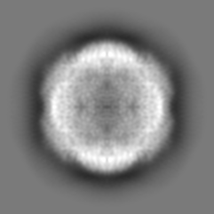
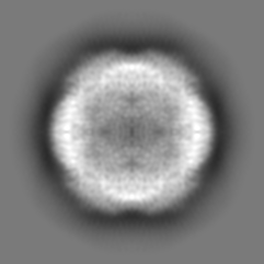
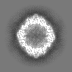
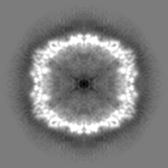
| Projections & slices | Image control
Images are generated by Spider. | ||||||||||||||||||||||||||||||||||||||||||||||||||||||||||||||||||||
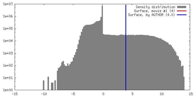
| Voxel size | X=Y=Z: 1.489 Å | ||||||||||||||||||||||||||||||||||||||||||||||||||||||||||||||||||||
| Density |
| ||||||||||||||||||||||||||||||||||||||||||||||||||||||||||||||||||||
| Symmetry | Space group: 1 | ||||||||||||||||||||||||||||||||||||||||||||||||||||||||||||||||||||
| Details | EMDB XML:
CCP4 map header:
| ||||||||||||||||||||||||||||||||||||||||||||||||||||||||||||||||||||
 Z (Sec.)
 Y (Row.)
 X (Col.)


-Supplemental data
- Sample components
-Entire : recombinant, virus-like particle of Penaeus stylirostris densovir...
| Entire | Name: recombinant, virus-like particle of Penaeus stylirostris densovirus (PstDNV) or infectious hypodermal and hematopoietic necrosis virus (IHHNV) |
|---|---|
| Components |
|
-Supramolecule #1000: recombinant, virus-like particle of Penaeus stylirostris densovir...
| Supramolecule | Name: recombinant, virus-like particle of Penaeus stylirostris densovirus (PstDNV) or infectious hypodermal and hematopoietic necrosis virus (IHHNV) type: sample / ID: 1000 Details: virus-like particles (VLP) self-assembled 60 copies of the recombinant 37.5-kD viral coat protein only Oligomeric state: 60 viral coat protein subunits form T1 icosahedral protein shell Number unique components: 1 |
|---|---|
| Molecular weight | Theoretical: 2.25 MDa |
-Supramolecule #1: Infectious hypodermal and hematopoietic necrosis virus
| Supramolecule | Name: Infectious hypodermal and hematopoietic necrosis virus type: virus / ID: 1 / Name.synonym: Penaeus stylirostris densovirus (PstDNV) Details: Virus-like particles (VLP) self-assembled from 60 monomers of the recombinant 37.5-kD viral coat protein NCBI-ID: 111792 Sci species name: Infectious hypodermal and hematopoietic necrosis virus Virus type: VIRUS-LIKE PARTICLE / Virus isolate: STRAIN / Virus enveloped: No / Virus empty: Yes / Syn species name: Penaeus stylirostris densovirus (PstDNV) |
|---|---|
| Host (natural) | Organism:  Litopenaeus stylirostris (blue shrimp) / synonym: INVERTEBRATES Litopenaeus stylirostris (blue shrimp) / synonym: INVERTEBRATES |
| Molecular weight | Theoretical: 2.25 MDa |
| Virus shell | Shell ID: 1 / Diameter: 225 Å / T number (triangulation number): 1 |
-Experimental details
-Structure determination
| Method | cryo EM |
|---|---|
 Processing Processing | single particle reconstruction |
| Aggregation state | particle |
-Sample preparation
| Concentration | 1. mg/mL |
|---|---|
| Buffer | pH: 7.5 / Details: 10mM Tris-HCl, 100mM NaCl, 1mM MgCl2, 1mM CaCl2 |
| Grid | Details: Holey carbon 400 mesh copper grid |
| Vitrification | Cryogen name: ETHANE / Instrument: HOMEMADE PLUNGER Details: Vitrification instrument: Guillotine-style plunge freezeing device Method: A small vial of ethane is placed inside a larger liquid nitrogen reservoir. The grid holding a few microliters of the sample is held in place at the bottom of a plunger by the means of fine ...Method: A small vial of ethane is placed inside a larger liquid nitrogen reservoir. The grid holding a few microliters of the sample is held in place at the bottom of a plunger by the means of fine tweezers. Once the ethane in the vial is completely frozen, it needs to be slightly melted. When the liquid ethane is ready, a piece of filter paper is then pressed against the sample to blot of excess buffer, sufficient to leave a thin layer on the grid. After a predetermined time, the filter paper is removed, and the plunger is allowed to drop into the liquid ethane. Once the grid enters the liquid ethane, the sample is rapidly frozen, and the grid is transferred under liquid nitrogen to a storage box immersed liquid nitrogen for later use in the microscope. |
- Electron microscopy
| Microscope | FEI/PHILIPS CM300FEG/T |
|---|---|
| Temperature | Average: 98 K |
| Alignment procedure | Legacy - Astigmatism: live FFT at 200K mag |
| Details | low dose imaging |
| Date | Sep 7, 2005 |
| Image recording | Category: FILM / Film or detector model: KODAK SO-163 FILM / Digitization - Scanner: ZEISS SCAI / Digitization - Sampling interval: 7 µm / Number real images: 9 / Average electron dose: 18.41 e/Å2 / Od range: 1.24 / Bits/pixel: 8 |
| Tilt angle max | 0 |
| Electron beam | Acceleration voltage: 300 kV / Electron source:  FIELD EMISSION GUN FIELD EMISSION GUN |
| Electron optics | Calibrated magnification: 47000 / Illumination mode: FLOOD BEAM / Imaging mode: BRIGHT FIELD / Cs: 2.0 mm / Nominal defocus max: 3.241 µm / Nominal defocus min: 1.362 µm / Nominal magnification: 45000 |
| Sample stage | Specimen holder: Eucentric / Specimen holder model: GATAN LIQUID NITROGEN / Tilt angle min: -0 |
-Image processing
| Details | The particles were selected using an automatic selection program. Selection of particles was subsequently refined interactively at the computer terminal. |
|---|---|
| CTF correction | Details: Each particle |
| Final reconstruction | Applied symmetry - Point group: I (icosahedral) / Algorithm: OTHER / Resolution.type: BY AUTHOR / Resolution: 6.17 Å / Resolution method: FSC 0.5 CUT-OFF / Software - Name: AUTO3DEM Details: final map includes data to 6.1 Ang resolution (fsc 0.2 cut-off) Number images used: 9009 |
