Movie
Movie Controller

[English] 日本語
 Yorodumi
- EMDB-15805: RecBCD-DNA in complex with the phage protein Abc2 and host PpiB -
+ Open data

- Basic information
| Entry |  | ||||||||||||
|---|---|---|---|---|---|---|---|---|---|---|---|---|---|
| Title | RecBCD-DNA in complex with the phage protein Abc2 and host PpiB | ||||||||||||
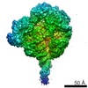
 Map data Map data | Final CryoEM map of the RecBCD-Abc2-DNA complex bound to the host PpiB protein | ||||||||||||
 Sample Sample |
| ||||||||||||
 Keywords Keywords | Homologous recombination / DNA repair / phage / Helicase / Nuclease / Inhibitor / Protein complex / Enzyme / DNA mimic / DNA BINDING PROTEIN | ||||||||||||
| Function / homology |  Function and homology information Function and homology informationexodeoxyribonuclease V / exodeoxyribonuclease V activity / exodeoxyribonuclease V complex / clearance of foreign intracellular DNA / DNA 5'-3' helicase / single-stranded DNA helicase activity / recombinational repair / 3'-5' DNA helicase activity / DNA 3'-5' helicase / DNA helicase activity ...exodeoxyribonuclease V / exodeoxyribonuclease V activity / exodeoxyribonuclease V complex / clearance of foreign intracellular DNA / DNA 5'-3' helicase / single-stranded DNA helicase activity / recombinational repair / 3'-5' DNA helicase activity / DNA 3'-5' helicase / DNA helicase activity / helicase activity / response to radiation / double-strand break repair via homologous recombination / DNA recombination / 5'-3' DNA helicase activity / DNA damage response / magnesium ion binding / ATP hydrolysis activity / DNA binding / ATP binding / cytosol Similarity search - Function | ||||||||||||
| Biological species |   Salmonella phage P22 (virus) Salmonella phage P22 (virus) | ||||||||||||
| Method | single particle reconstruction / cryo EM / Resolution: 3.8 Å | ||||||||||||
 Authors Authors | Wilkinson M / Wilkinson OJ / Feyerherm C / Fletcher EE / Wigley DB / Dillingham MS | ||||||||||||
| Funding support |  United Kingdom, 3 items United Kingdom, 3 items
| ||||||||||||
 Citation Citation | Journal: Elife / Year: 2022 Title: Structures of RecBCD in complex with phage-encoded inhibitor proteins reveal distinctive strategies for evasion of a bacterial immunity hub. Authors: Martin Wilkinson / Oliver J Wilkinson / Connie Feyerherm / Emma E Fletcher / Dale B Wigley / Mark S Dillingham / Abstract: Following infection of bacterial cells, bacteriophage modulate double-stranded DNA break repair pathways to protect themselves from host immunity systems and prioritise their own recombinases. Here, ...Following infection of bacterial cells, bacteriophage modulate double-stranded DNA break repair pathways to protect themselves from host immunity systems and prioritise their own recombinases. Here, we present biochemical and structural analysis of two phage proteins, gp5.9 and Abc2, which target the DNA break resection complex RecBCD. These exemplify two contrasting mechanisms for control of DNA break repair in which the RecBCD complex is either inhibited or co-opted for the benefit of the invading phage. Gp5.9 completely inhibits RecBCD by preventing it from binding to DNA. The RecBCD-gp5.9 structure shows that gp5.9 acts by substrate mimicry, binding predominantly to the RecB arm domain and competing sterically for the DNA binding site. Gp5.9 adopts a parallel coiled-coil architecture that is unprecedented for a natural DNA mimic protein. In contrast, binding of Abc2 does not substantially affect the biochemical activities of isolated RecBCD. The RecBCD-Abc2 structure shows that Abc2 binds to the Chi-recognition domains of the RecC subunit in a position that might enable it to mediate the loading of phage recombinases onto its single-stranded DNA products. | ||||||||||||
| History |
|
- Structure visualization
| Supplemental images |
|---|

- Downloads & links
-EMDB archive
| Map data | emd_15805.map.gz | 43.9 MB | EMDB map data format | |
|---|---|---|---|---|
| Header (meta data) | emd-15805-v30.xmlemd-15805.xml | 28.6 KB 28.6 KB | Display Display | EMDB header |
| Images |  emd_15805.png emd_15805.png | 111.6 KB | ||
| Filedesc metadata | emd-15805.cif.gz | 9.6 KB | ||
| Others | emd_15805_half_map_1.map.gzemd_15805_half_map_2.map.gz | 36.8 MB 36.8 MB | ||
| Archive directory |  http://ftp.pdbj.org/pub/emdb/structures/EMD-15805ftp://ftp.pdbj.org/pub/emdb/structures/EMD-15805 http://ftp.pdbj.org/pub/emdb/structures/EMD-15805ftp://ftp.pdbj.org/pub/emdb/structures/EMD-15805 | HTTPS FTP |
-Related structure data
| Related structure data |  8b1uMC  8b1rC  8b1tC C: citing same article ( M: atomic model generated by this map |
|---|---|
| Similar structure data |


-Links
| EMDB pages | EMDB (EBI/PDBe) / EMDataResource |
|---|---|
| Related items in Molecule of the Month |


-Map
| File | Download / File: emd_15805.map.gz / Format: CCP4 / Size: 47.6 MB / Type: IMAGE STORED AS FLOATING POINT NUMBER (4 BYTES) | ||||||||||||||||||||||||||||||||||||
|---|---|---|---|---|---|---|---|---|---|---|---|---|---|---|---|---|---|---|---|---|---|---|---|---|---|---|---|---|---|---|---|---|---|---|---|---|---|
| Annotation | Final CryoEM map of the RecBCD-Abc2-DNA complex bound to the host PpiB protein | ||||||||||||||||||||||||||||||||||||
| Projections & slices | Image control
Images are generated by Spider. | ||||||||||||||||||||||||||||||||||||
| Voxel size | X=Y=Z: 1.06 Å | ||||||||||||||||||||||||||||||||||||
| Density |
| ||||||||||||||||||||||||||||||||||||
| Symmetry | Space group: 1 | ||||||||||||||||||||||||||||||||||||
| Details | EMDB XML:
|
 Z (Sec.)
 Y (Row.)
 X (Col.)


-Supplemental data
-Half map: halfmap1
| File | emd_15805_half_map_1.map | ||||||||||||
|---|---|---|---|---|---|---|---|---|---|---|---|---|---|
| Annotation | halfmap1 | ||||||||||||
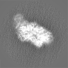
| Projections & Slices |
| ||||||||||||
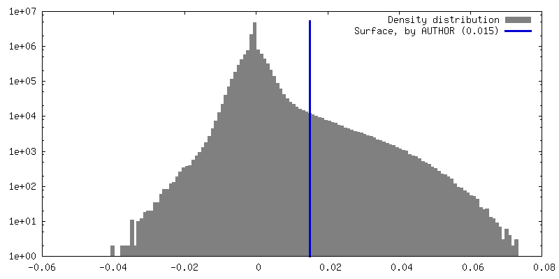
| Density Histograms |


-Half map: halfmap2
| File | emd_15805_half_map_2.map | ||||||||||||
|---|---|---|---|---|---|---|---|---|---|---|---|---|---|
| Annotation | halfmap2 | ||||||||||||
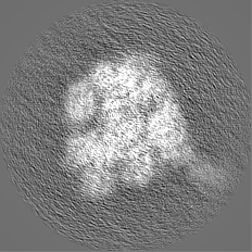
| Projections & Slices |
| ||||||||||||
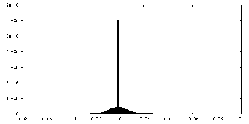
| Density Histograms |


- Sample components
+Entire : RecBCD-Abc2-PpiB-DNA complex
+Supramolecule #1: RecBCD-Abc2-PpiB-DNA complex
+Supramolecule #2: RecBCD enzyme subunits RecB, RecC and RecD
+Supramolecule #3: DNA (70-MER)
+Supramolecule #4: Anti-RecBCD protein 2
+Macromolecule #1: RecBCD enzyme subunit RecB
+Macromolecule #2: RecBCD enzyme subunit RecC
+Macromolecule #3: RecBCD enzyme subunit RecD
+Macromolecule #5: Anti-RecBCD protein 2
+Macromolecule #4: DNA (70-MER)
+Macromolecule #6: PHOSPHOAMINOPHOSPHONIC ACID-ADENYLATE ESTER

+Macromolecule #7: MAGNESIUM ION
-Experimental details
-Structure determination
| Method | cryo EM |
|---|---|
 Processing Processing | single particle reconstruction |
| Aggregation state | particle |
-Sample preparation
| Concentration | 0.1 mg/mL | ||||||||||||||||||
|---|---|---|---|---|---|---|---|---|---|---|---|---|---|---|---|---|---|---|---|
| Buffer | pH: 7.5 Component:
| ||||||||||||||||||
| Grid | Model: C-flat-2/1 / Material: COPPER / Mesh: 400 / Support film - Material: GRAPHENE OXIDE Details: Mixture of graphene oxide with 0.3 mM DDM detergent applied directly to grids twice before application of sample | ||||||||||||||||||
| Vitrification | Cryogen name: ETHANE / Chamber humidity: 90 % / Instrument: FEI VITROBOT MARK IV / Details: 1.5s blot time. | ||||||||||||||||||
| Details | RecBCD-Abc2 mixed with 1.5x molar excess of synthesised DNA substrate, 2 mM ADPNP and 5 mM MgCl2 prior to making grids |
- Electron microscopy
| Microscope | FEI TITAN KRIOS |
|---|---|
| Specialist optics | Energy filter - Slit width: 20 eV |
| Image recording | Film or detector model: GATAN K2 SUMMIT (4k x 4k) / Detector mode: COUNTING / Number grids imaged: 1 / Number real images: 2500 / Average exposure time: 12.0 sec. / Average electron dose: 56.0 e/Å2 |
| Electron beam | Acceleration voltage: 300 kV / Electron source:  FIELD EMISSION GUN FIELD EMISSION GUN |
| Electron optics | C2 aperture diameter: 50.0 µm / Illumination mode: FLOOD BEAM / Imaging mode: BRIGHT FIELD / Cs: 2.7 mm / Nominal defocus max: 2.4 µm / Nominal defocus min: 1.2 µm / Nominal magnification: 130000 |
| Sample stage | Specimen holder model: FEI TITAN KRIOS AUTOGRID HOLDER |
| Experimental equipment |  Model: Titan Krios / Image courtesy: FEI Company |