+ データを開く
- 基本情報
基本情報
| 登録情報 | データベース: EMDB / ID: EMD-1559 | |||||||||
|---|---|---|---|---|---|---|---|---|---|---|
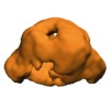
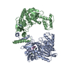
| タイトル | 3D structure of human endoglin | |||||||||
 マップデータ マップデータ | structure of human endoglin | |||||||||
 試料 試料 |
| |||||||||
 キーワード キーワード | Endoglin / CD105 / TGF-beta / HHT disorder / zona pellucida / preeclampsia | |||||||||
| 機能・相同性 | Zona pellucida domain / regulation of transforming growth factor beta receptor signaling pathway 機能・相同性情報 機能・相同性情報 | |||||||||
| 生物種 |  Homo sapiens (ヒト) Homo sapiens (ヒト) | |||||||||
| 手法 | 単粒子再構成法 / ネガティブ染色法 / 解像度: 25.0 Å | |||||||||
 データ登録者 データ登録者 | Llorca O / Trujillo A / Blanco FJ / Bernabeu C | |||||||||
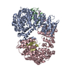
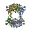
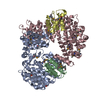
 引用 引用 |  ジャーナル: J Mol Biol / 年: 2007 ジャーナル: J Mol Biol / 年: 2007タイトル: Structural model of human endoglin, a transmembrane receptor responsible for hereditary hemorrhagic telangiectasia. 著者: Oscar Llorca / Arturo Trujillo / Francisco J Blanco / Carmelo Bernabeu /  要旨: Endoglin is a type I membrane protein expressed as a disulphide-linked homodimer on human vascular endothelial cells whose haploinsufficiency is responsible for the dominant vascular dysplasia known ...Endoglin is a type I membrane protein expressed as a disulphide-linked homodimer on human vascular endothelial cells whose haploinsufficiency is responsible for the dominant vascular dysplasia known as hereditary hemorrhagic telangiectasia (HHT). Structurally, endoglin belongs to the zona pellucida (ZP) family of proteins that share a ZP domain of approximately 260 amino acid residues at their extracellular region. Endoglin is a component of the TGF-beta receptor complex, interacts with the TGF-beta signalling receptors types I and II, and modulates cellular responses to TGF-beta. Here, we have determined for the first time the three-dimensional structure of the approximately 140 kDa extracellular domain of endoglin at 25 A resolution, using single-particle electron microscopy (EM). This reconstruction provides the general architecture of endoglin, which arranges as a dome made of antiparallel oriented monomers enclosing a cavity at one end. A high-resolution structure of endoglin has also been modelled de novo and found to be consistent with the experimental reconstruction. Each subunit comprises three well-defined domains, two of them corresponding to ZP regions, organised into an open U-shaped monomer. This domain arrangement was found to closely resemble the overall structure derived experimentally and the three modelled de novo domains were tentatively assigned to the domains observed in the EM reconstruction. This molecular model was further tested by tagging endoglin's C terminus with an IgG Fc fragment visible after 3D reconstruction of the labelled protein. Combined, these data provide the structural framework to interpret endoglin's functional domains and mutations found in HHT patients. | |||||||||
| 履歴 |
|
- 構造の表示
構造の表示
| ムービー |
 ムービービューア ムービービューア |
|---|---|
| 構造ビューア | EMマップ:  SurfView SurfView Molmil Molmil Jmol/JSmol Jmol/JSmol |
| 添付画像 |
- ダウンロードとリンク
-EMDBアーカイブ
| マップデータ |  emd_1559.map.gz emd_1559.map.gz | 345.7 KB |  EMDBマップデータ形式 EMDBマップデータ形式 | |
|---|---|---|---|---|
| ヘッダ (付随情報) |  emd-1559-v30.xml emd-1559-v30.xml emd-1559.xml emd-1559.xml | 8.9 KB 8.9 KB | 表示 表示 |  EMDBヘッダ EMDBヘッダ |
| 画像 |  1559.gif 1559.gif | 55.8 KB | ||
| アーカイブディレクトリ |  http://ftp.pdbj.org/pub/emdb/structures/EMD-1559 http://ftp.pdbj.org/pub/emdb/structures/EMD-1559 ftp://ftp.pdbj.org/pub/emdb/structures/EMD-1559 ftp://ftp.pdbj.org/pub/emdb/structures/EMD-1559 | HTTPS FTP |
-検証レポート
| 文書・要旨 |  emd_1559_validation.pdf.gz emd_1559_validation.pdf.gz | 190.6 KB | 表示 |  EMDB検証レポート EMDB検証レポート |
|---|---|---|---|---|
| 文書・詳細版 |  emd_1559_full_validation.pdf.gz emd_1559_full_validation.pdf.gz | 189.7 KB | 表示 | |
| XML形式データ |  emd_1559_validation.xml.gz emd_1559_validation.xml.gz | 5 KB | 表示 | |
| アーカイブディレクトリ |  https://ftp.pdbj.org/pub/emdb/validation_reports/EMD-1559 https://ftp.pdbj.org/pub/emdb/validation_reports/EMD-1559 ftp://ftp.pdbj.org/pub/emdb/validation_reports/EMD-1559 ftp://ftp.pdbj.org/pub/emdb/validation_reports/EMD-1559 | HTTPS FTP |
-関連構造データ
- リンク
リンク
| EMDBのページ |  EMDB (EBI/PDBe) / EMDB (EBI/PDBe) /  EMDataResource EMDataResource |
|---|
- マップ
マップ
| ファイル |  ダウンロード / ファイル: emd_1559.map.gz / 形式: CCP4 / 大きさ: 1.9 MB / タイプ: IMAGE STORED AS FLOATING POINT NUMBER (4 BYTES) ダウンロード / ファイル: emd_1559.map.gz / 形式: CCP4 / 大きさ: 1.9 MB / タイプ: IMAGE STORED AS FLOATING POINT NUMBER (4 BYTES) | ||||||||||||||||||||||||||||||||||||||||||||||||||||||||||||||||||||
|---|---|---|---|---|---|---|---|---|---|---|---|---|---|---|---|---|---|---|---|---|---|---|---|---|---|---|---|---|---|---|---|---|---|---|---|---|---|---|---|---|---|---|---|---|---|---|---|---|---|---|---|---|---|---|---|---|---|---|---|---|---|---|---|---|---|---|---|---|---|
| 注釈 | structure of human endoglin | ||||||||||||||||||||||||||||||||||||||||||||||||||||||||||||||||||||
| 投影像・断面図 | 画像のコントロール
画像は Spider により作成 | ||||||||||||||||||||||||||||||||||||||||||||||||||||||||||||||||||||
| ボクセルのサイズ | X=Y=Z: 2.3 Å | ||||||||||||||||||||||||||||||||||||||||||||||||||||||||||||||||||||
| 密度 |
| ||||||||||||||||||||||||||||||||||||||||||||||||||||||||||||||||||||
| 対称性 | 空間群: 1 | ||||||||||||||||||||||||||||||||||||||||||||||||||||||||||||||||||||
| 詳細 | EMDB XML:
CCP4マップ ヘッダ情報:
| ||||||||||||||||||||||||||||||||||||||||||||||||||||||||||||||||||||
-添付データ
- 試料の構成要素
-全体 : Extracellular region of human endoglin, from Glu26 to Leu587 residues.
| 全体 | 名称: Extracellular region of human endoglin, from Glu26 to Leu587 residues. |
|---|---|
| 要素 |
|
-超分子 #1000: Extracellular region of human endoglin, from Glu26 to Leu587 residues.
| 超分子 | 名称: Extracellular region of human endoglin, from Glu26 to Leu587 residues. タイプ: sample / ID: 1000 / 集合状態: One disulphide-linked homodimer / Number unique components: 1 |
|---|---|
| 分子量 | 実験値: 130 KDa / 理論値: 140 KDa / 手法: SDS-PAGE |
-分子 #1: Transmembrane receptor
| 分子 | 名称: Transmembrane receptor / タイプ: protein_or_peptide / ID: 1 / Name.synonym: Endoglin / コピー数: 2 / 集合状態: Homodimer / 組換発現: Yes |
|---|---|
| 由来(天然) | 生物種:  Homo sapiens (ヒト) / 別称: Human / 細胞中の位置: Plasma membrane Homo sapiens (ヒト) / 別称: Human / 細胞中の位置: Plasma membrane |
| 分子量 | 実験値: 130 KDa / 理論値: 140 KDa |
| 組換発現 | 生物種: Mouse myeloma cell line NS0 |
| 配列 | GO: regulation of transforming growth factor beta receptor signaling pathway InterPro: Zona pellucida domain |
-実験情報
-構造解析
| 手法 | ネガティブ染色法 |
|---|---|
 解析 解析 | 単粒子再構成法 |
| 試料の集合状態 | particle |
- 試料調製
試料調製
| 濃度 | 0.1 mg/mL |
|---|---|
| 緩衝液 | pH: 7 / 詳細: 150mM NaCl, 50mM Na2HPO4, 10% glycerol |
| 染色 | タイプ: NEGATIVE 詳細: EndoEC was applied to carbon-coated grids and negatively stained with 1% w/v uranyl acetate. |
| グリッド | 詳細: 40 mesh Copper/Palladium grid. |
| 凍結 | 凍結剤: NONE / 装置: OTHER |
- 電子顕微鏡法
| 顕微鏡 | JEOL 1230 |
|---|---|
| アライメント法 | Legacy - 非点収差: Correction with FFT and CCD camera |
| 詳細 | Microscope used JEOL-1230 |
| 撮影 | カテゴリ: FILM / フィルム・検出器のモデル: KODAK SO-163 FILM / デジタル化 - スキャナー: OTHER / デジタル化 - サンプリング間隔: 10.5 µm 詳細: Images scanned with a MINOLTA Dimage Scan Multi Pro scanner at 2400 dpi ビット/ピクセル: 16 |
| 電子線 | 加速電圧: 100 kV / 電子線源: TUNGSTEN HAIRPIN |
| 電子光学系 | 照射モード: FLOOD BEAM / 撮影モード: BRIGHT FIELD / Cs: 2.9 mm / 倍率(公称値): 50000 |
| 試料ステージ | 試料ホルダー: Eucentric / 試料ホルダーモデル: OTHER |
- 画像解析
画像解析
| 最終 再構成 | 想定した対称性 - 点群: C2 (2回回転対称) / アルゴリズム: OTHER / 解像度のタイプ: BY AUTHOR / 解像度: 25.0 Å / 解像度の算出法: FSC 0.5 CUT-OFF / ソフトウェア - 名称: EMAN / 使用した粒子像数: 2964 |
|---|---|
| 最終 2次元分類 | クラス数: 16 |
 ムービー
ムービー コントローラー


 UCSF Chimera


 Z (Sec.)
 Y (Row.)
 X (Col.)


