large ribosomal subunit / ribosome binding / transferase activity / 5S rRNA binding / ribosomal large subunit assembly / large ribosomal subunit rRNA binding / cytosolic large ribosomal subunit / cytoplasmic translation / tRNA binding / negative regulation of translation ...large ribosomal subunit / ribosome binding / transferase activity / 5S rRNA binding / ribosomal large subunit assembly / large ribosomal subunit rRNA binding / cytosolic large ribosomal subunit / cytoplasmic translation / tRNA binding / negative regulation of translation / rRNA binding / structural constituent of ribosome / ribosome / translation / ribonucleoprotein complex / GTPase activity / mRNA binding / GTP binding / RNA binding / metal ion binding / cytoplasm Similarity search - Function
GTPase HflX / GTPase HflX, N-terminal / HflX-type guanine nucleotide-binding (G) domain / GTP-binding protein, middle domain / GTPase HflX, N-terminal domain superfamily / GTP-binding GTPase N-terminal / GTP-binding GTPase Middle Region / HflX-type guanine nucleotide-binding (G) domain profile. / Ribosomal protein L31 type B / 50S ribosome-binding GTPase ...GTPase HflX / GTPase HflX, N-terminal / HflX-type guanine nucleotide-binding (G) domain / GTP-binding protein, middle domain / GTPase HflX, N-terminal domain superfamily / GTP-binding GTPase N-terminal / GTP-binding GTPase Middle Region / HflX-type guanine nucleotide-binding (G) domain profile. / Ribosomal protein L31 type B / 50S ribosome-binding GTPase / GTP binding domain / : / Ribosomal protein L11, bacterial-type / Ribosomal protein L31 signature. / Ribosomal protein L31 / Ribosomal protein L31 superfamily / Ribosomal protein L31 / Ribosomal protein L16 signature 1. / Ribosomal protein L21, conserved site / Ribosomal protein L21 signature. / Ribosomal protein L16 signature 2. / Ribosomal protein L16, conserved site / Ribosomal protein L6, conserved site / Ribosomal protein L6 signature 1. / : / Ribosomal protein L11, N-terminal / Ribosomal protein L11, N-terminal domain / Ribosomal protein L11/L12 / Ribosomal protein L11, C-terminal / Ribosomal protein L11, C-terminal domain superfamily / Ribosomal protein L11/L12, N-terminal domain superfamily / Ribosomal protein L11/L12 / Ribosomal protein L11, RNA binding domain / Ribosomal protein L17 signature. / Ribosomal protein L36 signature. / Ribosomal protein L32p, bacterial type / Ribosomal protein L28/L24 superfamily / : / Ribosomal protein L33, conserved site / Ribosomal protein L33 signature. / Ribosomal protein L35, conserved site / Ribosomal protein L35 signature. / Ribosomal protein L28 / Ribosomal protein L35, non-mitochondrial / Ribosomal protein L18, bacterial-type / : / Ribosomal protein L6, bacterial-type / Ribosomal protein L5, bacterial-type / Ribosomal protein L36 / Ribosomal protein L36 superfamily / Ribosomal protein L36 / Ribosomal protein L19, conserved site / Ribosomal protein L19 signature. / Ribosomal protein L27, conserved site / Ribosomal protein L27 signature. / Ribosomal protein L20 signature. / Ribosomal protein L22, bacterial/chloroplast-type / Ribosomal protein L14P, bacterial-type / Ribosomal protein L34, conserved site / Ribosomal protein L34 signature. / Ribosomal protein L2, bacterial/organellar-type / Ribosomal protein L35 / Ribosomal protein L35 superfamily / Ribosomal protein L35 / Ribosomal protein L33 / Ribosomal protein L18 / Ribosomal L18 of archaea, bacteria, mitoch. and chloroplast / Ribosomal protein L33 / Ribosomal L28 family / Ribosomal protein L33 superfamily / Ribosomal protein L16 / Ribosomal protein L28/L24 / Ribosomal protein L30, bacterial-type / L28p-like / : / Ribosomal protein L27 / Ribosomal L27 protein / Ribosomal protein L20 / Ribosomal L32p protein family / Ribosomal protein L19 / Ribosomal protein L20 / Ribosomal protein L20, C-terminal / Ribosomal protein L19 / Ribosomal protein L19 superfamily / Ribosomal protein L21 / Ribosomal protein L32p / Large ribosomal subunit protein uL24, C-terminal domain / Ribosomal protein L17 / Ribosomal protein L17 superfamily / Ribosomal protein L17 / Ribosomal protein L21-like / L21-like superfamily / Ribosomal prokaryotic L21 protein / Ribosomal protein L34 / Ribosomal protein L34 / Ribosomal protein L24 / Ribosomal protein L3, bacterial/organelle-type / Ribosomal protein L15, bacterial-type / 50S ribosomal protein uL4 / Ribosomal protein L13, bacterial-type Similarity search - Domain/homology
Large ribosomal subunit protein uL24 / Large ribosomal subunit protein bL19 / Large ribosomal subunit protein bL31B / Large ribosomal subunit protein bL35 / Large ribosomal subunit protein uL2 / Large ribosomal subunit protein uL4 / Large ribosomal subunit protein uL11 / Large ribosomal subunit protein bL20 / Large ribosomal subunit protein bL27 / Large ribosomal subunit protein bL28 ...Large ribosomal subunit protein uL24 / Large ribosomal subunit protein bL19 / Large ribosomal subunit protein bL31B / Large ribosomal subunit protein bL35 / Large ribosomal subunit protein uL2 / Large ribosomal subunit protein uL4 / Large ribosomal subunit protein uL11 / Large ribosomal subunit protein bL20 / Large ribosomal subunit protein bL27 / Large ribosomal subunit protein bL28 / Large ribosomal subunit protein uL29 / Large ribosomal subunit protein bL32B / Large ribosomal subunit protein bL33A / Large ribosomal subunit protein bL34 / Large ribosomal subunit protein bL36 / Large ribosomal subunit protein uL3 / Large ribosomal subunit protein uL23 / Large ribosomal subunit protein uL6 / Large ribosomal subunit protein uL18 / Large ribosomal subunit protein uL15 / Large ribosomal subunit protein bL17 / Large ribosomal subunit protein uL13 / Large ribosomal subunit protein bL21 / GTPase HflX / Large ribosomal subunit protein uL22 / Large ribosomal subunit protein uL16 / Large ribosomal subunit protein uL14 / Large ribosomal subunit protein uL5 / Large ribosomal subunit protein uL30 Similarity search - Component
Biological species
Listeria monocytogenes E (bacteria) / Listeria monocytogenes EGD-e (bacteria)
Method
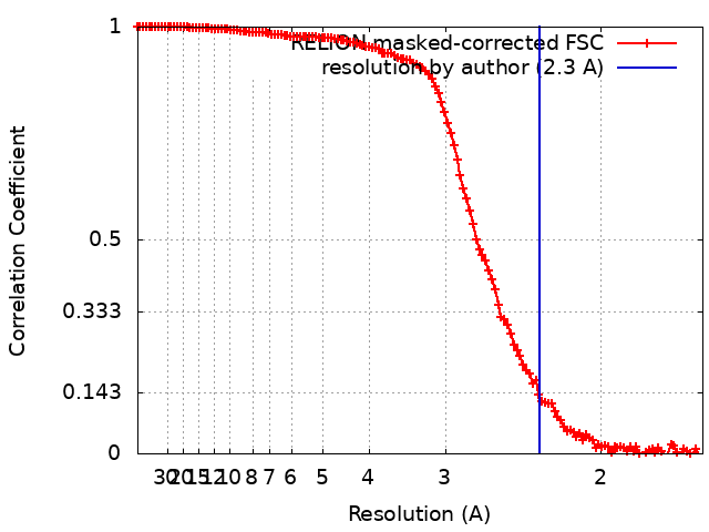
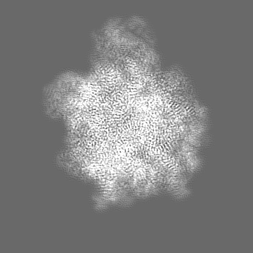
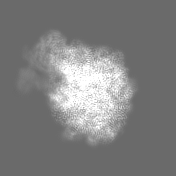
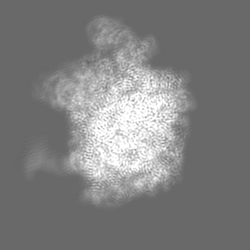
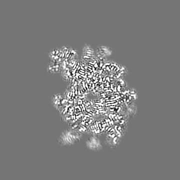
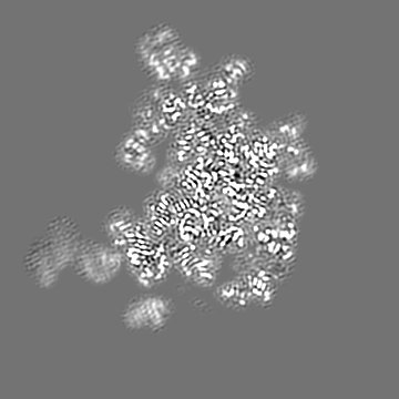
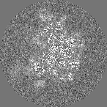
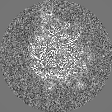
single particle reconstruction / cryo EM / Resolution: 2.3 Å
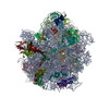
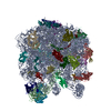
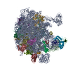
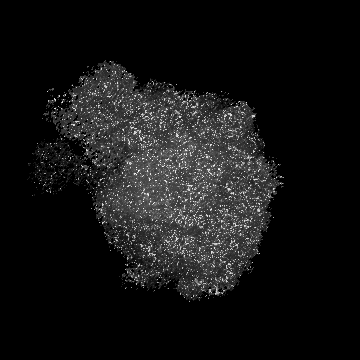
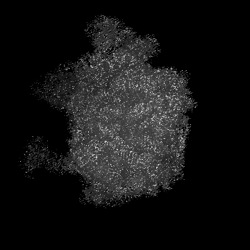
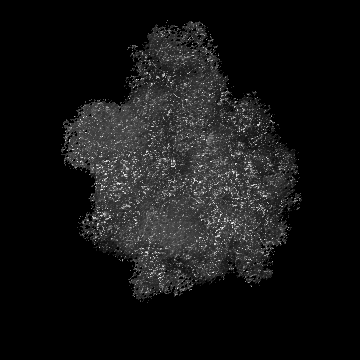
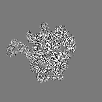
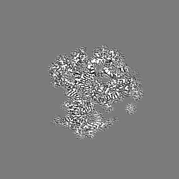
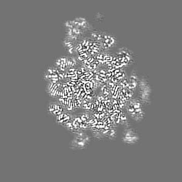
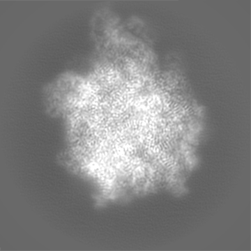
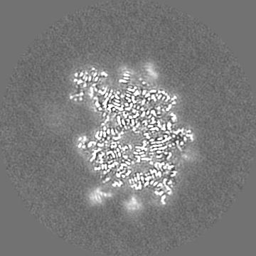
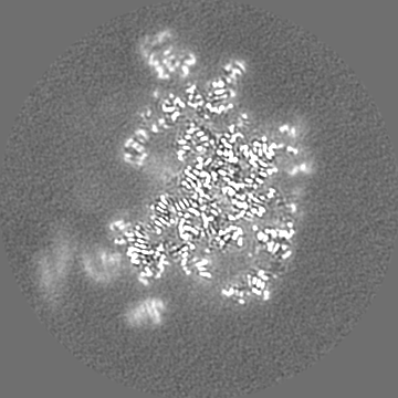
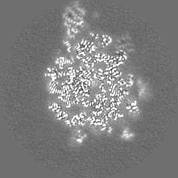
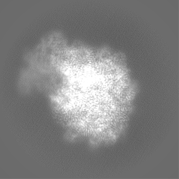
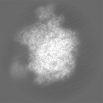
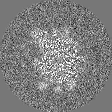
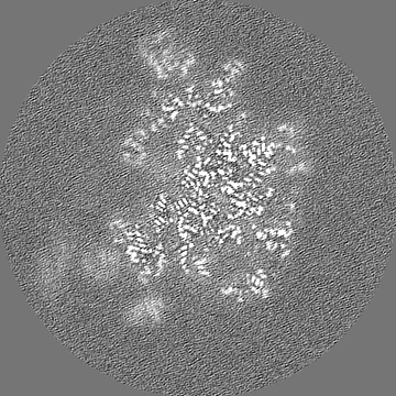
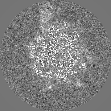
Journal: Nucleic Acids Res / Year: 2022 Title: Structural basis for HflXr-mediated antibiotic resistance in Listeria monocytogenes. Authors: Timm O Koller / Kathryn J Turnbull / Karolis Vaitkevicius / Caillan Crowe-McAuliffe / Mohammad Roghanian / Ondřej Bulvas / Jose A Nakamoto / Tatsuaki Kurata / Christina Julius / Gemma C ...Authors: Timm O Koller / Kathryn J Turnbull / Karolis Vaitkevicius / Caillan Crowe-McAuliffe / Mohammad Roghanian / Ondřej Bulvas / Jose A Nakamoto / Tatsuaki Kurata / Christina Julius / Gemma C Atkinson / Jörgen Johansson / Vasili Hauryliuk / Daniel N Wilson / Abstract: HflX is a ubiquitous bacterial GTPase that splits and recycles stressed ribosomes. In addition to HflX, Listeria monocytogenes contains a second HflX homolog, HflXr. Unlike HflX, HflXr confers ...HflX is a ubiquitous bacterial GTPase that splits and recycles stressed ribosomes. In addition to HflX, Listeria monocytogenes contains a second HflX homolog, HflXr. Unlike HflX, HflXr confers resistance to macrolide and lincosamide antibiotics by an experimentally unexplored mechanism. Here, we have determined cryo-EM structures of L. monocytogenes HflXr-50S and HflX-50S complexes as well as L. monocytogenes 70S ribosomes in the presence and absence of the lincosamide lincomycin. While the overall geometry of HflXr on the 50S subunit is similar to that of HflX, a loop within the N-terminal domain of HflXr, which is two amino acids longer than in HflX, reaches deeper into the peptidyltransferase center. Moreover, unlike HflX, the binding of HflXr induces conformational changes within adjacent rRNA nucleotides that would be incompatible with drug binding. These findings suggest that HflXr confers resistance using an allosteric ribosome protection mechanism, rather than by simply splitting and recycling antibiotic-stalled ribosomes.
Details: 0.05% Nikkol and 0.2 mg/mL FLAG peptide during elution.
Grid
Model: Quantifoil R2/2 / Material: COPPER / Support film - Material: CARBON / Support film - topology: CONTINUOUS / Support film - Film thickness: 2 / Pretreatment - Type: GLOW DISCHARGE
Vitrification
Cryogen name: ETHANE-PROPANE / Chamber humidity: 100 % / Chamber temperature: 277 K / Instrument: FEI VITROBOT MARK IV
Details
5 OD260/mL
-
Electron microscopy
Microscope
FEI TITAN KRIOS
Image recording
Film or detector model: GATAN K2 SUMMIT (4k x 4k) / Detector mode: COUNTING / Average electron dose: 35.022 e/Å2
Electron beam
Acceleration voltage: 300 kV / Electron source: FIELD EMISSION GUN
In the structure databanks used in Yorodumi, some data are registered as the other names, "COVID-19 virus" and "2019-nCoV". Here are the details of the virus and the list of structure data.
Jan 31, 2019. EMDB accession codes are about to change! (news from PDBe EMDB page)
EMDB accession codes are about to change! (news from PDBe EMDB page)
The allocation of 4 digits for EMDB accession codes will soon come to an end. Whilst these codes will remain in use, new EMDB accession codes will include an additional digit and will expand incrementally as the available range of codes is exhausted. The current 4-digit format prefixed with “EMD-” (i.e. EMD-XXXX) will advance to a 5-digit format (i.e. EMD-XXXXX), and so on. It is currently estimated that the 4-digit codes will be depleted around Spring 2019, at which point the 5-digit format will come into force.
The EM Navigator/Yorodumi systems omit the EMD- prefix.
Related info.:Q: What is EMD? / ID/Accession-code notation in Yorodumi/EM Navigator
Yorodumi is a browser for structure data from EMDB, PDB, SASBDB, etc.
This page is also the successor to EM Navigator detail page, and also detail information page/front-end page for Omokage search.
The word "yorodu" (or yorozu) is an old Japanese word meaning "ten thousand". "mi" (miru) is to see.
Related info.:EMDB / PDB / SASBDB / Comparison of 3 databanks / Yorodumi Search / Aug 31, 2016. New EM Navigator & Yorodumi / Yorodumi Papers / Jmol/JSmol / Function and homology information / Changes in new EM Navigator and Yorodumi
 Movie
Movie Controller

 Yorodumi
 Open data
 Basic information
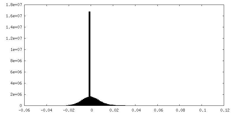
 Map data
 Sample
 Keywords
 Function and homology information
 Listeria monocytogenes E (bacteria) /
  Authors
 Germany,
  Sweden, 2 items
  Citation


 Structure visualization
 Downloads & links
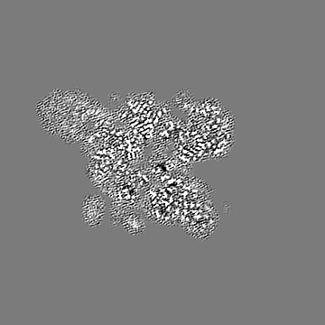
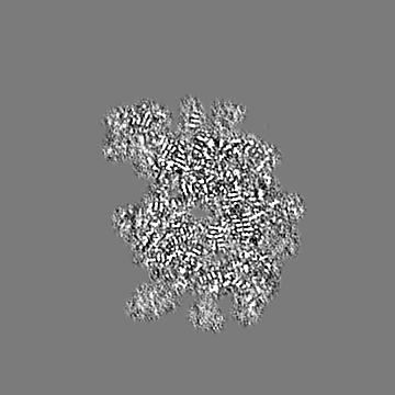
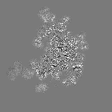
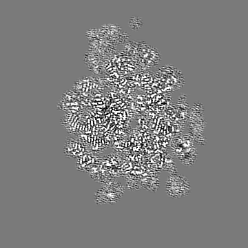
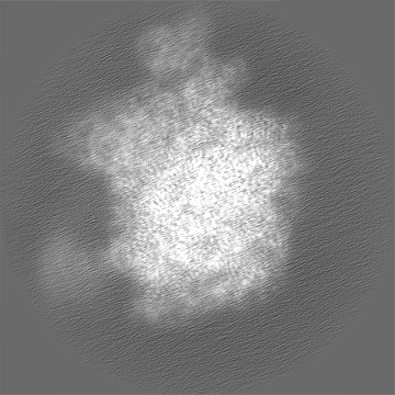
 emd_15161.png
 http://ftp.pdbj.org/pub/emdb/structures/EMD-15161


 Z (Sec.)
 Y (Row.)
 X (Col.)


 Sample components

 Processing
 Electron microscopy
 FIELD EMISSION GUN