[English] 日本語
Yorodumi
- PDB-8a5i: Cryo-EM structure of Lincomycin bound to the Listeria monocytogen... -

+
Open data


ID or keywords:

Loading...

-
Basic information

Entry
Database: PDB / ID: 8a5i
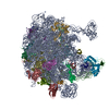
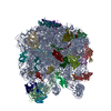
TitleCryo-EM structure of Lincomycin bound to the Listeria monocytogenes 50S ribosomal subunit.
Components
  • (50S ribosomal protein ...) x 27
  • 23S ribosomal RNA
  • 5S ribosomal RNA
KeywordsRIBOSOME / Listeria monocytogenes / Lincomycin / 50S / antibiotic
Function / homology
Function and homology information


large ribosomal subunit rRNA binding / large ribosomal subunit / cytoplasmic translation / 5S rRNA binding / cytosolic large ribosomal subunit / transferase activity / tRNA binding / negative regulation of translation / rRNA binding / ribosome ...large ribosomal subunit rRNA binding / large ribosomal subunit / cytoplasmic translation / 5S rRNA binding / cytosolic large ribosomal subunit / transferase activity / tRNA binding / negative regulation of translation / rRNA binding / ribosome / structural constituent of ribosome / ribonucleoprotein complex / translation / mRNA binding / RNA binding / cytoplasm
Similarity search - Function
Ribosomal protein L31 type B / Ribosomal protein L31 signature. / Ribosomal protein L31 / Ribosomal protein L31 superfamily / Ribosomal protein L31 / Ribosomal protein L21, conserved site / Ribosomal protein L21 signature. / Ribosomal protein L16 signature 1. / : / Ribosomal protein L6, conserved site ...Ribosomal protein L31 type B / Ribosomal protein L31 signature. / Ribosomal protein L31 / Ribosomal protein L31 superfamily / Ribosomal protein L31 / Ribosomal protein L21, conserved site / Ribosomal protein L21 signature. / Ribosomal protein L16 signature 1. / : / Ribosomal protein L6, conserved site / Ribosomal protein L6 signature 1. / Ribosomal protein L16, conserved site / Ribosomal protein L16 signature 2. / Ribosomal protein L17 signature. / Ribosomal protein L28/L24 superfamily / Ribosomal protein L36 signature. / Ribosomal protein L32p, bacterial type / Ribosomal protein L28 / Ribosomal protein L35, conserved site / Ribosomal protein L35 signature. / Ribosomal protein L33, conserved site / Ribosomal protein L33 signature. / Ribosomal protein L35, non-mitochondrial / Ribosomal protein L5, bacterial-type / Ribosomal protein L6, bacterial-type / Ribosomal protein L18, bacterial-type / Ribosomal protein L19, conserved site / Ribosomal protein L19 signature. / Ribosomal protein L36 / Ribosomal protein L36 superfamily / Ribosomal protein L36 / Ribosomal protein L20 signature. / Ribosomal protein L27, conserved site / Ribosomal protein L27 signature. / Ribosomal protein L14P, bacterial-type / Ribosomal protein L34, conserved site / Ribosomal protein L34 signature. / Ribosomal protein L22, bacterial/chloroplast-type / Ribosomal protein L35 / Ribosomal protein L35 superfamily / Ribosomal protein L2, bacterial/organellar-type / Ribosomal protein L35 / Ribosomal L28 family / Ribosomal protein L33 / Ribosomal protein L33 / Ribosomal protein L28/L24 / Ribosomal protein L33 superfamily / : / Ribosomal protein L30, bacterial-type / Ribosomal protein L16 / Ribosomal protein L18 / Ribosomal L18 of archaea, bacteria, mitoch. and chloroplast / L28p-like / Ribosomal protein L20 / Ribosomal protein L20 / Ribosomal protein L20, C-terminal / Ribosomal protein L21 / Ribosomal protein L27 / Ribosomal L27 protein / Ribosomal protein L19 / Ribosomal protein L19 superfamily / Ribosomal protein L19 / Ribosomal proteins 50S L24/mitochondrial 39S L24 / Ribosomal protein L17 / Ribosomal protein L17 superfamily / Ribosomal protein L17 / Ribosomal protein L21-like / L21-like superfamily / Ribosomal prokaryotic L21 protein / Ribosomal L32p protein family / Ribosomal protein L24 / Ribosomal protein L32p / Ribosomal protein L34 / Ribosomal protein L34 / Ribosomal protein L13, bacterial-type / Ribosomal protein L3, bacterial/organelle-type / Ribosomal protein L15, bacterial-type / 50S ribosomal protein uL4 / Ribosomal protein L30, conserved site / Ribosomal protein L5, conserved site / Ribosomal protein L29, conserved site / Ribosomal protein L30 signature. / : / Ribosomal protein L5, N-terminal / Ribosomal protein L5 signature. / Ribosomal protein L15, conserved site / Ribosomal protein L5, C-terminal / Ribosomal protein L5 / Ribosomal protein L5 domain superfamily / Ribosomal protein L2, conserved site / Ribosomal protein L2 signature. / Ribosomal protein L10e/L16 / Ribosomal protein L10e/L16 superfamily / Ribosomal protein L29 signature. / Ribosomal protein L5 / Ribosomal protein L6, alpha-beta domain / Ribosomal protein L6 / ribosomal L5P family C-terminus / Ribosomal protein L6, alpha-beta domain superfamily / Ribosomal protein L16p/L10e
Similarity search - Domain/homology
LINCOMYCIN / : / 1,4-DIAMINOBUTANE / SPERMIDINE / : / : / RNA / RNA (> 10) / RNA (> 100) / RNA (> 1000) ...LINCOMYCIN / : / 1,4-DIAMINOBUTANE / SPERMIDINE / : / : / RNA / RNA (> 10) / RNA (> 100) / RNA (> 1000) / Large ribosomal subunit protein uL24 / Large ribosomal subunit protein bL19 / Large ribosomal subunit protein bL31B / Large ribosomal subunit protein bL35 / Large ribosomal subunit protein uL2 / Large ribosomal subunit protein uL4 / Large ribosomal subunit protein bL20 / Large ribosomal subunit protein bL27 / Large ribosomal subunit protein bL28 / Large ribosomal subunit protein uL29 / Large ribosomal subunit protein bL32B / Large ribosomal subunit protein bL33A / Large ribosomal subunit protein bL34 / Large ribosomal subunit protein bL36 / Large ribosomal subunit protein uL3 / Large ribosomal subunit protein uL23 / Large ribosomal subunit protein uL6 / Large ribosomal subunit protein uL18 / Large ribosomal subunit protein uL15 / Large ribosomal subunit protein bL17 / Large ribosomal subunit protein uL13 / Large ribosomal subunit protein bL21 / Large ribosomal subunit protein uL22 / Large ribosomal subunit protein uL16 / Large ribosomal subunit protein uL14 / Large ribosomal subunit protein uL5 / Large ribosomal subunit protein uL30
Similarity search - Component
Biological speciesListeria monocytogenes EGD-e (bacteria)
MethodELECTRON MICROSCOPY / single particle reconstruction / cryo EM / Resolution: 2.3 Å
AuthorsKoller, T.O. / Crowe-McAuliffe, C. / Wilson, D.N.
Funding support Sweden, 2items
OrganizationGrant numberCountry
Other governmentDLR01KI1820
Swedish Research Council2018-00956 Sweden
CitationJournal: Nucleic Acids Res / Year: 2022
Title: Structural basis for HflXr-mediated antibiotic resistance in Listeria monocytogenes.
Authors: Timm O Koller / Kathryn J Turnbull / Karolis Vaitkevicius / Caillan Crowe-McAuliffe / Mohammad Roghanian / Ondřej Bulvas / Jose A Nakamoto / Tatsuaki Kurata / Christina Julius / Gemma C ...Authors: Timm O Koller / Kathryn J Turnbull / Karolis Vaitkevicius / Caillan Crowe-McAuliffe / Mohammad Roghanian / Ondřej Bulvas / Jose A Nakamoto / Tatsuaki Kurata / Christina Julius / Gemma C Atkinson / Jörgen Johansson / Vasili Hauryliuk / Daniel N Wilson /
Abstract: HflX is a ubiquitous bacterial GTPase that splits and recycles stressed ribosomes. In addition to HflX, Listeria monocytogenes contains a second HflX homolog, HflXr. Unlike HflX, HflXr confers ...HflX is a ubiquitous bacterial GTPase that splits and recycles stressed ribosomes. In addition to HflX, Listeria monocytogenes contains a second HflX homolog, HflXr. Unlike HflX, HflXr confers resistance to macrolide and lincosamide antibiotics by an experimentally unexplored mechanism. Here, we have determined cryo-EM structures of L. monocytogenes HflXr-50S and HflX-50S complexes as well as L. monocytogenes 70S ribosomes in the presence and absence of the lincosamide lincomycin. While the overall geometry of HflXr on the 50S subunit is similar to that of HflX, a loop within the N-terminal domain of HflXr, which is two amino acids longer than in HflX, reaches deeper into the peptidyltransferase center. Moreover, unlike HflX, the binding of HflXr induces conformational changes within adjacent rRNA nucleotides that would be incompatible with drug binding. These findings suggest that HflXr confers resistance using an allosteric ribosome protection mechanism, rather than by simply splitting and recycling antibiotic-stalled ribosomes.
History
DepositionJun 15, 2022Deposition site: PDBE / Processing site: PDBE
Revision 1.0Nov 2, 2022Provider: repository / Type: Initial release
Revision 1.1Nov 9, 2022Group: Database references / Category: citation / citation_author
Item: _citation.pdbx_database_id_PubMed / _citation.title ..._citation.pdbx_database_id_PubMed / _citation.title / _citation_author.identifier_ORCID / _citation_author.name
Revision 1.2Nov 16, 2022Group: Advisory / Database references / Derived calculations
Category: citation / pdbx_validate_close_contact ...citation / pdbx_validate_close_contact / struct_conn / struct_conn_type
Item: _citation.journal_volume / _citation.page_first / _citation.page_last

-
Structure visualization

Structure viewerMolecule:
MolmilJmol/JSmol

Downloads & links

-
Assembly

Deposited unit
1: 50S ribosomal protein L28
2: 50S ribosomal protein L29
3: 50S ribosomal protein L30
4: 50S ribosomal protein L31 type B
5: 50S ribosomal protein L32-2
6: 50S ribosomal protein L33 1
7: 50S ribosomal protein L34
8: 50S ribosomal protein L35
9: 50S ribosomal protein L36
A: 23S ribosomal RNA
B: 5S ribosomal RNA
G: 50S ribosomal protein L2
H: 50S ribosomal protein L3
I: 50S ribosomal protein L4
J: 50S ribosomal protein L5
K: 50S ribosomal protein L6
M: 50S ribosomal protein L13
N: 50S ribosomal protein L14
O: 50S ribosomal protein L15
P: 50S ribosomal protein L16
Q: 50S ribosomal protein L17
R: 50S ribosomal protein L18
S: 50S ribosomal protein L19
T: 50S ribosomal protein L20
U: 50S ribosomal protein L21
V: 50S ribosomal protein L22
W: 50S ribosomal protein L23
X: 50S ribosomal protein L24
Z: 50S ribosomal protein L27
hetero molecules


Theoretical massNumber of molelcules
Total (without water)1,341,564201
Polymers1,336,65629
Non-polymers4,908172
Water24,8791381
1


  • Idetical with deposited unit
  • defined by author
  • Evidence: electron microscopy
TypeNameSymmetry operationNumber
identity operation1_5551

-
Components

+
50S ribosomal protein ... , 27 types, 27 molecules 123456789GHIJKMNOPQRSTUVWXZ

#1: Protein 50S ribosomal protein L28 /


Mass: 7010.383 Da / Num. of mol.: 1 / Source method: isolated from a natural source / Source: (natural) Listeria monocytogenes EGD-e (bacteria) / Strain: ATCC BAA-679 / EGD-e / References: UniProt: P66144
#2: Protein 50S ribosomal protein L29 /


Mass: 7414.548 Da / Num. of mol.: 1 / Source method: isolated from a natural source / Source: (natural) Listeria monocytogenes EGD-e (bacteria) / Strain: ATCC BAA-679 / EGD-e / References: UniProt: P66166
#3: Protein 50S ribosomal protein L30 /


Mass: 6504.753 Da / Num. of mol.: 1 / Source method: isolated from a natural source / Source: (natural) Listeria monocytogenes EGD-e (bacteria) / Strain: ATCC BAA-679 / EGD-e / References: UniProt: Q927M5
#4: Protein 50S ribosomal protein L31 type B / Ribosome


Mass: 9274.360 Da / Num. of mol.: 1 / Source method: isolated from a natural source / Source: (natural) Listeria monocytogenes EGD-e (bacteria) / Strain: ATCC BAA-679 / EGD-e / References: UniProt: P0A485
#5: Protein 50S ribosomal protein L32-2 / Ribosome


Mass: 6534.564 Da / Num. of mol.: 1 / Source method: isolated from a natural source / Source: (natural) Listeria monocytogenes EGD-e (bacteria) / Strain: ATCC BAA-679 / EGD-e / References: UniProt: P66207
#6: Protein/peptide 50S ribosomal protein L33 1 / Ribosome


Mass: 6021.034 Da / Num. of mol.: 1 / Source method: isolated from a natural source / Source: (natural) Listeria monocytogenes EGD-e (bacteria) / Strain: ATCC BAA-679 / EGD-e / References: UniProt: P66219
#7: Protein/peptide 50S ribosomal protein L34 /


Mass: 5319.457 Da / Num. of mol.: 1 / Source method: isolated from a natural source / Source: (natural) Listeria monocytogenes EGD-e (bacteria) / Strain: ATCC BAA-679 / EGD-e / References: UniProt: P66248
#8: Protein 50S ribosomal protein L35 /


Mass: 7746.438 Da / Num. of mol.: 1 / Source method: isolated from a natural source / Source: (natural) Listeria monocytogenes EGD-e (bacteria) / Strain: ATCC BAA-679 / EGD-e / References: UniProt: P0A491
#9: Protein/peptide 50S ribosomal protein L36 /


Mass: 4336.460 Da / Num. of mol.: 1 / Source method: isolated from a natural source / Source: (natural) Listeria monocytogenes EGD-e (bacteria) / Strain: ATCC BAA-679 / EGD-e / References: UniProt: P66290
#12: Protein 50S ribosomal protein L2 /


Mass: 30568.367 Da / Num. of mol.: 1 / Source method: isolated from a natural source / Source: (natural) Listeria monocytogenes EGD-e (bacteria) / Strain: ATCC BAA-679 / EGD-e / References: UniProt: P60426
#13: Protein 50S ribosomal protein L3 /


Mass: 22863.459 Da / Num. of mol.: 1 / Source method: isolated from a natural source / Source: (natural) Listeria monocytogenes EGD-e (bacteria) / Strain: ATCC BAA-679 / EGD-e / References: UniProt: Q8Y440
#14: Protein 50S ribosomal protein L4 /


Mass: 22640.262 Da / Num. of mol.: 1 / Source method: isolated from a natural source / Source: (natural) Listeria monocytogenes EGD-e (bacteria) / Strain: ATCC BAA-679 / EGD-e / References: UniProt: P61055
#15: Protein 50S ribosomal protein L5 /


Mass: 20023.248 Da / Num. of mol.: 1 / Source method: isolated from a natural source / Source: (natural) Listeria monocytogenes EGD-e (bacteria) / Strain: ATCC BAA-679 / EGD-e / References: UniProt: Q927L9
#16: Protein 50S ribosomal protein L6 /


Mass: 19434.365 Da / Num. of mol.: 1 / Source method: isolated from a natural source / Source: (natural) Listeria monocytogenes EGD-e (bacteria) / Strain: ATCC BAA-679 / EGD-e / References: UniProt: Q8Y444
#17: Protein 50S ribosomal protein L13 /


Mass: 16227.671 Da / Num. of mol.: 1 / Source method: isolated from a natural source / Source: (natural) Listeria monocytogenes EGD-e (bacteria) / Strain: ATCC BAA-679 / EGD-e / References: UniProt: Q8Y458
#18: Protein 50S ribosomal protein L14 /


Mass: 13248.427 Da / Num. of mol.: 1 / Source method: isolated from a natural source / Source: (natural) Listeria monocytogenes EGD-e (bacteria) / Strain: ATCC BAA-679 / EGD-e / References: UniProt: Q927L7
#19: Protein 50S ribosomal protein L15 /


Mass: 15812.118 Da / Num. of mol.: 1 / Source method: isolated from a natural source / Source: (natural) Listeria monocytogenes EGD-e (bacteria) / Strain: ATCC BAA-679 / EGD-e / References: UniProt: Q8Y447
#20: Protein 50S ribosomal protein L16 /


Mass: 16171.043 Da / Num. of mol.: 1 / Source method: isolated from a natural source / Source: (natural) Listeria monocytogenes EGD-e (bacteria) / Strain: ATCC BAA-679 / EGD-e / References: UniProt: Q927L4
#21: Protein 50S ribosomal protein L17 /


Mass: 15246.793 Da / Num. of mol.: 1 / Source method: isolated from a natural source / Source: (natural) Listeria monocytogenes EGD-e (bacteria) / Strain: ATCC BAA-679 / EGD-e / References: UniProt: Q8Y450
#22: Protein 50S ribosomal protein L18 /


Mass: 13121.936 Da / Num. of mol.: 1 / Source method: isolated from a natural source / Source: (natural) Listeria monocytogenes EGD-e (bacteria) / Strain: ATCC BAA-679 / EGD-e / References: UniProt: Q8Y445
#23: Protein 50S ribosomal protein L19 /


Mass: 13146.463 Da / Num. of mol.: 1 / Source method: isolated from a natural source / Source: (natural) Listeria monocytogenes EGD-e (bacteria) / Strain: ATCC BAA-679 / EGD-e / References: UniProt: O53083
#24: Protein 50S ribosomal protein L20 /


Mass: 13719.350 Da / Num. of mol.: 1 / Source method: isolated from a natural source / Source: (natural) Listeria monocytogenes EGD-e (bacteria) / Strain: ATCC BAA-679 / EGD-e / References: UniProt: P66103
#25: Protein 50S ribosomal protein L21 /


Mass: 11234.029 Da / Num. of mol.: 1 / Source method: isolated from a natural source / Source: (natural) Listeria monocytogenes EGD-e (bacteria) / Strain: ATCC BAA-679 / EGD-e / References: UniProt: Q8Y6Y9
#26: Protein 50S ribosomal protein L22 /


Mass: 12895.945 Da / Num. of mol.: 1 / Source method: isolated from a natural source / Source: (natural) Listeria monocytogenes EGD-e (bacteria) / Strain: ATCC BAA-679 / EGD-e / References: UniProt: Q927L2
#27: Protein 50S ribosomal protein L23 /


Mass: 10935.654 Da / Num. of mol.: 1 / Source method: isolated from a natural source / Source: (natural) Listeria monocytogenes EGD-e (bacteria) / Strain: ATCC BAA-679 / EGD-e / References: UniProt: Q8Y441
#28: Protein 50S ribosomal protein L24 /


Mass: 11205.304 Da / Num. of mol.: 1 / Source method: isolated from a natural source / Source: (natural) Listeria monocytogenes EGD-e (bacteria) / References: UniProt: A0A0P6T4V2
#29: Protein 50S ribosomal protein L27 /


Mass: 10571.930 Da / Num. of mol.: 1 / Source method: isolated from a natural source / Source: (natural) Listeria monocytogenes EGD-e (bacteria) / Strain: ATCC BAA-679 / EGD-e / References: UniProt: P66125

-
RNA chain , 2 types, 2 molecules AB

#10: RNA chain 23S ribosomal RNA /


Mass: 950710.438 Da / Num. of mol.: 1 / Source method: isolated from a natural source / Source: (natural) Listeria monocytogenes EGD-e (bacteria) / References: GenBank: 523836572
#11: RNA chain 5S ribosomal RNA /


Mass: 36716.730 Da / Num. of mol.: 1 / Source method: isolated from a natural source / Source: (natural) Listeria monocytogenes EGD-e (bacteria) / References: GenBank: 1182963193

-
Non-polymers , 7 types, 1553 molecules
#30: Chemical ChemComp-ZN / ZINC ION


Mass: 65.409 Da / Num. of mol.: 2 / Source method: obtained synthetically / Formula: Zn
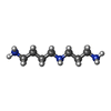
#31: Chemical ChemComp-SPD / SPERMIDINE / N-(2-AMINO-PROPYL)-1,4-DIAMINOBUTANE / PA(34) / Spermidine


Mass: 145.246 Da / Num. of mol.: 1 / Source method: obtained synthetically / Formula: C7H19N3
#32: Chemical...
ChemComp-MG / MAGNESIUM ION


Mass: 24.305 Da / Num. of mol.: 165 / Source method: isolated from a natural source / Formula: Mg
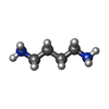
#33: Chemical ChemComp-PUT / 1,4-DIAMINOBUTANE / PUTRESCINE / Putrescine


Mass: 88.151 Da / Num. of mol.: 2 / Source method: obtained synthetically / Formula: C4H12N2
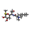
#34: Chemical ChemComp-3QB / LINCOMYCIN / methyl (5R)-5-[(1R,2R)-2-hydroxy-1-{[(4R)-1-methyl-4-propyl-L-prolyl]amino}propyl]-1-thio-beta-L-arabinopyranoside / Lincomycin


Mass: 406.537 Da / Num. of mol.: 1 / Source method: obtained synthetically / Formula: C18H34N2O6S / Feature type: SUBJECT OF INVESTIGATION / Comment: antibiotic*YM
#35: Chemical ChemComp-K / POTASSIUM ION


Mass: 39.098 Da / Num. of mol.: 1 / Source method: isolated from a natural source / Formula: K
#36: Water ChemComp-HOH / water / Water


Mass: 18.015 Da / Num. of mol.: 1381 / Source method: isolated from a natural source / Formula: H2O

-
Details

Has ligand of interestY

-
Experimental details

-
Experiment

ExperimentMethod: ELECTRON MICROSCOPY
EM experimentAggregation state: PARTICLE / 3D reconstruction method: single particle reconstruction

-
Sample preparation

ComponentName: Lincomycin bound to the Listeria monocytogenes 50S ribosomal subunit.
Type: RIBOSOME / Entity ID: #1-#29 / Source: NATURAL
Molecular weightValue: 1 MDa / Experimental value: NO
Source (natural)Organism: Listeria monocytogenes E (bacteria)
Buffer solutionpH: 7.5 / Details: 0.05% Nikkol
Buffer component
IDConc.NameFormulaBuffer-ID
120 mMHEPESC8H18N2O4S1
215 mMMagnesium acetateMg(OAc)21
395 mMPotassium chlorideKCl1
45 mMAmmonium chlorideNH4Cl1
50.5 mMCalcium chlorideCaCl21
68 mMPutrescineC4H12N21
71 mMSpermidineC7H19N31
80.5 mMEDTAEthylenediaminetetraacetic acidC10H16N2O81
SpecimenEmbedding applied: NO / Shadowing applied: NO / Staining applied: NO / Vitrification applied: YES / Details: 5 OD260/mL
Specimen supportGrid material: COPPER / Grid type: Quantifoil R2/2
VitrificationInstrument: FEI VITROBOT MARK IV / Cryogen name: ETHANE-PROPANE / Humidity: 100 % / Chamber temperature: 277 K

-
Electron microscopy imaging

Experimental equipment
Model: Titan Krios / Image courtesy: FEI Company
MicroscopyModel: FEI TITAN KRIOS
Electron gunElectron source: FIELD EMISSION GUN / Accelerating voltage: 300 kV / Illumination mode: FLOOD BEAM
Electron lensMode: BRIGHT FIELDBright-field microscopy / Nominal magnification: 270000 X / Nominal defocus max: 1400 nm / Nominal defocus min: 400 nm
Image recordingElectron dose: 40.3 e/Å2 / Detector mode: COUNTING / Film or detector model: GATAN K2 SUMMIT (4k x 4k)

-
Processing

EM software
IDNameVersionCategory
2EPU2.12.1image acquisition
4CTFFIND4.1.14CTF correction
10RELION3.1.3initial Euler assignment
11RELION3.1.3final Euler assignment
12RELION3.1.3classification
CTF correctionType: PHASE FLIPPING AND AMPLITUDE CORRECTION
Particle selectionNum. of particles selected: 506262
3D reconstructionResolution: 2.3 Å / Resolution method: FSC 0.143 CUT-OFF / Num. of particles: 285330 / Algorithm: FOURIER SPACE / Num. of class averages: 1 / Symmetry type: POINT

+
About Yorodumi

-
News

-
Feb 9, 2022. New format data for meta-information of EMDB entries

New format data for meta-information of EMDB entries

  • Version 3 of the EMDB header file is now the official format.
  • The previous official version 1.9 will be removed from the archive.

Related info.:EMDB header

External links:wwPDB to switch to version 3 of the EMDB data model

-
Aug 12, 2020. Covid-19 info

Covid-19 info

URL: https://pdbj.org/emnavi/covid19.php

New page: Covid-19 featured information page in EM Navigator.

Related info.:Covid-19 info / Mar 5, 2020. Novel coronavirus structure data

+
Mar 5, 2020. Novel coronavirus structure data

Novel coronavirus structure data

Related info.:Yorodumi Speices / Aug 12, 2020. Covid-19 info

External links:COVID-19 featured content - PDBj / Molecule of the Month (242):Coronavirus Proteases

+
Jan 31, 2019. EMDB accession codes are about to change! (news from PDBe EMDB page)

EMDB accession codes are about to change! (news from PDBe EMDB page)

  • The allocation of 4 digits for EMDB accession codes will soon come to an end. Whilst these codes will remain in use, new EMDB accession codes will include an additional digit and will expand incrementally as the available range of codes is exhausted. The current 4-digit format prefixed with “EMD-” (i.e. EMD-XXXX) will advance to a 5-digit format (i.e. EMD-XXXXX), and so on. It is currently estimated that the 4-digit codes will be depleted around Spring 2019, at which point the 5-digit format will come into force.
  • The EM Navigator/Yorodumi systems omit the EMD- prefix.

Related info.:Q: What is EMD? / ID/Accession-code notation in Yorodumi/EM Navigator

External links:EMDB Accession Codes are Changing Soon! / Contact to PDBj

+
Jul 12, 2017. Major update of PDB

Major update of PDB

  • wwPDB released updated PDB data conforming to the new PDBx/mmCIF dictionary.
  • This is a major update changing the version number from 4 to 5, and with Remediation, in which all the entries are updated.
  • In this update, many items about electron microscopy experimental information are reorganized (e.g. em_software).
  • Now, EM Navigator and Yorodumi are based on the updated data.

External links:wwPDB Remediation / Enriched Model Files Conforming to OneDep Data Standards Now Available in the PDB FTP Archive

-
Yorodumi

Thousand views of thousand structures

  • Yorodumi is a browser for structure data from EMDB, PDB, SASBDB, etc.
  • This page is also the successor to EM Navigator detail page, and also detail information page/front-end page for Omokage search.
  • The word "yorodu" (or yorozu) is an old Japanese word meaning "ten thousand". "mi" (miru) is to see.

Related info.:EMDB / PDB / SASBDB / Comparison of 3 databanks / Yorodumi Search / Aug 31, 2016. New EM Navigator & Yorodumi / Yorodumi Papers / Jmol/JSmol / Function and homology information / Changes in new EM Navigator and Yorodumi

Read more