 ムービー
ムービー コントローラー


+ データを開く

- 基本情報
基本情報
| 登録情報 | データベース: EMDB / ID: EMD-1506 | |||||||||
|---|---|---|---|---|---|---|---|---|---|---|
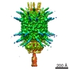
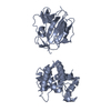
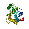
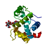
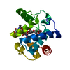
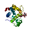
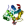
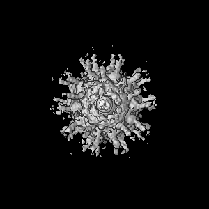
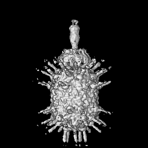
| タイトル | Crystal and cryoEM structural studies of a cell wall degrading enzyme in the bacteriophage phi29 tail | |||||||||
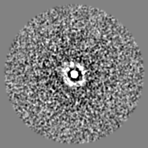
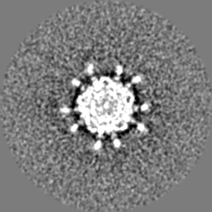
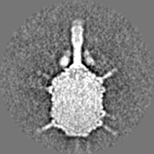
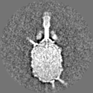
 マップデータ マップデータ | This is an image of a surface rendered top-view of bacteriophage phi29 mutant sus13(330) | |||||||||
 試料 試料 |
| |||||||||
 キーワード キーワード | Cell wall / phi29 / hydrolase / infection / structure | |||||||||
| 生物種 |   Bacillus phage phi29 (ファージ) Bacillus phage phi29 (ファージ) | |||||||||
| 手法 | 単粒子再構成法 / クライオ電子顕微鏡法 / 解像度: 30.0 Å | |||||||||
 データ登録者 データ登録者 | Xiang Y / Morais MC / Cohen DN / Bowman VD / Anderson DL / Rossmann MG | |||||||||
 引用 引用 | ジャーナル: Proc Natl Acad Sci U S A / 年: 2008 タイトル: Crystal and cryoEM structural studies of a cell wall degrading enzyme in the bacteriophage phi29 tail. 著者: Ye Xiang / Marc C Morais / Daniel N Cohen / Valorie D Bowman / Dwight L Anderson / Michael G Rossmann /  要旨: The small bacteriophage phi29 must penetrate the approximately 250-A thick external peptidoglycan cell wall and cell membrane of the Gram-positive Bacillus subtilis, before ejecting its dsDNA genome ...The small bacteriophage phi29 must penetrate the approximately 250-A thick external peptidoglycan cell wall and cell membrane of the Gram-positive Bacillus subtilis, before ejecting its dsDNA genome through its tail into the bacterial cytoplasm. The tail of bacteriophage phi29 is noncontractile and approximately 380 A long. A 1.8-A resolution crystal structure of gene product 13 (gp13) shows that this tail protein has spatially well separated N- and C-terminal domains, whose structures resemble lysozyme-like enzymes and metallo-endopeptidases, respectively. CryoEM reconstructions of the WT bacteriophage and mutant bacteriophages missing some or most of gp13 shows that this enzyme is located at the distal end of the phi29 tail knob. This finding suggests that gp13 functions as a tail-associated, peptidoglycan-degrading enzyme able to cleave both the polysaccharide backbone and peptide cross-links of the peptidoglycan cell wall. Comparisons of the gp13(-) mutants with the phi29 mature and emptied phage structures suggest the sequence of events that occur during the penetration of the tail through the peptidoglycan layer. | |||||||||
| 履歴 |
|
- 構造の表示
構造の表示
| ムービー |
 ムービービューア ムービービューア |
|---|---|
| 構造ビューア | EMマップ: SurfViewMolmilJmol/JSmol |
| 添付画像 |

- ダウンロードとリンク
-EMDBアーカイブ
| マップデータ | emd_1506.map.gz | 76.9 MB | EMDBマップデータ形式 | |
|---|---|---|---|---|
| ヘッダ (付随情報) | emd-1506-v30.xmlemd-1506.xml | 9.7 KB 9.7 KB | 表示 表示 | EMDBヘッダ |
| 画像 |  1506.gif 1506.gif | 53.1 KB | ||
| アーカイブディレクトリ |  http://ftp.pdbj.org/pub/emdb/structures/EMD-1506ftp://ftp.pdbj.org/pub/emdb/structures/EMD-1506 http://ftp.pdbj.org/pub/emdb/structures/EMD-1506ftp://ftp.pdbj.org/pub/emdb/structures/EMD-1506 | HTTPS FTP |
-検証レポート
| 文書・要旨 | emd_1506_validation.pdf.gz | 253.2 KB | 表示 | EMDB検証レポート |
|---|---|---|---|---|
| 文書・詳細版 | emd_1506_full_validation.pdf.gz | 252.3 KB | 表示 | |
| XML形式データ | emd_1506_validation.xml.gz | 6.6 KB | 表示 | |
| アーカイブディレクトリ | https://ftp.pdbj.org/pub/emdb/validation_reports/EMD-1506ftp://ftp.pdbj.org/pub/emdb/validation_reports/EMD-1506 | HTTPS FTP |
-関連構造データ
-リンク
| EMDBのページ | EMDB (EBI/PDBe) / EMDataResource |
|---|
-マップ
| ファイル | ダウンロード / ファイル: emd_1506.map.gz / 形式: CCP4 / 大きさ: 100.6 MB / タイプ: IMAGE STORED AS FLOATING POINT NUMBER (4 BYTES) | ||||||||||||||||||||||||||||||||||||||||||||||||||||||||||||||||||||
|---|---|---|---|---|---|---|---|---|---|---|---|---|---|---|---|---|---|---|---|---|---|---|---|---|---|---|---|---|---|---|---|---|---|---|---|---|---|---|---|---|---|---|---|---|---|---|---|---|---|---|---|---|---|---|---|---|---|---|---|---|---|---|---|---|---|---|---|---|---|
| 注釈 | This is an image of a surface rendered top-view of bacteriophage phi29 mutant sus13(330) | ||||||||||||||||||||||||||||||||||||||||||||||||||||||||||||||||||||
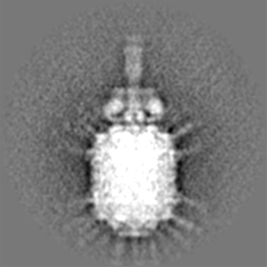
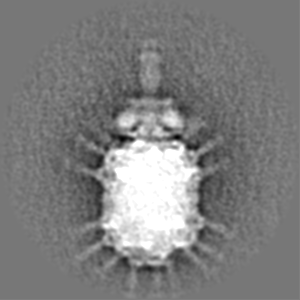
| 投影像・断面図 | 画像のコントロール
画像は Spider により作成 | ||||||||||||||||||||||||||||||||||||||||||||||||||||||||||||||||||||
| ボクセルのサイズ | X=Y=Z: 4.24 Å | ||||||||||||||||||||||||||||||||||||||||||||||||||||||||||||||||||||
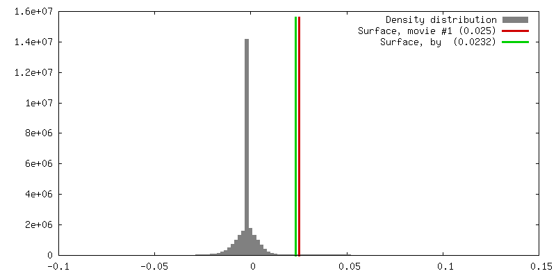
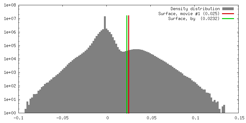
| 密度 |
| ||||||||||||||||||||||||||||||||||||||||||||||||||||||||||||||||||||
| 対称性 | 空間群: 1 | ||||||||||||||||||||||||||||||||||||||||||||||||||||||||||||||||||||
| 詳細 | EMDB XML:
CCP4マップ ヘッダ情報:
| ||||||||||||||||||||||||||||||||||||||||||||||||||||||||||||||||||||
 Z (Sec.)
 Y (Row.)
 X (Col.)


-添付データ
- 試料の構成要素
-全体 : Bacteriophage phi29 mutant sus13(330)
| 全体 | 名称: Bacteriophage phi29 mutant sus13(330) |
|---|---|
| 要素 |
|
-超分子 #1000: Bacteriophage phi29 mutant sus13(330)
| 超分子 | 名称: Bacteriophage phi29 mutant sus13(330) / タイプ: sample / ID: 1000 集合状態: capsid protein forms t3 q5 prolate icosahedron Number unique components: 9 |
|---|---|
| 分子量 | 理論値: 35.2 MDa |
-超分子 #1: Bacillus phage phi29
| 超分子 | 名称: Bacillus phage phi29 / タイプ: virus / ID: 1 詳細: Fibered bacteriophage phi29 mutant sus13(330). A large fragment of gene product 13 could be detected in the mutant particles. NCBI-ID: 10756 / 生物種: Bacillus phage phi29 / ウイルスタイプ: VIRUS-LIKE PARTICLE / ウイルス・単離状態: SPECIES / ウイルス・エンベロープ: No / ウイルス・中空状態: No |
|---|---|
| 宿主 | 生物種:  |
| 分子量 | 理論値: 35.2 MDa |
| ウイルス殻 | Shell ID: 1 / 名称: Phi29 sus13330 / 直径: 530 Å / T番号(三角分割数): 3 |
-実験情報
-構造解析
| 手法 | クライオ電子顕微鏡法 |
|---|---|
 解析 解析 | 単粒子再構成法 |
| 試料の集合状態 | particle |
-試料調製
| 濃度 | 1 mg/mL |
|---|---|
| 緩衝液 | pH: 7.8 / 詳細: 25mM Tris-HCl pH 7.8, 50mM NaCl, 5mM MgCl2 |
| グリッド | 詳細: holey carbon |
| 凍結 | 凍結剤: ETHANE / チャンバー内温度: 113 K / 装置: OTHER |
- 電子顕微鏡法
| 顕微鏡 | FEI/PHILIPS CM300FEG/T |
|---|---|
| 撮影 | デジタル化 - スキャナー: ZEISS SCAI / デジタル化 - サンプリング間隔: 7 µm / 実像数: 70 / 平均電子線量: 20 e/Å2 / 詳細: after scanning, images binned by a factor of 2 / ビット/ピクセル: 8 |
| 電子線 | 加速電圧: 300 kV / 電子線源:  FIELD EMISSION GUN FIELD EMISSION GUN |
| 電子光学系 | 照射モード: FLOOD BEAM / 撮影モード: BRIGHT FIELD / 最大 デフォーカス(公称値): 3.3 µm / 最小 デフォーカス(公称値): 1.5 µm |
| 試料ステージ | 試料ホルダー: Side entry liquid nitrogen-cooled cryo specimen holder 試料ホルダーモデル: GATAN LIQUID NITROGEN |
-画像解析
| CTF補正 | 詳細: phase flip |
|---|---|
| 最終 再構成 | 想定した対称性 - 点群: C1 (非対称) / アルゴリズム: OTHER / 解像度のタイプ: BY AUTHOR / 解像度: 30.0 Å / 解像度の算出法: FSC 0.5 CUT-OFF / ソフトウェア - 名称: EMAN / 詳細: asymmetric / 使用した粒子像数: 5188 |
| 最終 2次元分類 | クラス数: 250 |
-原子モデル構築 1
| 初期モデル | PDB ID: Chain - Chain ID: A |
|---|---|
| ソフトウェア | 名称: emfit |
| 詳細 | PDBEntryID_givenInChain. Protocol: Rigid Body. Two possible biological functional units were fitted into the em map |
| 精密化 | 空間: REAL / プロトコル: RIGID BODY FIT / 当てはまり具合の基準: Density distribution |
