ムービー
ムービー コントローラー


+ データを開く

- 基本情報
基本情報
| 登録情報 |  | |||||||||
|---|---|---|---|---|---|---|---|---|---|---|
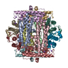
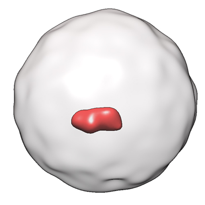
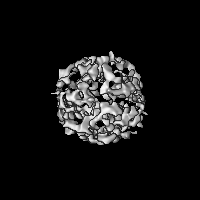
| タイトル | Dps nanocage with the "dumbbell" mineral core | |||||||||
 マップデータ マップデータ | ||||||||||
 試料 試料 |
| |||||||||
| 生物種 |  | |||||||||
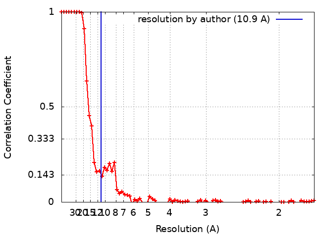
| 手法 | 単粒子再構成法 / クライオ電子顕微鏡法 / 解像度: 10.9 Å | |||||||||
 データ登録者 データ登録者 | Chesnokov YM / Kamyshinsky RA | |||||||||
| 資金援助 |  ロシア, 1件 ロシア, 1件
| |||||||||
 引用 引用 | ジャーナル: Int J Mol Sci / 年: 2022 タイトル: Structural Insights into Iron Ions Accumulation in Dps Nanocage. 著者: Yury Chesnokov / Andrey Mozhaev / Roman Kamyshinsky / Alexander Gordienko / Liubov Dadinova / 要旨: Dps (DNA-binding protein from starved cells) is well known for the structural protection of bacterial DNA by the formation of highly ordered intracellular assemblies under stress conditions. ...Dps (DNA-binding protein from starved cells) is well known for the structural protection of bacterial DNA by the formation of highly ordered intracellular assemblies under stress conditions. Moreover, this ferritin-like protein can perform fast oxidation of ferrous ions and subsequently accumulate clusters of ferric ions in its nanocages, thus providing the bacterium with physical and chemical protection. Here, cryo-electron microscopy was used to study the accumulation of iron ions in the nanocage of a Dps protein from . We demonstrate that Fe concentration in the solution and incubation time have an insignificant effect on the volume and the morphology of iron minerals formed in Dps nanocages. However, an increase in the Fe level leads to an increase in the proportion of larger clusters and the clusters themselves are composed of discrete ~1-1.5 nm subunits. | |||||||||
| 履歴 |
|
- 構造の表示
構造の表示
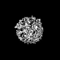
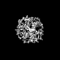
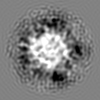
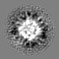
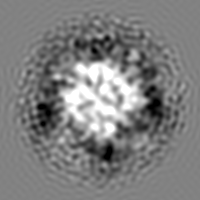
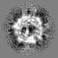
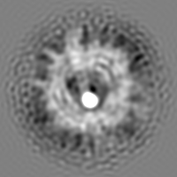
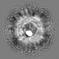
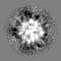
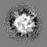
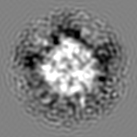
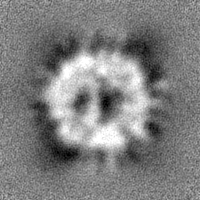
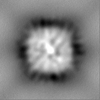
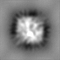
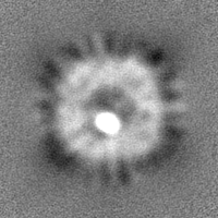
| 添付画像 |
|---|
- ダウンロードとリンク
-EMDBアーカイブ
| マップデータ | emd_14949.map.gz | 28.5 MB |  EMDBマップデータ形式 EMDBマップデータ形式 | |
|---|---|---|---|---|
| ヘッダ (付随情報) | emd-14949-v30.xmlemd-14949.xml | 17.1 KB 17.1 KB | 表示 表示 | EMDBヘッダ |
| FSC (解像度算出) | emd_14949_fsc.xml | 9.3 KB | 表示 | FSCデータファイル |
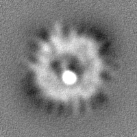
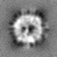
| 画像 |  emd_14949.png emd_14949.png | 68.4 KB | ||
| マスクデータ | emd_14949_msk_1.map | 30.5 MB | マスクマップ | |
| その他 | emd_14949_half_map_1.map.gzemd_14949_half_map_2.map.gz | 28.3 MB 28.3 MB | ||
| アーカイブディレクトリ |  http://ftp.pdbj.org/pub/emdb/structures/EMD-14949ftp://ftp.pdbj.org/pub/emdb/structures/EMD-14949 http://ftp.pdbj.org/pub/emdb/structures/EMD-14949ftp://ftp.pdbj.org/pub/emdb/structures/EMD-14949 | HTTPS FTP |
-検証レポート
| 文書・要旨 | emd_14949_validation.pdf.gz | 657.3 KB | 表示 | EMDB検証レポート |
|---|---|---|---|---|
| 文書・詳細版 | emd_14949_full_validation.pdf.gz | 656.9 KB | 表示 | |
| XML形式データ | emd_14949_validation.xml.gz | 12.6 KB | 表示 | |
| CIF形式データ | emd_14949_validation.cif.gz | 17.6 KB | 表示 | |
| アーカイブディレクトリ | https://ftp.pdbj.org/pub/emdb/validation_reports/EMD-14949ftp://ftp.pdbj.org/pub/emdb/validation_reports/EMD-14949 | HTTPS FTP |
-関連構造データ


-リンク
| EMDBのページ | EMDB (EBI/PDBe) / EMDataResource |
|---|
-マップ
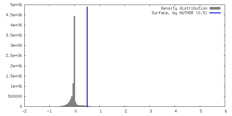
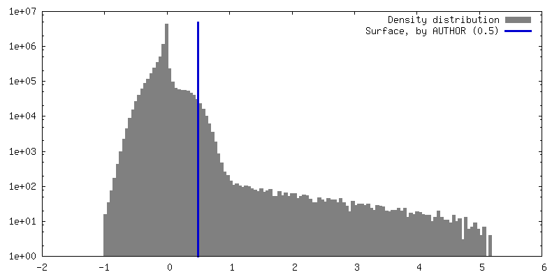
| ファイル | ダウンロード / ファイル: emd_14949.map.gz / 形式: CCP4 / 大きさ: 30.5 MB / タイプ: IMAGE STORED AS FLOATING POINT NUMBER (4 BYTES) | ||||||||||||||||||||||||||||||||||||
|---|---|---|---|---|---|---|---|---|---|---|---|---|---|---|---|---|---|---|---|---|---|---|---|---|---|---|---|---|---|---|---|---|---|---|---|---|---|
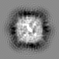
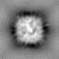
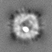
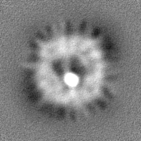
| 投影像・断面図 | 画像のコントロール
画像は Spider により作成 | ||||||||||||||||||||||||||||||||||||
| ボクセルのサイズ | X=Y=Z: 0.86 Å | ||||||||||||||||||||||||||||||||||||
| 密度 |
| ||||||||||||||||||||||||||||||||||||
| 対称性 | 空間群: 1 | ||||||||||||||||||||||||||||||||||||
| 詳細 | EMDB XML:
|
 Z (Sec.)
 Y (Row.)
 X (Col.)


-添付データ
-マスク #1
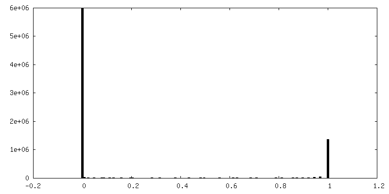
| ファイル | emd_14949_msk_1.map | ||||||||||||
|---|---|---|---|---|---|---|---|---|---|---|---|---|---|
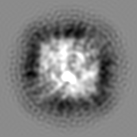
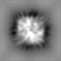
| 投影像・断面図 |
| ||||||||||||
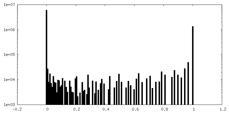
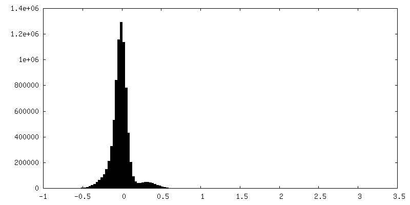
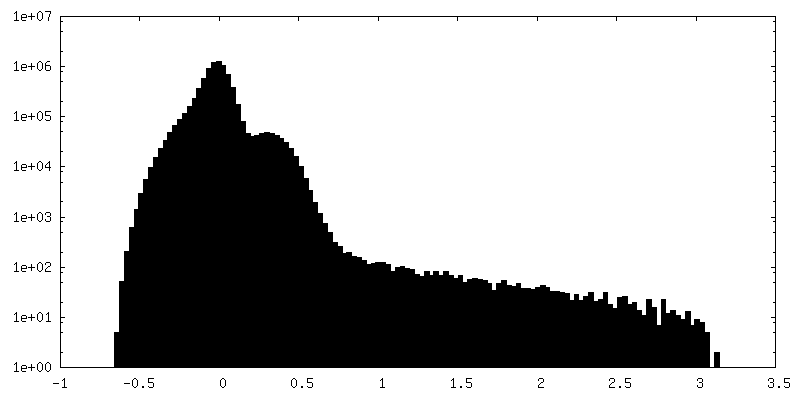
| 密度ヒストグラム |


-ハーフマップ: #1
| ファイル | emd_14949_half_map_1.map | ||||||||||||
|---|---|---|---|---|---|---|---|---|---|---|---|---|---|
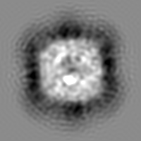
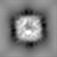
| 投影像・断面図 |
| ||||||||||||
| 密度ヒストグラム |

-ハーフマップ: #2
| ファイル | emd_14949_half_map_2.map | ||||||||||||
|---|---|---|---|---|---|---|---|---|---|---|---|---|---|
| 投影像・断面図 |
| ||||||||||||
| 密度ヒストグラム |


- 試料の構成要素
-全体 : Dps control
| 全体 | 名称: Dps control |
|---|---|
| 要素 |
|
-超分子 #1: Dps control
| 超分子 | 名称: Dps control / タイプ: organelle_or_cellular_component / ID: 1 / 親要素: 0 |
|---|---|
| 由来(天然) | 生物種: |
| 分子量 | 理論値: 224 KDa |
| 組換発現 | 生物種: |
-実験情報
-構造解析
| 手法 | クライオ電子顕微鏡法 |
|---|---|
 解析 解析 | 単粒子再構成法 |
| 試料の集合状態 | particle |
-試料調製
| 濃度 | 3.5 mg/mL | |||||||||||||||
|---|---|---|---|---|---|---|---|---|---|---|---|---|---|---|---|---|
| 緩衝液 | pH: 8 構成要素:
| |||||||||||||||
| 凍結 | 凍結剤: ETHANE / チャンバー内湿度: 100 % / チャンバー内温度: 293 K / 装置: FEI VITROBOT MARK IV / 詳細: blotting for 2.5 seconds. |
- 電子顕微鏡法
| 顕微鏡 | FEI TITAN KRIOS |
|---|---|
| 温度 | 最低: 78.0 K / 最高: 90.0 K |
| 特殊光学系 | 球面収差補正装置: Cs corrector by CEOS |
| 撮影 | フィルム・検出器のモデル: FEI FALCON II (4k x 4k) 検出モード: INTEGRATING / デジタル化 - サイズ - 横: 4096 pixel / デジタル化 - サイズ - 縦: 4096 pixel / デジタル化 - サンプリング間隔: 14.0 µm / デジタル化 - 画像ごとのフレーム数: 1-40 / 撮影したグリッド数: 1 / 実像数: 1419 / 平均露光時間: 2.0 sec. / 平均電子線量: 80.0 e/Å2 |
| 電子線 | 加速電圧: 300 kV / 電子線源:  FIELD EMISSION GUN FIELD EMISSION GUN |
| 電子光学系 | C2レンズ絞り径: 100.0 µm / 最大 デフォーカス(補正後): 2.0 µm / 最小 デフォーカス(補正後): 0.8 µm / 倍率(補正後): 162790 / 照射モード: FLOOD BEAM / 撮影モード: BRIGHT FIELD / Cs: 0.001 mm / 最大 デフォーカス(公称値): 2.0 µm / 最小 デフォーカス(公称値): 0.8 µm / 倍率(公称値): 75000 |
| 試料ステージ | 試料ホルダーモデル: FEI TITAN KRIOS AUTOGRID HOLDER ホルダー冷却材: NITROGEN |
| 実験機器 |  モデル: Titan Krios / 画像提供: FEI Company |
+画像解析
-原子モデル構築 1
| 初期モデル | PDB ID: |
|---|---|
| 精密化 | 空間: REAL / プロトコル: RIGID BODY FIT |