 Movie
Movie Controller

[English] 日本語
 Yorodumi
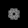
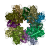
- EMDB-14625: Subtomogram averaging of Rubisco from Cyanobium carboxysome (oute... -
+ Open data

- Basic information
| Entry | 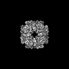 | ||||||||||||
|---|---|---|---|---|---|---|---|---|---|---|---|---|---|
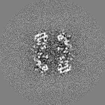
| Title | Subtomogram averaging of Rubisco from Cyanobium carboxysome (outerlayer) | ||||||||||||
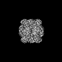
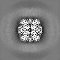
 Map data Map data | main map from cisTEM reconstruction within emClarity package, postprocessed with relion | ||||||||||||
 Sample Sample |
| ||||||||||||
| Biological species |  Cyanobium sp. PCC 7001 (bacteria) Cyanobium sp. PCC 7001 (bacteria) | ||||||||||||
| Method | subtomogram averaging / cryo EM / Resolution: 4.28 Å | ||||||||||||
 Authors Authors | Ni T / Zhu Y / Seaton-Burn W / Zhang P | ||||||||||||
| Funding support |  United Kingdom, European Union, 3 items United Kingdom, European Union, 3 items
| ||||||||||||
 Citation Citation | Journal: Nat Commun / Year: 2022 Title: Structure and assembly of cargo Rubisco in two native α-carboxysomes. Authors: Tao Ni / Yaqi Sun / Will Burn / Monsour M J Al-Hazeem / Yanan Zhu / Xiulian Yu / Lu-Ning Liu / Peijun Zhang /  Abstract: Carboxysomes are a family of bacterial microcompartments in cyanobacteria and chemoautotrophs. They encapsulate Ribulose 1,5-bisphosphate carboxylase/oxygenase (Rubisco) and carbonic anhydrase ...Carboxysomes are a family of bacterial microcompartments in cyanobacteria and chemoautotrophs. They encapsulate Ribulose 1,5-bisphosphate carboxylase/oxygenase (Rubisco) and carbonic anhydrase catalyzing carbon fixation inside a proteinaceous shell. How Rubisco complexes pack within the carboxysomes is unknown. Using cryo-electron tomography, we determine the distinct 3D organization of Rubisco inside two distant α-carboxysomes from a marine α-cyanobacterium Cyanobium sp. PCC 7001 where Rubiscos are organized in three concentric layers, and from a chemoautotrophic bacterium Halothiobacillus neapolitanus where they form intertwining spirals. We further resolve the structures of native Rubisco as well as its higher-order assembly at near-atomic resolutions by subtomogram averaging. The structures surprisingly reveal that the authentic intrinsically disordered linker protein CsoS2 interacts with Rubiscos in native carboxysomes but functions distinctively in the two α-carboxysomes. In contrast to the uniform Rubisco-CsoS2 association in the Cyanobium α-carboxysome, CsoS2 binds only to the Rubiscos close to the shell in the Halo α-carboxysome. Our findings provide critical knowledge of the assembly principles of α-carboxysomes, which may aid in the rational design and repurposing of carboxysome structures for new functions. | ||||||||||||
| History |
|
- Structure visualization
| Supplemental images |
|---|

- Downloads & links
-EMDB archive
| Map data | emd_14625.map.gz | 31 MB |  EMDB map data format EMDB map data format | |
|---|---|---|---|---|
| Header (meta data) | emd-14625-v30.xmlemd-14625.xml | 14.2 KB 14.2 KB | Display Display | EMDB header |
| FSC (resolution estimation) | emd_14625_fsc.xml | 7.4 KB | Display | FSC data file |
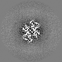
| Images |  emd_14625.png emd_14625.png | 99.4 KB | ||
| Others | emd_14625_half_map_1.map.gzemd_14625_half_map_2.map.gz | 20.2 MB 20.2 MB | ||
| Archive directory |  http://ftp.pdbj.org/pub/emdb/structures/EMD-14625ftp://ftp.pdbj.org/pub/emdb/structures/EMD-14625 http://ftp.pdbj.org/pub/emdb/structures/EMD-14625ftp://ftp.pdbj.org/pub/emdb/structures/EMD-14625 | HTTPS FTP |
-Validation report
| Summary document | emd_14625_validation.pdf.gz | 779.9 KB | Display | EMDB validaton report |
|---|---|---|---|---|
| Full document | emd_14625_full_validation.pdf.gz | 779.4 KB | Display | |
| Data in XML | emd_14625_validation.xml.gz | 13.2 KB | Display | |
| Data in CIF | emd_14625_validation.cif.gz | 18.4 KB | Display | |
| Arichive directory | https://ftp.pdbj.org/pub/emdb/validation_reports/EMD-14625ftp://ftp.pdbj.org/pub/emdb/validation_reports/EMD-14625 | HTTPS FTP |
-Related structure data


-Links
| EMDB pages | EMDB (EBI/PDBe) / EMDataResource |
|---|
-Map
| File | Download / File: emd_14625.map.gz / Format: CCP4 / Size: 34.3 MB / Type: IMAGE STORED AS FLOATING POINT NUMBER (4 BYTES) | ||||||||||||||||||||||||||||||||||||
|---|---|---|---|---|---|---|---|---|---|---|---|---|---|---|---|---|---|---|---|---|---|---|---|---|---|---|---|---|---|---|---|---|---|---|---|---|---|
| Annotation | main map from cisTEM reconstruction within emClarity package, postprocessed with relion | ||||||||||||||||||||||||||||||||||||
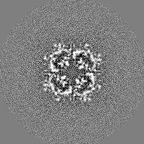
| Projections & slices | Image control
Images are generated by Spider. | ||||||||||||||||||||||||||||||||||||
| Voxel size | X=Y=Z: 1.34 Å | ||||||||||||||||||||||||||||||||||||
| Density |
| ||||||||||||||||||||||||||||||||||||
| Symmetry | Space group: 1 | ||||||||||||||||||||||||||||||||||||
| Details | EMDB XML:
|
 Z (Sec.)
 Y (Row.)
 X (Col.)


-Supplemental data
-Half map: emClarity half map 1 from cisTEM reconstruction
| File | emd_14625_half_map_1.map | ||||||||||||
|---|---|---|---|---|---|---|---|---|---|---|---|---|---|
| Annotation | emClarity half map 1 from cisTEM reconstruction | ||||||||||||
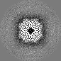
| Projections & Slices |
| ||||||||||||
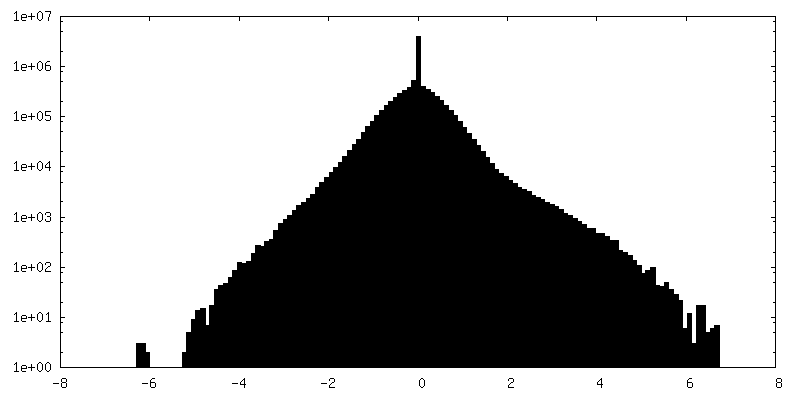
| Density Histograms |


-Half map: emClarity half map 1 from cisTEM reconstruction
| File | emd_14625_half_map_2.map | ||||||||||||
|---|---|---|---|---|---|---|---|---|---|---|---|---|---|
| Annotation | emClarity half map 1 from cisTEM reconstruction | ||||||||||||
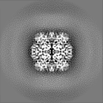
| Projections & Slices |
| ||||||||||||
| Density Histograms |


- Sample components
-Entire : Structure of Rubisco within Cyanobium carboxysome
| Entire | Name: Structure of Rubisco within Cyanobium carboxysome |
|---|---|
| Components |
|
-Supramolecule #1: Structure of Rubisco within Cyanobium carboxysome
| Supramolecule | Name: Structure of Rubisco within Cyanobium carboxysome / type: complex / ID: 1 / Chimera: Yes / Parent: 0 / Macromolecule list: #1-#2 |
|---|---|
| Source (natural) | Organism: Cyanobium sp. PCC 7001 (bacteria) |
-Experimental details
-Structure determination
| Method | cryo EM |
|---|---|
 Processing Processing | subtomogram averaging |
| Aggregation state | particle |
-Sample preparation
| Buffer | pH: 8 |
|---|---|
| Vitrification | Cryogen name: ETHANE |
- Electron microscopy
| Microscope | FEI TITAN KRIOS |
|---|---|
| Image recording | Film or detector model: GATAN K3 (6k x 4k) / Average electron dose: 3.65 e/Å2 |
| Electron beam | Acceleration voltage: 300 kV / Electron source:  FIELD EMISSION GUN FIELD EMISSION GUN |
| Electron optics | Illumination mode: FLOOD BEAM / Imaging mode: BRIGHT FIELD / Nominal defocus max: 5.0 µm / Nominal defocus min: 2.5 µm |
| Experimental equipment |  Model: Titan Krios / Image courtesy: FEI Company |
-Image processing
| Final reconstruction | Applied symmetry - Point group: D4 (2x4 fold dihedral) / Algorithm: FOURIER SPACE / Resolution.type: BY AUTHOR / Resolution: 4.28 Å / Resolution method: FSC 0.143 CUT-OFF / Software: (Name: emClarity, cisTEM) / Number subtomograms used: 87185 |
|---|---|
| Extraction | Number tomograms: 139 / Number images used: 152317 |
| Final angle assignment | Type: OTHER / Software - Name: emClarity / Details: cross correlation |
| FSC plot (resolution estimation) |  |
-Atomic model buiding 1
| Refinement | Space: REAL / Protocol: AB INITIO MODEL |
|---|
