 ムービー
ムービー コントローラー


+ データを開く

- 基本情報
基本情報
| 登録情報 | データベース: EMDB / ID: EMD-1461 | |||||||||
|---|---|---|---|---|---|---|---|---|---|---|
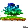
| タイトル | Near-atomic resolution using electron cryomicroscopy and single-particle reconstruction. | |||||||||
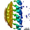
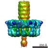
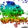
 マップデータ マップデータ | Map of rotavirus VP6 protein, after icosahedral averaging and 13-fold non-icosahedral averaging | |||||||||
 試料 試料 |
| |||||||||
| 生物種 |  Bovine rotavirus (ウイルス) Bovine rotavirus (ウイルス) | |||||||||
| 手法 | 単粒子再構成法 / クライオ電子顕微鏡法 / ネガティブ染色法 / 解像度: 3.8 Å | |||||||||
 データ登録者 データ登録者 | Zhang X / Settembre E / Xu C / Dormitzer PR / Bellamy R / Harrison SC / Grigorieff N | |||||||||
 引用 引用 | ジャーナル: Proc Natl Acad Sci U S A / 年: 2008 タイトル: Near-atomic resolution using electron cryomicroscopy and single-particle reconstruction. 著者: Xing Zhang / Ethan Settembre / Chen Xu / Philip R Dormitzer / Richard Bellamy / Stephen C Harrison / Nikolaus Grigorieff /  要旨: Electron cryomicroscopy (cryo-EM) yields images of macromolecular assemblies and their components, from which 3D structures can be determined, by using an image processing method commonly known as ...Electron cryomicroscopy (cryo-EM) yields images of macromolecular assemblies and their components, from which 3D structures can be determined, by using an image processing method commonly known as "single-particle reconstruction." During the past two decades, this technique has become an important tool for 3D structure determination, but it generally has not been possible to determine atomic models. In principle, individual molecular images contain high-resolution information contaminated by a much higher level of noise. In practice, it has been unclear whether current averaging methods are adequate to extract this information from the background. We present here a reconstruction, obtained by using recently developed image processing methods, of the rotavirus inner capsid particle ("double-layer particle" or DLP) at a resolution suitable for interpretation by an atomic model. The result establishes single-particle reconstruction as a high-resolution technique. We show by direct comparison that the cryo-EM reconstruction of viral protein 6 (VP6) of the rotavirus DLP is similar in clarity to a 3.8-A resolution map obtained from x-ray crystallography. At this resolution, most of the amino acid side chains produce recognizable density. The icosahedral symmetry of the particle was an important factor in achieving this resolution in the cryo-EM analysis, but as the size of recordable datasets increases, single-particle reconstruction also is likely to yield structures at comparable resolution from samples of much lower symmetry. This potential has broad implications for structural cell biology. | |||||||||
| 履歴 |
|
- 構造の表示
構造の表示
| ムービー |
 ムービービューア ムービービューア |
|---|---|
| 構造ビューア | EMマップ: SurfViewMolmilJmol/JSmol |
| 添付画像 |

- ダウンロードとリンク
-EMDBアーカイブ
| マップデータ | emd_1461.map.gz | 1.6 MB | EMDBマップデータ形式 | |
|---|---|---|---|---|
| ヘッダ (付随情報) | emd-1461-v30.xmlemd-1461.xml | 9.7 KB 9.7 KB | 表示 表示 | EMDBヘッダ |
| 画像 |  1461.gif 1461.gif | 34.9 KB | ||
| アーカイブディレクトリ |  http://ftp.pdbj.org/pub/emdb/structures/EMD-1461ftp://ftp.pdbj.org/pub/emdb/structures/EMD-1461 http://ftp.pdbj.org/pub/emdb/structures/EMD-1461ftp://ftp.pdbj.org/pub/emdb/structures/EMD-1461 | HTTPS FTP |
-関連構造データ

-リンク
| EMDBのページ | EMDB (EBI/PDBe) / EMDataResource |
|---|
-マップ
| ファイル | ダウンロード / ファイル: emd_1461.map.gz / 形式: CCP4 / 大きさ: 6.5 MB / タイプ: IMAGE STORED AS FLOATING POINT NUMBER (4 BYTES) | ||||||||||||||||||||||||||||||||||||||||||||||||||||||||||||||||||||
|---|---|---|---|---|---|---|---|---|---|---|---|---|---|---|---|---|---|---|---|---|---|---|---|---|---|---|---|---|---|---|---|---|---|---|---|---|---|---|---|---|---|---|---|---|---|---|---|---|---|---|---|---|---|---|---|---|---|---|---|---|---|---|---|---|---|---|---|---|---|
| 注釈 | Map of rotavirus VP6 protein, after icosahedral averaging and 13-fold non-icosahedral averaging | ||||||||||||||||||||||||||||||||||||||||||||||||||||||||||||||||||||
| 投影像・断面図 | 画像のコントロール
画像は Spider により作成 これらの図は立方格子座標系で作成されたものです | ||||||||||||||||||||||||||||||||||||||||||||||||||||||||||||||||||||
| ボクセルのサイズ | X=Y=Z: 1.23 Å | ||||||||||||||||||||||||||||||||||||||||||||||||||||||||||||||||||||
| 密度 |
| ||||||||||||||||||||||||||||||||||||||||||||||||||||||||||||||||||||
| 対称性 | 空間群: 1 | ||||||||||||||||||||||||||||||||||||||||||||||||||||||||||||||||||||
| 詳細 | EMDB XML:
CCP4マップ ヘッダ情報:
| ||||||||||||||||||||||||||||||||||||||||||||||||||||||||||||||||||||
 Z (Sec.)
 X (Row.)
 Y (Col.)


-添付データ
- 試料の構成要素
-全体 : Rotavirus VP6 protein
| 全体 | 名称: Rotavirus VP6 protein |
|---|---|
| 要素 |
|
-超分子 #1000: Rotavirus VP6 protein
| 超分子 | 名称: Rotavirus VP6 protein / タイプ: sample / ID: 1000 詳細: VP6 is one component of rotavirus DLP, which has been deposited separately (accession number EMD-1460) 集合状態: 780 molecules of VP6 form a DLP particle with 12 molecules of VP1, 120 molecules of VP2, 12 molecules of VP3 and 11 dsRNA molecules Number unique components: 1 |
|---|---|
| 分子量 | 理論値: 41 KDa |
-分子 #1: VP6
| 分子 | 名称: VP6 / タイプ: protein_or_peptide / ID: 1 / Name.synonym: Virus protein 6 / 組換発現: No |
|---|---|
| 由来(天然) | 生物種: Bovine rotavirus (ウイルス) |
| 分子量 | 実験値: 41 KDa |
-実験情報
-構造解析
| 手法 | ネガティブ染色法, クライオ電子顕微鏡法 |
|---|---|
 解析 解析 | 単粒子再構成法 |
| 試料の集合状態 | particle |
-試料調製
| 濃度 | 5 mg/mL |
|---|---|
| 緩衝液 | pH: 7.4 |
| 染色 | タイプ: NEGATIVE / 詳細: Ice |
| グリッド | 詳細: Lacy carbon and C-flat |
| 凍結 | 凍結剤: ETHANE / チャンバー内湿度: 30 % / 装置: HOMEMADE PLUNGER 詳細: Vitrification instrument: Home-made. Vitrification carried out in air at room temperature 手法: Blot for 3 seconds before plunging |
- 電子顕微鏡法
| 顕微鏡 | FEI TECNAI F30 |
|---|---|
| 温度 | 平均: 90 K |
| アライメント法 | Legacy - 非点収差: Objective lens astigmatism was corrected Legacy - Electron beam tilt params: 0 |
| 日付 | 2007年6月1日 |
| 撮影 | カテゴリ: FILM / フィルム・検出器のモデル: KODAK SO-163 FILM / デジタル化 - スキャナー: ZEISS SCAI / デジタル化 - サンプリング間隔: 7 µm / 実像数: 386 / 平均電子線量: 15 e/Å2 / Od range: 1 / ビット/ピクセル: 8 |
| Tilt angle min | 0 |
| Tilt angle max | 0 |
| 電子線 | 加速電圧: 300 kV / 電子線源:  FIELD EMISSION GUN FIELD EMISSION GUN |
| 電子光学系 | 倍率(補正後): 56540 / 照射モード: FLOOD BEAM / 撮影モード: BRIGHT FIELD / Cs: 2 mm / 最大 デフォーカス(公称値): 3.5 µm / 最小 デフォーカス(公称値): 1.1 µm / 倍率(公称値): 59000 |
| 試料ステージ | 試料ホルダー: Eucentric, side-entry / 試料ホルダーモデル: GATAN LIQUID NITROGEN |
| 実験機器 |  モデル: Tecnai F30 / 画像提供: FEI Company |
-画像解析
| 詳細 | Particles were selected using the computer program SIGNATURE |
|---|---|
| CTF補正 | 詳細: Each particle |
| 最終 再構成 | 想定した対称性 - 点群: I (正20面体型対称) / アルゴリズム: OTHER / 解像度のタイプ: BY AUTHOR / 解像度: 3.8 Å / 解像度の算出法: OTHER / ソフトウェア - 名称: FREALIGN 詳細: The FREALIGN reconstruction was followed by non-icosahedral averaging using the Uppsala program package for X-ray crystallography 使用した粒子像数: 8400 |
