 Movie
Movie Controller

[English] 日本語
 Yorodumi
- EMDB-1576: Structural analysis of the interaction of human adenovirus 5 with... -
+ Open data

- Basic information
| Entry | Database: EMDB / ID: EMD-1576 | |||||||||
|---|---|---|---|---|---|---|---|---|---|---|
| Title | Structural analysis of the interaction of human adenovirus 5 with erythrocytes. | |||||||||
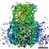
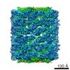
 Map data Map data | This is a map of the adenovirus fivefold vertex interacting directly with the surface of a human red blood cell ghost. | |||||||||
 Sample Sample |
| |||||||||
 Keywords Keywords | Adenovirus / erythrocyte / complement receptor / coxsackie-adenovirus receptor / penton base | |||||||||
| Biological species |  Homo sapiens (human) / Adenovirus serotype 5 Homo sapiens (human) / Adenovirus serotype 5 | |||||||||
| Method | single particle reconstruction / cryo EM / Resolution: 40.0 Å | |||||||||
 Authors Authors | Carlisle RC / Di Y / Cerny AM / Sonnen AF-P / Sim RB / Green NK / Subr V / Ulbrich K / Gilbert RJC / Fisher KD ...Carlisle RC / Di Y / Cerny AM / Sonnen AF-P / Sim RB / Green NK / Subr V / Ulbrich K / Gilbert RJC / Fisher KD / Finberg RW / Seymour LW | |||||||||
 Citation Citation | Journal: Blood / Year: 2009 Title: Human erythrocytes bind and inactivate type 5 adenovirus by presenting Coxsackie virus-adenovirus receptor and complement receptor 1. Authors: Robert C Carlisle / Ying Di / Anna M Cerny / Andreas F-P Sonnen / Robert B Sim / Nicola K Green / Vladimir Subr / Karel Ulbrich / Robert J C Gilbert / Kerry D Fisher / Robert W Finberg / Leonard W Seymour /  Abstract: Type 5 adenovirus (Ad5) is a human pathogen that has been widely developed for therapeutic uses, with only limited success to date. We report here the novel finding that human erythrocytes present ...Type 5 adenovirus (Ad5) is a human pathogen that has been widely developed for therapeutic uses, with only limited success to date. We report here the novel finding that human erythrocytes present Coxsackie virus-adenovirus receptor (CAR) providing an Ad5 sequestration mechanism that protects against systemic infection. Interestingly, erythrocytes from neither mice nor rhesus macaques present CAR. Excess Ad5 fiber protein or anti-CAR antibody inhibits the binding of Ad5 to human erythrocytes and cryo-electron microscopy shows attachment via the fiber protein of Ad5, leading to close juxtaposition with the erythrocyte membrane. Human, but not murine, erythrocytes also present complement receptor (CR1), which binds Ad5 in the presence of antibodies and complement. Transplantation of human erythrocytes into nonobese diabetic/severe combined immunodeficiency mice extends blood circulation of intravenous Ad5 but decreases its extravasation into human xenograft tumors. Ad5 also shows extended circulation in transgenic mice presenting CAR on their erythrocytes, although it clears rapidly in transgenic mice presenting erythrocyte CR1. Hepatic infection is inhibited in both transgenic models. Erythrocytes may therefore restrict Ad5 infection (natural and therapeutic) in humans, independent of antibody status, presenting a formidable challenge to Ad5 therapeutics. "Stealthing" of Ad5 using hydrophilic polymers may enable circumvention of these natural virus traps. | |||||||||
| History |
|
- Structure visualization
| Movie |
 Movie viewer Movie viewer |
|---|---|
| Structure viewer | EM map: SurfViewMolmilJmol/JSmol |
| Supplemental images |


- Downloads & links
-EMDB archive
| Map data | emd_1576.map.gz | 2 MB | EMDB map data format | |
|---|---|---|---|---|
| Header (meta data) | emd-1576-v30.xmlemd-1576.xml | 10.1 KB 10.1 KB | Display Display | EMDB header |
| Images |  1576.gif 1576.gif | 90.8 KB | ||
| Others | IMAGE_DETAILSemd_1576_1.tifemd_1576_2.tifemd_1576_3.tifemd_1576_4.tifemd_1576_5.tifemd_1576_6.tifemd_1576_7.tif | 771 B 807.1 KB 688.3 KB 833.2 KB 805.4 KB 818 KB 703.4 KB 88 KB | ||
| Archive directory |  http://ftp.pdbj.org/pub/emdb/structures/EMD-1576ftp://ftp.pdbj.org/pub/emdb/structures/EMD-1576 http://ftp.pdbj.org/pub/emdb/structures/EMD-1576ftp://ftp.pdbj.org/pub/emdb/structures/EMD-1576 | HTTPS FTP |
-Related structure data
| Similar structure data |
|---|
-Links
| EMDB pages | EMDB (EBI/PDBe) / EMDataResource |
|---|
-Map
| File | Download / File: emd_1576.map.gz / Format: CCP4 / Size: 29.8 MB / Type: IMAGE STORED AS FLOATING POINT NUMBER (4 BYTES) | ||||||||||||||||||||||||||||||||||||||||||||||||||||||||||||||||||||
|---|---|---|---|---|---|---|---|---|---|---|---|---|---|---|---|---|---|---|---|---|---|---|---|---|---|---|---|---|---|---|---|---|---|---|---|---|---|---|---|---|---|---|---|---|---|---|---|---|---|---|---|---|---|---|---|---|---|---|---|---|---|---|---|---|---|---|---|---|---|
| Annotation | This is a map of the adenovirus fivefold vertex interacting directly with the surface of a human red blood cell ghost. | ||||||||||||||||||||||||||||||||||||||||||||||||||||||||||||||||||||
| Projections & slices | Image control
Images are generated by Spider. | ||||||||||||||||||||||||||||||||||||||||||||||||||||||||||||||||||||
| Voxel size | X=Y=Z: 2.6 Å | ||||||||||||||||||||||||||||||||||||||||||||||||||||||||||||||||||||
| Density |
| ||||||||||||||||||||||||||||||||||||||||||||||||||||||||||||||||||||
| Symmetry | Space group: 1 | ||||||||||||||||||||||||||||||||||||||||||||||||||||||||||||||||||||
| Details | EMDB XML:
CCP4 map header:
| ||||||||||||||||||||||||||||||||||||||||||||||||||||||||||||||||||||
 Z (Sec.)
 X (Row.)
 Y (Col.)


-Supplemental data
-Others
| Details | [IMAGE_DETAILS] First six images are of the interaction, and will be published in the Blood manuscript with the reconstruction. The last one is an aligned average, showing the virus, membrane and fibre attaching. The images are of Ad5 interacting with erythrocyte ghost membranes via its fibre that projects from the fivefold vertex represent the stage in virus-membrane interaction prior to the one reconstructed in the deposited map. The interaction is via the coxsackie-adenovirus receptor (CAR), which binds to the end of the fibre. The main focus of the manuscript describing this work was the demonstration that CAR is found on human erythrocytes, along with complement receptor 1 (CR1) and that this has major implications for the use of Ad5 as a vector in immunisation. |
|---|---|
| Image | |
| Image | |
| Image | |
| Image | |
| Image | |
| Image | |
| Image |
- Sample components
-Entire : Human adenovirus serotype 5 interacting with the surface of human...
| Entire | Name: Human adenovirus serotype 5 interacting with the surface of human erythrocyte-derived ghosts. |
|---|---|
| Components |
|
-Supramolecule #1000: Human adenovirus serotype 5 interacting with the surface of human...
| Supramolecule | Name: Human adenovirus serotype 5 interacting with the surface of human erythrocyte-derived ghosts. type: sample / ID: 1000 Oligomeric state: One penton vertex binds to the erythrocyte surface Number unique components: 2 |
|---|
-Supramolecule #1: Adenovirus serotype 5
| Supramolecule | Name: Adenovirus serotype 5 / type: virus / ID: 1 / Name.synonym: Adenovirus Details: The adenovirus was mixed with erythrocyte ghosts, to which they bound. Sci species name: Adenovirus serotype 5 / Virus type: VIRION / Virus isolate: SEROTYPE / Virus enveloped: No / Virus empty: Yes / Syn species name: Adenovirus |
|---|---|
| Host (natural) | Organism: Homo sapiens (human) / synonym: VERTEBRATES |
-Supramolecule #2: Erythrocyte ghost
| Supramolecule | Name: Erythrocyte ghost / type: organelle_or_cellular_component / ID: 2 / Name.synonym: red blood cell ghost Details: The ghosts were prepared from fresh human blood using osmotic shock and sonication. Recombinant expression: No |
|---|---|
| Source (natural) | Organism: Homo sapiens (human) / synonym: Human / Tissue: Blood / Cell: Erythrocyte / Location in cell: Plasma membrane |
-Experimental details
-Structure determination
| Method | cryo EM |
|---|---|
 Processing Processing | single particle reconstruction |
| Aggregation state | particle |
-Sample preparation
| Buffer | pH: 7.4 / Details: PBS |
|---|---|
| Grid | Details: 300 mesh copper grid with lacey carbon film |
| Vitrification | Cryogen name: ETHANE / Instrument: HOMEMADE PLUNGER / Details: Vitrification instrument: Homemade plunger / Method: Blot for 1-2 seconds before plunging. |
- Electron microscopy
| Microscope | FEI TECNAI F30 |
|---|---|
| Image recording | Category: FILM / Film or detector model: KODAK SO-163 FILM / Digitization - Scanner: ZEISS SCAI / Digitization - Sampling interval: 7 µm / Number real images: 4 / Bits/pixel: 8 |
| Electron beam | Acceleration voltage: 300 kV / Electron source:  FIELD EMISSION GUN FIELD EMISSION GUN |
| Electron optics | Illumination mode: FLOOD BEAM / Imaging mode: BRIGHT FIELD / Cs: 2 mm / Nominal defocus max: 5.0 µm / Nominal defocus min: 1.0 µm / Nominal magnification: 27000 |
| Sample stage | Specimen holder: Eucentric / Specimen holder model: GATAN LIQUID NITROGEN |
| Experimental equipment |  Model: Tecnai F30 / Image courtesy: FEI Company |
-Image processing
| CTF correction | Details: Per micrograph |
|---|---|
| Final reconstruction | Applied symmetry - Point group: C1 (asymmetric) / Algorithm: OTHER / Resolution.type: BY AUTHOR / Resolution: 40.0 Å / Resolution method: FSC 0.5 CUT-OFF / Software - Name: SPIDER Details: Final map was calculated using imposed fivefold symmetry orthogonal to the axis of view. Number images used: 27 |
| Final angle assignment | Details: Angles around y axis, since viewed perpendicular to fivefold vertex, using Euler convention of SPIDER and XPLOR |
