 Movie
Movie Controller

[English] 日本語
 Yorodumi
- EMDB-14331: Core-binding domain of fungal E3-binding domain bound to the pyru... -
+ Open data

- Basic information
| Entry |  | |||||||||
|---|---|---|---|---|---|---|---|---|---|---|
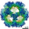
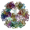
| Title | Core-binding domain of fungal E3-binding domain bound to the pyruvate dehydrogenase E2 core | |||||||||
 Map data Map data | Post-processed | |||||||||
 Sample Sample |
| |||||||||
 Keywords Keywords | Metabolism / dehydrogenase / E3-binding / acetyl-transferase / TRANSFERASE | |||||||||
| Function / homology |  Function and homology information Function and homology informationdihydrolipoyllysine-residue acetyltransferase / dihydrolipoyllysine-residue acetyltransferase activity / pyruvate decarboxylation to acetyl-CoA / pyruvate dehydrogenase complex / acyltransferase activity / mitochondrial matrix / structural molecule activity / mitochondrion Similarity search - Function | |||||||||
| Biological species |  Neurospora crassa (fungus) Neurospora crassa (fungus) | |||||||||
| Method | single particle reconstruction / cryo EM / Resolution: 3.3 Å | |||||||||
 Authors Authors | Forsberg BO | |||||||||
| Funding support |  Sweden, 1 items Sweden, 1 items
| |||||||||
 Citation Citation | Journal: Commun Biol / Year: 2023 Title: The structure and evolutionary diversity of the fungal E3-binding protein. Authors: Bjoern O Forsberg /  Abstract: The pyruvate dehydrogenase complex (PDC) is a central metabolic enzyme in all living cells composed majorly of E1, E2, and E3. Tight coupling of their reactions makes each component essential, so ...The pyruvate dehydrogenase complex (PDC) is a central metabolic enzyme in all living cells composed majorly of E1, E2, and E3. Tight coupling of their reactions makes each component essential, so that any loss impacts oxidative metabolism pathologically. E3 retention is mediated by the E3-binding protein (E3BP), which is here resolved within the PDC core from N.crassa, resolved to 3.2Å. Fungal and mammalian E3BP are shown to be orthologs, arguing E3BP as a broadly eukaryotic gene. Fungal E3BP architectures predicted from sequence data and computational models further bridge the evolutionary distance between N.crassa and humans, and suggest discriminants for E3-specificity. This is confirmed by similarities in their respective E3-binding domains, where an interaction previously not described is also predicted. This provides evolutionary parallels for a crucial interaction human metabolism, an interaction specific to fungi that can be targeted, and an example of protein evolution following gene neofunctionalization. | |||||||||
| History |
|
- Structure visualization
| Supplemental images |
|---|

- Downloads & links
-EMDB archive
| Map data | emd_14331.map.gz | 7.2 MB | EMDB map data format | |
|---|---|---|---|---|
| Header (meta data) | emd-14331-v30.xmlemd-14331.xml | 18.1 KB 18.1 KB | Display Display | EMDB header |
| Images | 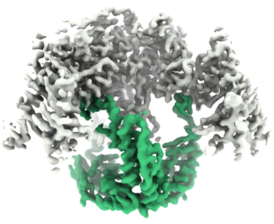 emd_14331.png emd_14331.png | 145.2 KB | ||
| Masks | emd_14331_msk_1.map | 64 MB | Mask map | |
| Filedesc metadata | emd-14331.cif.gz | 5.5 KB | ||
| Others | emd_14331_additional_1.map.gzemd_14331_half_map_1.map.gzemd_14331_half_map_2.map.gz | 56.6 MB 50.1 MB 50 MB | ||
| Archive directory |  http://ftp.pdbj.org/pub/emdb/structures/EMD-14331ftp://ftp.pdbj.org/pub/emdb/structures/EMD-14331 http://ftp.pdbj.org/pub/emdb/structures/EMD-14331ftp://ftp.pdbj.org/pub/emdb/structures/EMD-14331 | HTTPS FTP |
-Related structure data
| Related structure data |  7r5mMC  8ohsC M: atomic model generated by this map C: citing same article ( |
|---|---|
| Similar structure data |
-Links
| EMDB pages | EMDB (EBI/PDBe) / EMDataResource |
|---|---|
| Related items in Molecule of the Month |


-Map
| File | Download / File: emd_14331.map.gz / Format: CCP4 / Size: 64 MB / Type: IMAGE STORED AS FLOATING POINT NUMBER (4 BYTES) | ||||||||||||||||||||||||||||||||||||
|---|---|---|---|---|---|---|---|---|---|---|---|---|---|---|---|---|---|---|---|---|---|---|---|---|---|---|---|---|---|---|---|---|---|---|---|---|---|
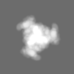
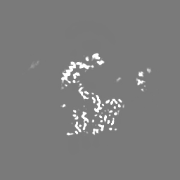
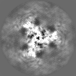
| Annotation | Post-processed | ||||||||||||||||||||||||||||||||||||
| Projections & slices | Image control
Images are generated by Spider. | ||||||||||||||||||||||||||||||||||||
| Voxel size | X=Y=Z: 0.86 Å | ||||||||||||||||||||||||||||||||||||
| Density |
| ||||||||||||||||||||||||||||||||||||
| Symmetry | Space group: 1 | ||||||||||||||||||||||||||||||||||||
| Details | EMDB XML:
|
 Z (Sec.)
 Y (Row.)
 X (Col.)


-Supplemental data
-Mask #1
| File | emd_14331_msk_1.map | ||||||||||||
|---|---|---|---|---|---|---|---|---|---|---|---|---|---|
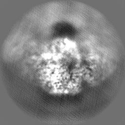
| Projections & Slices |
| ||||||||||||
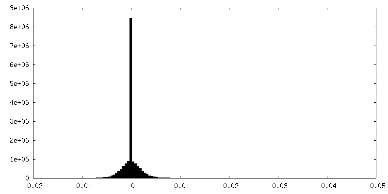
| Density Histograms |


-Additional map: Deepemhancer
| File | emd_14331_additional_1.map | ||||||||||||
|---|---|---|---|---|---|---|---|---|---|---|---|---|---|
| Annotation | Deepemhancer | ||||||||||||
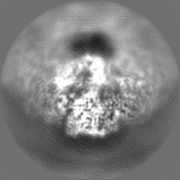
| Projections & Slices |
| ||||||||||||
| Density Histograms |


-Half map: Half-map 1
| File | emd_14331_half_map_1.map | ||||||||||||
|---|---|---|---|---|---|---|---|---|---|---|---|---|---|
| Annotation | Half-map 1 | ||||||||||||
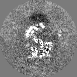
| Projections & Slices |
| ||||||||||||
| Density Histograms |


-Half map: Half-map 2
| File | emd_14331_half_map_2.map | ||||||||||||
|---|---|---|---|---|---|---|---|---|---|---|---|---|---|
| Annotation | Half-map 2 | ||||||||||||
| Projections & Slices |
| ||||||||||||
| Density Histograms |


- Sample components
-Entire : Fungal E3BP bound to PDC E2
| Entire | Name: Fungal E3BP bound to PDC E2 |
|---|---|
| Components |
|
-Supramolecule #1: Fungal E3BP bound to PDC E2
| Supramolecule | Name: Fungal E3BP bound to PDC E2 / type: complex / ID: 1 / Parent: 0 / Macromolecule list: all Details: Trimer of the core-binding domain of N.crassa E3-binding protein, interior to icosahedral core assembly of E2 catalytic domains. |
|---|---|
| Source (natural) | Organism: Neurospora crassa (fungus) |
-Macromolecule #1: Dihydrolipoyllysine-residue acetyltransferase component of pyruva...
| Macromolecule | Name: Dihydrolipoyllysine-residue acetyltransferase component of pyruvate dehydrogenase complex, mitochondrial type: protein_or_peptide / ID: 1 / Number of copies: 6 / Enantiomer: LEVO / EC number: dihydrolipoyllysine-residue acetyltransferase |
|---|---|
| Source (natural) | Organism: Neurospora crassa (fungus) Strain: ATCC 24698 / 74-OR23-1A / CBS 708.71 / DSM 1257 / FGSC 987 |
| Molecular weight | Theoretical: 25.194967 KDa |
| Recombinant expression | Organism:  |
| Sequence | String: MAAYTDVPIS GMRKTIAARL KESVTENPHF FVSTNLSVSK LLKLRQALNS SADGRYKLSV NDFLIKAMGI ASKRVPTVNS SWRDGVIRQ FETVDVSVAV ATPNGLITPI VKGVEGKGLE SISAAVKELA KKARDGKLKP EEYQGGSISI SNMGMNPAVQ S FTAIINPP ...String: MAAYTDVPIS GMRKTIAARL KESVTENPHF FVSTNLSVSK LLKLRQALNS SADGRYKLSV NDFLIKAMGI ASKRVPTVNS SWRDGVIRQ FETVDVSVAV ATPNGLITPI VKGVEGKGLE SISAAVKELA KKARDGKLKP EEYQGGSISI SNMGMNPAVQ S FTAIINPP QAAILAVGAP QKVAVPVENE DGTTGVSWDE QIIVTASFDH KVVDGAVGAE WIRELKKVIE NPLELLL UniProtKB: Dihydrolipoyllysine-residue acetyltransferase component of pyruvate dehydrogenase complex, mitochondrial |
-Macromolecule #2: Pyruvate dehydrogenase X component
| Macromolecule | Name: Pyruvate dehydrogenase X component / type: protein_or_peptide / ID: 2 / Number of copies: 3 / Enantiomer: LEVO |
|---|---|
| Source (natural) | Organism: Neurospora crassa (fungus) Strain: ATCC 24698 / 74-OR23-1A / CBS 708.71 / DSM 1257 / FGSC 987 |
| Molecular weight | Theoretical: 20.476209 KDa |
| Recombinant expression | Organism: |
| Sequence | String: HHHHHHSQDP NSSPSSENLY FQSPAPPPVA VVTAPISLSA AIDVQNKLHK TIGVFLPLST FITRATEIAN QKLPLPANYQ PTADELFNQ VLGLDKVTRK ESRGSYTPTF GSFVAPQRAA RKADIIDILA APSTRVAASA QSKSAAPGLT TSGPNVFSLQ V PKSEEKRA QAFLQKMKLV LEQEPDKLVR A UniProtKB: Pyruvate dehydrogenase X component |
-Experimental details
-Structure determination
| Method | cryo EM |
|---|---|
 Processing Processing | single particle reconstruction |
| Aggregation state | particle |
-Sample preparation
| Buffer | pH: 7.5 |
|---|---|
| Vitrification | Cryogen name: ETHANE |
- Electron microscopy
| Microscope | FEI TITAN KRIOS |
|---|---|
| Image recording | Film or detector model: GATAN K2 SUMMIT (4k x 4k) / Average electron dose: 31.4 e/Å2 |
| Electron beam | Acceleration voltage: 300 kV / Electron source:  FIELD EMISSION GUN FIELD EMISSION GUN |
| Electron optics | Illumination mode: FLOOD BEAM / Imaging mode: BRIGHT FIELD / Nominal defocus max: 2.0 µm / Nominal defocus min: 0.5 µm |
| Experimental equipment |  Model: Titan Krios / Image courtesy: FEI Company |
-Image processing
| Startup model | Type of model: PDB ENTRY PDB model - PDB ID: |
|---|---|
| Final reconstruction | Applied symmetry - Point group: C3 (3 fold cyclic) / Algorithm: FOURIER SPACE / Resolution.type: BY AUTHOR / Resolution: 3.3 Å / Resolution method: FSC 0.143 CUT-OFF / Software - Name: RELION (ver. 3.1) / Number images used: 792478 |
| Initial angle assignment | Type: MAXIMUM LIKELIHOOD / Software - Name: RELION (ver. 3.1) |
| Final angle assignment | Type: MAXIMUM LIKELIHOOD / Software - Name: RELION (ver. 3.1) |
-Atomic model buiding 1
| Refinement | Space: REAL |
|---|---|
| Output model | PDB-7r5m: |
