[English] 日本語
 Yorodumi
- EMDB-1431: The three-dimensional structure of genomic RNA in bacteriophage M... -
+ Open data
- Basic information
| Entry | Database: EMDB / ID: EMD-1431 | |||||||||
|---|---|---|---|---|---|---|---|---|---|---|
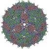
| Title | The three-dimensional structure of genomic RNA in bacteriophage MS2: implications for assembly. | |||||||||
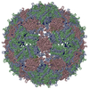
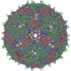
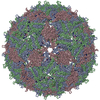
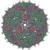
 Map data Map data | CryoEM map of the intact virion of bacteriophage MS2 | |||||||||
 Sample Sample |
| |||||||||
| Biological species |  Enterobacterio phage MS2 (virus) Enterobacterio phage MS2 (virus) | |||||||||
| Method | single particle reconstruction / cryo EM / Resolution: 9.0 Å | |||||||||
 Authors Authors | Toropova K / Basnak G / Twarock R / Stockley PG / Ranson NA | |||||||||
 Citation Citation |  Journal: J Mol Biol / Year: 2008 Journal: J Mol Biol / Year: 2008Title: The three-dimensional structure of genomic RNA in bacteriophage MS2: implications for assembly. Authors: Katerina Toropova / Gabriella Basnak / Reidun Twarock / Peter G Stockley / Neil A Ranson /  Abstract: Using cryo-electron microscopy, single particle image processing and three-dimensional reconstruction with icosahedral averaging, we have determined the three-dimensional solution structure of ...Using cryo-electron microscopy, single particle image processing and three-dimensional reconstruction with icosahedral averaging, we have determined the three-dimensional solution structure of bacteriophage MS2 capsids reassembled from recombinant protein in the presence of short oligonucleotides. We have also significantly extended the resolution of the previously reported structure of the wild-type MS2 virion. The structures of recombinant MS2 capsids reveal clear density for bound RNA beneath the coat protein binding sites on the inner surface of the T=3 MS2 capsid, and show that a short extension of the minimal assembly initiation sequence that promotes an increase in the efficiency of assembly, interacts with the protein capsid forming a network of bound RNA. The structure of the wild-type MS2 virion at approximately 9 A resolution reveals icosahedrally ordered density encompassing approximately 90% of the single-stranded RNA genome. The genome in the wild-type virion is arranged as two concentric shells of density, connected along the 5-fold symmetry axes of the particle. This novel RNA fold provides new constraints for models of viral assembly. | |||||||||
| History |
|
- Structure visualization
| Movie |
 Movie viewer Movie viewer |
|---|---|
| Structure viewer | EM map:  SurfView SurfView Molmil Molmil Jmol/JSmol Jmol/JSmol |
| Supplemental images |
- Downloads & links
-EMDB archive
| Map data |  emd_1431.map.gz emd_1431.map.gz | 56.4 MB |  EMDB map data format EMDB map data format | |
|---|---|---|---|---|
| Header (meta data) |  emd-1431-v30.xml emd-1431-v30.xml emd-1431.xml emd-1431.xml | 7.7 KB 7.7 KB | Display Display |  EMDB header EMDB header |
| Images |  1431.gif 1431.gif | 94.2 KB | ||
| Archive directory |  http://ftp.pdbj.org/pub/emdb/structures/EMD-1431 http://ftp.pdbj.org/pub/emdb/structures/EMD-1431 ftp://ftp.pdbj.org/pub/emdb/structures/EMD-1431 ftp://ftp.pdbj.org/pub/emdb/structures/EMD-1431 | HTTPS FTP |
-Validation report
| Summary document |  emd_1431_validation.pdf.gz emd_1431_validation.pdf.gz | 252.3 KB | Display |  EMDB validaton report EMDB validaton report |
|---|---|---|---|---|
| Full document |  emd_1431_full_validation.pdf.gz emd_1431_full_validation.pdf.gz | 251.4 KB | Display | |
| Data in XML |  emd_1431_validation.xml.gz emd_1431_validation.xml.gz | 6.6 KB | Display | |
| Arichive directory |  https://ftp.pdbj.org/pub/emdb/validation_reports/EMD-1431 https://ftp.pdbj.org/pub/emdb/validation_reports/EMD-1431 ftp://ftp.pdbj.org/pub/emdb/validation_reports/EMD-1431 ftp://ftp.pdbj.org/pub/emdb/validation_reports/EMD-1431 | HTTPS FTP |
-Related structure data
- Links
Links
| EMDB pages |  EMDB (EBI/PDBe) / EMDB (EBI/PDBe) /  EMDataResource EMDataResource |
|---|
- Map
Map
| File |  Download / File: emd_1431.map.gz / Format: CCP4 / Size: 62.5 MB / Type: IMAGE STORED AS FLOATING POINT NUMBER (4 BYTES) Download / File: emd_1431.map.gz / Format: CCP4 / Size: 62.5 MB / Type: IMAGE STORED AS FLOATING POINT NUMBER (4 BYTES) | ||||||||||||||||||||||||||||||||||||||||||||||||||||||||||||||||||||
|---|---|---|---|---|---|---|---|---|---|---|---|---|---|---|---|---|---|---|---|---|---|---|---|---|---|---|---|---|---|---|---|---|---|---|---|---|---|---|---|---|---|---|---|---|---|---|---|---|---|---|---|---|---|---|---|---|---|---|---|---|---|---|---|---|---|---|---|---|---|
| Annotation | CryoEM map of the intact virion of bacteriophage MS2 | ||||||||||||||||||||||||||||||||||||||||||||||||||||||||||||||||||||
| Projections & slices | Image control
Images are generated by Spider. | ||||||||||||||||||||||||||||||||||||||||||||||||||||||||||||||||||||
| Voxel size | X=Y=Z: 1.26 Å | ||||||||||||||||||||||||||||||||||||||||||||||||||||||||||||||||||||
| Density |
| ||||||||||||||||||||||||||||||||||||||||||||||||||||||||||||||||||||
| Symmetry | Space group: 1 | ||||||||||||||||||||||||||||||||||||||||||||||||||||||||||||||||||||
| Details | EMDB XML:
CCP4 map header:
| ||||||||||||||||||||||||||||||||||||||||||||||||||||||||||||||||||||
-Supplemental data
- Sample components
-Entire : Bacteriophage MS2
| Entire | Name:  Bacteriophage MS2 (virus) Bacteriophage MS2 (virus) |
|---|---|
| Components |
|
-Supramolecule #1000: Bacteriophage MS2
| Supramolecule | Name: Bacteriophage MS2 / type: sample / ID: 1000 / Number unique components: 1 |
|---|
-Supramolecule #1: Enterobacterio phage MS2
| Supramolecule | Name: Enterobacterio phage MS2 / type: virus / ID: 1 / Name.synonym: MS2 / NCBI-ID: 12022 / Sci species name: Enterobacterio phage MS2 / Virus type: VIRION / Virus isolate: STRAIN / Virus enveloped: No / Virus empty: No / Syn species name: MS2 |
|---|---|
| Host (natural) | Organism:  |
| Virus shell | Shell ID: 1 / Name: capsid / Diameter: 285 Å / T number (triangulation number): 3 |
-Experimental details
-Structure determination
| Method | cryo EM |
|---|---|
 Processing Processing | single particle reconstruction |
| Aggregation state | particle |
- Sample preparation
| Vitrification | Cryogen name: ETHANE / Chamber humidity: 50 % / Chamber temperature: 22 K / Instrument: HOMEMADE PLUNGER Details: Vitrification instrument: double sided pneumatic blotter Method: 1.4s blot |
|---|
- Electron microscopy
| Microscope | FEI TECNAI F20 |
|---|---|
| Image recording | Category: FILM / Film or detector model: KODAK SO-163 FILM / Digitization - Scanner: OTHER / Average electron dose: 15 e/Å2 |
| Tilt angle min | 0 |
| Tilt angle max | 0 |
| Electron beam | Acceleration voltage: 200 kV / Electron source:  FIELD EMISSION GUN FIELD EMISSION GUN |
| Electron optics | Calibrated magnification: 50400 / Illumination mode: FLOOD BEAM / Imaging mode: BRIGHT FIELD / Cs: 2.0 mm / Nominal magnification: 50000 |
| Sample stage | Specimen holder: side entry / Specimen holder model: GATAN LIQUID NITROGEN |
| Experimental equipment |  Model: Tecnai F20 / Image courtesy: FEI Company |
- Image processing
| CTF correction | Details: phase flipping each particle |
|---|---|
| Final reconstruction | Applied symmetry - Point group: I (icosahedral) / Resolution.type: BY AUTHOR / Resolution: 9.0 Å / Resolution method: FSC 0.5 CUT-OFF / Software - Name: SPIDER |
 Movie
Movie Controller






 Z (Sec.)
 Y (Row.)
 X (Col.)


