+ Open data
- Basic information
| Entry |  | |||||||||
|---|---|---|---|---|---|---|---|---|---|---|
| Title | Proteasome-ZFAND5 Complex Z+E state | |||||||||
 Map data Map data | ZFAND5-proteasome complex Z E state | |||||||||
 Sample Sample |
| |||||||||
 Keywords Keywords | proteasome / ZFAND5 / Activation / STRUCTURAL PROTEIN | |||||||||
| Function / homology |  Function and homology information Function and homology informationImpaired BRCA2 translocation to the nucleus / Impaired BRCA2 binding to SEM1 (DSS1) / host-mediated perturbation of viral transcription / Hydrolases; Acting on peptide bonds (peptidases); Omega peptidases / integrator complex / proteasome accessory complex / meiosis I / purine ribonucleoside triphosphate binding / proteasome regulatory particle / cytosolic proteasome complex ...Impaired BRCA2 translocation to the nucleus / Impaired BRCA2 binding to SEM1 (DSS1) / host-mediated perturbation of viral transcription / Hydrolases; Acting on peptide bonds (peptidases); Omega peptidases / integrator complex / proteasome accessory complex / meiosis I / purine ribonucleoside triphosphate binding / proteasome regulatory particle / cytosolic proteasome complex / positive regulation of proteasomal protein catabolic process / proteasome-activating activity / proteasome regulatory particle, lid subcomplex / proteasome regulatory particle, base subcomplex / protein K63-linked deubiquitination / metal-dependent deubiquitinase activity / Regulation of ornithine decarboxylase (ODC) / Proteasome assembly / Cross-presentation of soluble exogenous antigens (endosomes) / proteasome core complex / Homologous DNA Pairing and Strand Exchange / Defective homologous recombination repair (HRR) due to BRCA1 loss of function / Defective HDR through Homologous Recombination Repair (HRR) due to PALB2 loss of BRCA1 binding function / Defective HDR through Homologous Recombination Repair (HRR) due to PALB2 loss of BRCA2/RAD51/RAD51C binding function / Somitogenesis / Resolution of D-loop Structures through Synthesis-Dependent Strand Annealing (SDSA) / K63-linked deubiquitinase activity / Resolution of D-loop Structures through Holliday Junction Intermediates / proteasome binding / Impaired BRCA2 binding to RAD51 / regulation of protein catabolic process / myofibril / proteasome storage granule / Presynaptic phase of homologous DNA pairing and strand exchange / polyubiquitin modification-dependent protein binding / protein deubiquitination / blastocyst development / immune system process / NF-kappaB binding / proteasome endopeptidase complex / proteasome core complex, beta-subunit complex / endopeptidase activator activity / proteasome assembly / threonine-type endopeptidase activity / mRNA export from nucleus / proteasome core complex, alpha-subunit complex / enzyme regulator activity / regulation of proteasomal protein catabolic process / proteasome complex / : / sarcomere / Regulation of activated PAK-2p34 by proteasome mediated degradation / Autodegradation of Cdh1 by Cdh1:APC/C / APC/C:Cdc20 mediated degradation of Securin / N-glycan trimming in the ER and Calnexin/Calreticulin cycle / stem cell differentiation / Asymmetric localization of PCP proteins / SCF-beta-TrCP mediated degradation of Emi1 / NIK-->noncanonical NF-kB signaling / Ubiquitin-dependent degradation of Cyclin D / TNFR2 non-canonical NF-kB pathway / AUF1 (hnRNP D0) binds and destabilizes mRNA / Vpu mediated degradation of CD4 / Assembly of the pre-replicative complex / Ubiquitin-Mediated Degradation of Phosphorylated Cdc25A / negative regulation of inflammatory response to antigenic stimulus / Degradation of DVL / Dectin-1 mediated noncanonical NF-kB signaling / P-body / Cdc20:Phospho-APC/C mediated degradation of Cyclin A / Degradation of AXIN / lipopolysaccharide binding / Hh mutants are degraded by ERAD / Activation of NF-kappaB in B cells / Degradation of GLI1 by the proteasome / G2/M Checkpoints / Hedgehog ligand biogenesis / Autodegradation of the E3 ubiquitin ligase COP1 / Defective CFTR causes cystic fibrosis / GSK3B and BTRC:CUL1-mediated-degradation of NFE2L2 / Regulation of RUNX3 expression and activity / Negative regulation of NOTCH4 signaling / Hedgehog 'on' state / Vif-mediated degradation of APOBEC3G / APC/C:Cdh1 mediated degradation of Cdc20 and other APC/C:Cdh1 targeted proteins in late mitosis/early G1 / FBXL7 down-regulates AURKA during mitotic entry and in early mitosis / double-strand break repair via homologous recombination / Degradation of GLI2 by the proteasome / GLI3 is processed to GLI3R by the proteasome / MAPK6/MAPK4 signaling / Degradation of beta-catenin by the destruction complex / Oxygen-dependent proline hydroxylation of Hypoxia-inducible Factor Alpha / double-strand break repair via nonhomologous end joining / ABC-family proteins mediated transport / HDR through Homologous Recombination (HRR) / CDK-mediated phosphorylation and removal of Cdc6 / CLEC7A (Dectin-1) signaling / SCF(Skp2)-mediated degradation of p27/p21 / response to virus / FCERI mediated NF-kB activation Similarity search - Function | |||||||||
| Biological species |  Homo sapiens (human) Homo sapiens (human) | |||||||||
| Method | single particle reconstruction / cryo EM / Resolution: 4.4 Å | |||||||||
 Authors Authors | Zhu Y / Lu Y | |||||||||
| Funding support | 1 items
| |||||||||
 Citation Citation |  Journal: Mol Cell / Year: 2023 Journal: Mol Cell / Year: 2023Title: Molecular mechanism for activation of the 26S proteasome by ZFAND5. Authors: Donghoon Lee / Yanan Zhu / Louis Colson / Xiaorong Wang / Siyi Chen / Emre Tkacik / Lan Huang / Qi Ouyang / Alfred L Goldberg / Ying Lu /   Abstract: Various hormones, kinases, and stressors (fasting, heat shock) stimulate 26S proteasome activity. To understand how its capacity to degrade ubiquitylated proteins can increase, we studied mouse ...Various hormones, kinases, and stressors (fasting, heat shock) stimulate 26S proteasome activity. To understand how its capacity to degrade ubiquitylated proteins can increase, we studied mouse ZFAND5, which promotes protein degradation during muscle atrophy. Cryo-electron microscopy showed that ZFAND5 induces large conformational changes in the 19S regulatory particle. ZFAND5's AN1 Zn-finger domain interacts with the Rpt5 ATPase and its C terminus with Rpt1 ATPase and Rpn1, a ubiquitin-binding subunit. Upon proteasome binding, ZFAND5 widens the entrance of the substrate translocation channel, yet it associates only transiently with the proteasome. Dissociation of ZFAND5 then stimulates opening of the 20S proteasome gate. Using single-molecule microscopy, we showed that ZFAND5 binds ubiquitylated substrates, prolongs their association with proteasomes, and increases the likelihood that bound substrates undergo degradation, even though ZFAND5 dissociates before substrate deubiquitylation. These changes in proteasome conformation and reaction cycle can explain the accelerated degradation and suggest how other proteasome activators may stimulate proteolysis. | |||||||||
| History |
|
- Structure visualization
| Supplemental images |
|---|
- Downloads & links
-EMDB archive
| Map data |  emd_14205.map.gz emd_14205.map.gz | 114.5 MB |  EMDB map data format EMDB map data format | |
|---|---|---|---|---|
| Header (meta data) |  emd-14205-v30.xml emd-14205-v30.xml emd-14205.xml emd-14205.xml | 49.6 KB 49.6 KB | Display Display |  EMDB header EMDB header |
| Images |  emd_14205.png emd_14205.png | 116.1 KB | ||
| Filedesc metadata |  emd-14205.cif.gz emd-14205.cif.gz | 14.1 KB | ||
| Archive directory |  http://ftp.pdbj.org/pub/emdb/structures/EMD-14205 http://ftp.pdbj.org/pub/emdb/structures/EMD-14205 ftp://ftp.pdbj.org/pub/emdb/structures/EMD-14205 ftp://ftp.pdbj.org/pub/emdb/structures/EMD-14205 | HTTPS FTP |
-Validation report
| Summary document |  emd_14205_validation.pdf.gz emd_14205_validation.pdf.gz | 640.1 KB | Display |  EMDB validaton report EMDB validaton report |
|---|---|---|---|---|
| Full document |  emd_14205_full_validation.pdf.gz emd_14205_full_validation.pdf.gz | 639.9 KB | Display | |
| Data in XML |  emd_14205_validation.xml.gz emd_14205_validation.xml.gz | 6.5 KB | Display | |
| Data in CIF |  emd_14205_validation.cif.gz emd_14205_validation.cif.gz | 7.6 KB | Display | |
| Arichive directory |  https://ftp.pdbj.org/pub/emdb/validation_reports/EMD-14205 https://ftp.pdbj.org/pub/emdb/validation_reports/EMD-14205 ftp://ftp.pdbj.org/pub/emdb/validation_reports/EMD-14205 ftp://ftp.pdbj.org/pub/emdb/validation_reports/EMD-14205 | HTTPS FTP |
-Related structure data
| Related structure data | 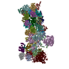 7qxxMC 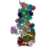 7qxnC 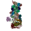 7qxpC 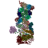 7qxuC 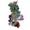 7qxwC  7qy7C 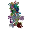 7qyaC 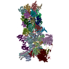 7qybC M: atomic model generated by this map C: citing same article ( |
|---|---|
| Similar structure data | Similarity search - Function & homology  F&H Search F&H Search |
- Links
Links
| EMDB pages |  EMDB (EBI/PDBe) / EMDB (EBI/PDBe) /  EMDataResource EMDataResource |
|---|---|
| Related items in Molecule of the Month |
- Map
Map
| File |  Download / File: emd_14205.map.gz / Format: CCP4 / Size: 125 MB / Type: IMAGE STORED AS FLOATING POINT NUMBER (4 BYTES) Download / File: emd_14205.map.gz / Format: CCP4 / Size: 125 MB / Type: IMAGE STORED AS FLOATING POINT NUMBER (4 BYTES) | ||||||||||||||||||||||||||||||||||||
|---|---|---|---|---|---|---|---|---|---|---|---|---|---|---|---|---|---|---|---|---|---|---|---|---|---|---|---|---|---|---|---|---|---|---|---|---|---|
| Annotation | ZFAND5-proteasome complex Z E state | ||||||||||||||||||||||||||||||||||||
| Projections & slices | Image control
Images are generated by Spider. | ||||||||||||||||||||||||||||||||||||
| Voxel size | X=Y=Z: 1.37 Å | ||||||||||||||||||||||||||||||||||||
| Density |
| ||||||||||||||||||||||||||||||||||||
| Symmetry | Space group: 1 | ||||||||||||||||||||||||||||||||||||
| Details | EMDB XML:
|
-Supplemental data
- Sample components
+Entire : human proteasome zfand5 complex Z+B state
+Supramolecule #1: human proteasome zfand5 complex Z+B state
+Macromolecule #1: 26S proteasome non-ATPase regulatory subunit 1
+Macromolecule #2: 26S proteasome non-ATPase regulatory subunit 3
+Macromolecule #3: 26S proteasome non-ATPase regulatory subunit 12
+Macromolecule #4: 26S proteasome non-ATPase regulatory subunit 11
+Macromolecule #5: 26S proteasome non-ATPase regulatory subunit 6
+Macromolecule #6: 26S proteasome non-ATPase regulatory subunit 7
+Macromolecule #7: 26S proteasome non-ATPase regulatory subunit 13
+Macromolecule #8: 26S proteasome non-ATPase regulatory subunit 4
+Macromolecule #9: 26S proteasome non-ATPase regulatory subunit 14
+Macromolecule #10: 26S proteasome non-ATPase regulatory subunit 8
+Macromolecule #11: 26S proteasome complex subunit SEM1
+Macromolecule #12: 26S proteasome non-ATPase regulatory subunit 2
+Macromolecule #13: 26S protease regulatory subunit 7
+Macromolecule #14: 26S proteasome regulatory subunit 4
+Macromolecule #15: 26S proteasome regulatory subunit 8
+Macromolecule #16: 26S protease regulatory subunit 6B
+Macromolecule #17: 26S proteasome regulatory subunit 10B
+Macromolecule #18: 26S protease regulatory subunit 6A
+Macromolecule #19: Proteasome subunit alpha type-6
+Macromolecule #20: Proteasome subunit alpha type-2
+Macromolecule #21: Proteasome subunit alpha type-4
+Macromolecule #22: Proteasome subunit alpha type-7
+Macromolecule #23: Proteasome subunit alpha type-5
+Macromolecule #24: Isoform Long of Proteasome subunit alpha type-1
+Macromolecule #25: Proteasome subunit alpha type-3
+Macromolecule #26: Proteasome subunit beta type-6
+Macromolecule #27: Proteasome subunit beta type-7
+Macromolecule #28: Proteasome subunit beta type-3
+Macromolecule #29: Proteasome subunit beta type-2
+Macromolecule #30: Proteasome subunit beta type-5
+Macromolecule #31: Proteasome subunit beta type-1
+Macromolecule #32: Proteasome subunit beta type-4
+Macromolecule #33: ZINC ION
+Macromolecule #34: ADENOSINE-5'-TRIPHOSPHATE
+Macromolecule #35: MAGNESIUM ION
+Macromolecule #36: ADENOSINE-5'-DIPHOSPHATE
-Experimental details
-Structure determination
| Method | cryo EM |
|---|---|
 Processing Processing | single particle reconstruction |
| Aggregation state | particle |
- Sample preparation
| Concentration | 2 mg/mL |
|---|---|
| Buffer | pH: 7 |
| Vitrification | Cryogen name: ETHANE |
- Electron microscopy
| Microscope | FEI TITAN KRIOS |
|---|---|
| Image recording | Film or detector model: GATAN K2 QUANTUM (4k x 4k) / Average electron dose: 46.6 e/Å2 |
| Electron beam | Acceleration voltage: 300 kV / Electron source:  FIELD EMISSION GUN FIELD EMISSION GUN |
| Electron optics | Illumination mode: FLOOD BEAM / Imaging mode: BRIGHT FIELD / Nominal defocus max: 2.5 µm / Nominal defocus min: 0.8 µm |
| Experimental equipment |  Model: Titan Krios / Image courtesy: FEI Company |
 Movie
Movie Controller


 Z (Sec.)
 Y (Row.)
 X (Col.)


