 ムービー
ムービー コントローラー


+ データを開く

- 基本情報
基本情報
| 登録情報 | データベース: EMDB / ID: EMD-1313 | |||||||||
|---|---|---|---|---|---|---|---|---|---|---|
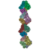
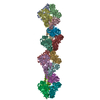
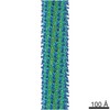
| タイトル | Post-translational cleavage of recombinantly expressed nitrilase from Rhodococcus rhodochrous J1 yields a stable, active helical form. | |||||||||
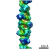
 マップデータ マップデータ | Map of the carboxy-terminal truncated nitrilase helix from Rhodococcus rhodochrous J1 | |||||||||
 試料 試料 |
| |||||||||
| 機能・相同性 | nitrilase activity / Carbon-nitrogen hydrolase 機能・相同性情報 機能・相同性情報 | |||||||||
| 生物種 |  Rhodococcus rhodochrous (バクテリア) Rhodococcus rhodochrous (バクテリア) | |||||||||
| 手法 | らせん対称体再構成法 / ネガティブ染色法 / 解像度: 18.0 Å | |||||||||
 データ登録者 データ登録者 | Thuku RN / Weber BW / Varsani A / Sewell BT | |||||||||
 引用 引用 | ジャーナル: FEBS J / 年: 2007 タイトル: Post-translational cleavage of recombinantly expressed nitrilase from Rhodococcus rhodochrous J1 yields a stable, active helical form. 著者: R Ndoria Thuku / Brandon W Weber / Arvind Varsani / B Trevor Sewell /  要旨: Nitrilases convert nitriles to the corresponding carboxylic acids and ammonia. The nitrilase from Rhodococcus rhodochrous J1 is known to be inactive as a dimer but to become active on oligomerization. ...Nitrilases convert nitriles to the corresponding carboxylic acids and ammonia. The nitrilase from Rhodococcus rhodochrous J1 is known to be inactive as a dimer but to become active on oligomerization. The recombinant enzyme undergoes post-translational cleavage at approximately residue 327, resulting in the formation of active, helical homo-oligomers. Determining the 3D structure of these helices using electron microscopy, followed by fitting the stain envelope with a model based on homology with other members of the nitrilase superfamily, enables the interacting surfaces to be identified. This also suggests that the reason for formation of the helices is related to the removal of steric hindrance arising from the 39 C-terminal amino acids from the wild-type protein. The helical form can be generated by expressing only residues 1-327. | |||||||||
| 履歴 |
|
- 構造の表示
構造の表示
| ムービー |
ムービービューア |
|---|---|
| 構造ビューア | EMマップ: SurfViewMolmilJmol/JSmol |
| 添付画像 |
 UCSF Chimera
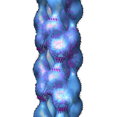
- ダウンロードとリンク
-EMDBアーカイブ
| マップデータ | emd_1313.map.gz | 609.9 KB | EMDBマップデータ形式 | |
|---|---|---|---|---|
| ヘッダ (付随情報) | emd-1313-v30.xmlemd-1313.xml | 10.3 KB 10.3 KB | 表示 表示 | EMDBヘッダ |
| 画像 |  1313.gif 1313.gif | 60.7 KB | ||
| アーカイブディレクトリ |  http://ftp.pdbj.org/pub/emdb/structures/EMD-1313ftp://ftp.pdbj.org/pub/emdb/structures/EMD-1313 http://ftp.pdbj.org/pub/emdb/structures/EMD-1313ftp://ftp.pdbj.org/pub/emdb/structures/EMD-1313 | HTTPS FTP |
-検証レポート
| 文書・要旨 | emd_1313_validation.pdf.gz | 200.1 KB | 表示 | EMDB検証レポート |
|---|---|---|---|---|
| 文書・詳細版 | emd_1313_full_validation.pdf.gz | 199.2 KB | 表示 | |
| XML形式データ | emd_1313_validation.xml.gz | 4.6 KB | 表示 | |
| アーカイブディレクトリ | https://ftp.pdbj.org/pub/emdb/validation_reports/EMD-1313ftp://ftp.pdbj.org/pub/emdb/validation_reports/EMD-1313 | HTTPS FTP |
-関連構造データ


-リンク
| EMDBのページ | EMDB (EBI/PDBe) / EMDataResource |
|---|
-マップ
| ファイル | ダウンロード / ファイル: emd_1313.map.gz / 形式: CCP4 / 大きさ: 7.8 MB / タイプ: IMAGE STORED AS FLOATING POINT NUMBER (4 BYTES) | ||||||||||||||||||||||||||||||||||||||||||||||||||||||||||||||||||||
|---|---|---|---|---|---|---|---|---|---|---|---|---|---|---|---|---|---|---|---|---|---|---|---|---|---|---|---|---|---|---|---|---|---|---|---|---|---|---|---|---|---|---|---|---|---|---|---|---|---|---|---|---|---|---|---|---|---|---|---|---|---|---|---|---|---|---|---|---|---|
| 注釈 | Map of the carboxy-terminal truncated nitrilase helix from Rhodococcus rhodochrous J1 | ||||||||||||||||||||||||||||||||||||||||||||||||||||||||||||||||||||
| 投影像・断面図 | 画像のコントロール
画像は Spider により作成 | ||||||||||||||||||||||||||||||||||||||||||||||||||||||||||||||||||||
| ボクセルのサイズ | X=Y=Z: 4 Å | ||||||||||||||||||||||||||||||||||||||||||||||||||||||||||||||||||||
| 密度 |
| ||||||||||||||||||||||||||||||||||||||||||||||||||||||||||||||||||||
| 対称性 | 空間群: 1 | ||||||||||||||||||||||||||||||||||||||||||||||||||||||||||||||||||||
| 詳細 | EMDB XML:
CCP4マップ ヘッダ情報:
| ||||||||||||||||||||||||||||||||||||||||||||||||||||||||||||||||||||
 Z (Sec.)
 Y (Row.)
 X (Col.)


-添付データ
- 試料の構成要素
-全体 : Carboxy-terminal truncated nitrilase from Rhodococcus rhodochrous J1
| 全体 | 名称: Carboxy-terminal truncated nitrilase from Rhodococcus rhodochrous J1 |
|---|---|
| 要素 |
|
-超分子 #1000: Carboxy-terminal truncated nitrilase from Rhodococcus rhodochrous J1
| 超分子 | 名称: Carboxy-terminal truncated nitrilase from Rhodococcus rhodochrous J1 タイプ: sample / ID: 1000 / Number unique components: 1 |
|---|---|
| 分子量 | 実験値: 36 KDa / 理論値: 36 KDa / 手法: SDS-PAGE MALDI-TOF mass spectrometry |
-分子 #1: J1 nitrilase 1-327
| 分子 | 名称: J1 nitrilase 1-327 / タイプ: protein_or_peptide / ID: 1 / 集合状態: helix / 組換発現: Yes |
|---|---|
| 由来(天然) | 生物種: Rhodococcus rhodochrous (バクテリア) / 株: J1 |
| 分子量 | 実験値: 36 KDa / 理論値: 36 KDa |
| 組換発現 | 生物種: |
| 配列 | GO: nitrilase activity / InterPro: Carbon-nitrogen hydrolase |
-実験情報
-構造解析
| 手法 | ネガティブ染色法 |
|---|---|
 解析 解析 | らせん対称体再構成法 |
| 試料の集合状態 | filament |
-試料調製
| 濃度 | 0.5 mg/mL |
|---|---|
| 緩衝液 | pH: 7.8 / 詳細: 100mM KH2PO4, 400mM KCl, 10% (v/v) EtOH |
| 染色 | タイプ: NEGATIVE 詳細: Sample was applied to glow discharged carbon films which were then washed twice with distilled water and then stained with 2% w/v uranyl acetate |
| グリッド | 詳細: 300 mesh |
| 凍結 | 凍結剤: NONE |
- 電子顕微鏡法
| 顕微鏡 | JEOL 2000EX |
|---|---|
| 撮影 | カテゴリ: FILM / フィルム・検出器のモデル: KODAK SO-163 FILM / デジタル化 - スキャナー: OTHER / デジタル化 - サンプリング間隔: 10 µm / 実像数: 50 / 平均電子線量: 30 e/Å2 / ビット/ピクセル: 16 |
| 電子線 | 加速電圧: 120 kV / 電子線源: TUNGSTEN HAIRPIN |
| 電子光学系 | 倍率(補正後): 50000 / 照射モード: FLOOD BEAM / 撮影モード: BRIGHT FIELD / 倍率(公称値): 50000 |
| 試料ステージ | 試料ホルダー: standard / 試料ホルダーモデル: OTHER |
-画像解析
| 詳細 | Helices were formed by autolysis of the wild-type protein after storage for one month at 4 degrees C |
|---|---|
| 最終 再構成 | 想定した対称性 - らせんパラメータ - Δz: 15.8 Å 想定した対称性 - らせんパラメータ - ΔΦ: 73.65 ° 想定した対称性 - らせんパラメータ - 軸対称性: D1 (2回x1回 2面回転対称) アルゴリズム: OTHER / 解像度のタイプ: BY AUTHOR / 解像度: 18.0 Å / 解像度の算出法: FSC 0.5 CUT-OFF / ソフトウェア - 名称: SPIDER / 詳細: D1 symmetry was imposed on map after convergence |
-原子モデル構築 1
| ソフトウェア | 名称: Situs |
|---|---|
| 詳細 | The helical symmetry was applied to a homology model to generate a helix containing 9 dimers. This was fitted to the map using CoLoRes. A two dimensional search of radial distance and azimuthal angle was conducted to find the best fit. |
| 精密化 | 空間: REAL / プロトコル: RIGID BODY FIT |
