Limulus polyphemus hemocyanin: 10 A cryo-EM structure, sequence analysis, molecular modelling and rigid-body fitting reveal the interfaces between the eight hexamers.
マップデータ
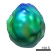
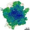
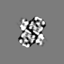
This is a map of the 8x6mer hemocyanin from the arthropod Limulus polyphemus. Mass correlated threshold: 0.001.
ジャーナル: J Mol Biol / 年: 2007 タイトル: Limulus polyphemus hemocyanin: 10 A cryo-EM structure, sequence analysis, molecular modelling and rigid-body fitting reveal the interfaces between the eight hexamers. 著者: Andreas G Martin / Frank Depoix / Michael Stohr / Ulrich Meissner / Silke Hagner-Holler / Kada Hammouti / Thorsten Burmester / Jochen Heyd / Willy Wriggers / Jürgen Markl / 要旨: The blue copper protein hemocyanin from the horseshoe crab Limulus polyphemus is among the largest respiratory proteins found in nature (3.5 MDa) and exhibits a highly cooperative oxygen binding. Its ...The blue copper protein hemocyanin from the horseshoe crab Limulus polyphemus is among the largest respiratory proteins found in nature (3.5 MDa) and exhibits a highly cooperative oxygen binding. Its 48 subunits are arranged as eight hexamers (1x6mers) that form the native 8x6mer in a nested hierarchy of 2x6mers and 4x6mers. This quaternary structure is established by eight subunit types (termed I, IIA, II, IIIA, IIIB, IV, V, and VI), of which only type II has been sequenced. Crystal structures of the 1x6mer are available, but for the 8x6mer only a 40 A 3D reconstruction exists. Consequently, the structural parameters of the 8x6mer are not firmly established, and the molecular interfaces between the eight hexamers are still to be defined. This, however, is crucial for understanding how allosteric transitions are mediated between the different levels of hierarchy. Here, we show the 10 A structure (FSC(1/2-bit) criterion) of the oxygenated 8x6mer from cryo-electron microscopy (cryo-EM) and single-particle analysis. Moreover, we show its molecular model as obtained by DNA sequencing of subunits II, IIIA, IV and VI, and molecular modelling and rigid-body fitting of all subunit types. Remarkably, the latter enabled us to improve the resolution of the cryo-EM structure from 11 A to the final 10 A. The 10 A structure allows firm assessment of various structural parameters of the 8x6mer, the 4x6mer and the 2x6mer, and reveals a total of 46 inter-hexamer bridges. These group as 11 types of interface: four at the 2x6mer level (II-II, II-IV, V-VI, IV-VI), three form the 4x6mer (V-V, V-VI, VI-IIIB/IV/V), and four are required to assemble the 8x6mer (IIIA-IIIA, IIIA-IIIB, II-IV, IV-IV). The molecular model shows the amino acid residues involved, and reveals that several of the interfaces are intriguingly histidine-rich and likely to transfer allosteric signals between the different levels of the nested hierarchy.
 ムービー
ムービー コントローラー


 データを開く
 基本情報
基本情報 マップデータ
 試料
試料 Limulus polyphemus (カブトガニ)
 データ登録者
 引用
引用
 構造の表示
構造の表示 ムービービューア
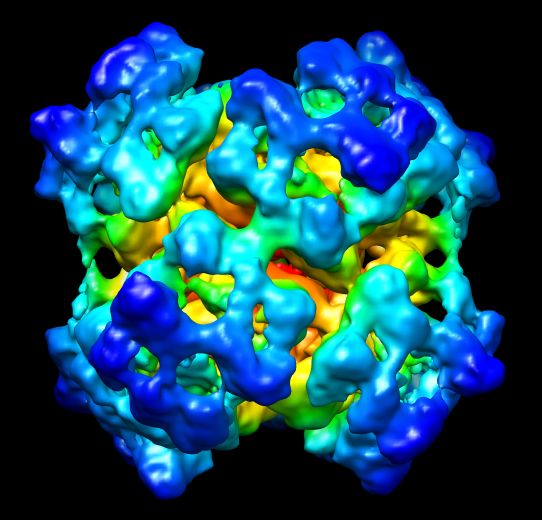
 ダウンロードとリンク
 EMD-1304.png
 http://ftp.pdbj.org/pub/emdb/structures/EMD-1304
 Z (Sec.)
 Y (Row.)
 X (Col.)


 試料の構成要素
 解析
解析 電子顕微鏡法
 FIELD EMISSION GUN
