[English] 日本語
Yorodumi
- EMDB-12809: MAT in complex with SAMH -

+
Open data


ID or keywords:

Loading...

-
Basic information

Entry
Database: EMDB / ID: EMD-12809
TitleMAT in complex with SAMH
Map dataS-adenosylmethionine hydrolase (SAMH) in complex with methionine S-adenosyltransferase(MAT)
Sample
  • Complex: filaments of METK in complex with SAM hydrolase
    • Complex: S-adenosylmethionine synthase
      • Protein or peptide: S-adenosylmethionine synthase
    • Complex: SAM hydrolase
      • Protein or peptide: SAM hydrolase
Function / homology
Function and homology information


S-adenosyl-L-methionine lyase / S-adenosyl-L-methionine lyase activity / methionine adenosyltransferase / methionine adenosyltransferase activity / S-adenosylmethionine biosynthetic process / one-carbon metabolic process / lyase activity / magnesium ion binding / ATP binding / cytoplasm
Similarity search - Function
S-adenosyl-L-methionine hydrolase / S-adenosylmethionine synthetase / S-adenosylmethionine synthetase, N-terminal / S-adenosylmethionine synthetase, central domain / S-adenosylmethionine synthetase, C-terminal / S-adenosylmethionine synthetase, conserved site / S-adenosylmethionine synthetase superfamily / S-adenosylmethionine synthetase, N-terminal domain / S-adenosylmethionine synthetase, central domain / S-adenosylmethionine synthetase, C-terminal domain ...S-adenosyl-L-methionine hydrolase / S-adenosylmethionine synthetase / S-adenosylmethionine synthetase, N-terminal / S-adenosylmethionine synthetase, central domain / S-adenosylmethionine synthetase, C-terminal / S-adenosylmethionine synthetase, conserved site / S-adenosylmethionine synthetase superfamily / S-adenosylmethionine synthetase, N-terminal domain / S-adenosylmethionine synthetase, central domain / S-adenosylmethionine synthetase, C-terminal domain / S-adenosylmethionine synthase signature 1. / S-adenosylmethionine synthase signature 2.
Similarity search - Domain/homology
S-adenosylmethionine synthase / S-Adenosylmethionine lyase
Similarity search - Component
Biological speciesEscherichia coli K-12 (bacteria) / Escherichia virus T3 / Escherichia coli (strain K12) (bacteria)
Methodsingle particle reconstruction / cryo EM / Resolution: 3.6 Å
AuthorsSimon H / Kleiner D / Shmulevich F / Zarivach R / Zalk R / Tang H / Ding F / Bershtein S
Funding support Israel, 1 items
OrganizationGrant numberCountry
Israel Science Foundation1630/15 Israel
Citation
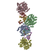
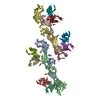
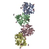
Journal: mBio / Year: 2021
Title: SAMase of Bacteriophage T3 Inactivates Escherichia coli's Methionine -Adenosyltransferase by Forming Heteropolymers.
Authors: Hadas Simon-Baram / Daniel Kleiner / Fannia Shmulevich / Raz Zarivach / Ran Zalk / Huayuan Tang / Feng Ding / Shimon Bershtein /
Abstract: -Adenosylmethionine lyase (SAMase) of bacteriophage T3 degrades the intracellular SAM pools of the host Escherichia coli cells, thereby inactivating a crucial metabolite involved in a plethora of ...-Adenosylmethionine lyase (SAMase) of bacteriophage T3 degrades the intracellular SAM pools of the host Escherichia coli cells, thereby inactivating a crucial metabolite involved in a plethora of cellular functions, including DNA methylation. SAMase is the first viral protein expressed upon infection, and its activity prevents methylation of the T3 genome. Maintenance of the phage genome in a fully unmethylated state has a profound effect on the infection strategy. It allows T3 to shift from a lytic infection under normal growth conditions to a transient lysogenic infection under glucose starvation. Using single-particle cryoelectron microscopy (cryo-EM) and biochemical assays, we demonstrate that SAMase performs its function by not only degrading SAM but also by interacting with and efficiently inhibiting the host's methionine -adenosyltransferase (MAT), the enzyme that produces SAM. Specifically, SAMase triggers open-ended head-to-tail assembly of E. coli MAT into an unusual linear filamentous structure in which adjacent MAT tetramers are joined by two SAMase dimers. Molecular dynamics simulations together with normal mode analyses suggest that the entrapment of MAT tetramers within filaments leads to an allosteric inhibition of MAT activity due to a shift to low-frequency, high-amplitude active-site-deforming modes. The amplification of uncorrelated motions between active-site residues weakens MAT's substrate binding affinity, providing a possible explanation for the observed loss of function. We propose that the dual function of SAMase as an enzyme that degrades SAM and as an inhibitor of MAT activity has emerged to achieve an efficient depletion of the intracellular SAM pools. Self-assembly of enzymes into filamentous structures in response to specific metabolic cues has recently emerged as a widespread strategy of metabolic regulation. In many instances, filamentation of metabolic enzymes occurs in response to starvation and leads to functional inactivation. Here, we report that bacteriophage T3 modulates the metabolism of the host E. coli cells by recruiting a similar strategy: silencing a central metabolic enzyme by subjecting it to phage-mediated polymerization. This observation points to an intriguing possibility that virus-induced polymerization of the host metabolic enzymes is a common mechanism implemented by viruses to metabolically reprogram and subdue infected cells.
History
DepositionApr 27, 2021-
Header (metadata) releaseJul 21, 2021-
Map releaseJul 21, 2021-
UpdateOct 13, 2021-
Current statusOct 13, 2021Processing site: PDBe / Status: Released

-
Structure visualization
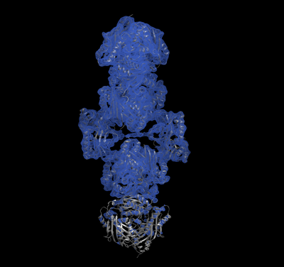
Movie
  • Surface view with section colored by density value
  • Surface level: 0.25
  • Imaged by UCSF Chimera
  • Download
  • Surface view colored by cylindrical radius
  • Surface level: 0.25
  • Imaged by UCSF Chimera
  • Download
  • Surface view with fitted model
  • Atomic models: PDB-7ock
  • Surface level: 0.25
  • Imaged by UCSF Chimera
  • Download
Movie viewer
Structure viewerEM map:
SurfViewMolmilJmol/JSmol
Supplemental images

Downloads & links

-
Map

FileDownload / File: emd_12809.map.gz / Format: CCP4 / Size: 103 MB / Type: IMAGE STORED AS FLOATING POINT NUMBER (4 BYTES)
AnnotationS-adenosylmethionine hydrolase (SAMH) in complex with methionine S-adenosyltransferase(MAT)
Voxel sizeX=Y=Z: 1.1 Å
Density
Contour LevelBy AUTHOR: 0.12 / Movie #1: 0.25
Minimum - Maximum-0.37523338 - 0.89323926
Average (Standard dev.)0.0014410599 (±0.04181811)
SymmetrySpace group: 1
Details

EMDB XML:

Map geometry
Axis orderXYZ
Origin000
Dimensions300300300
Spacing300300300
CellA=B=C: 330.0 Å
α=β=γ: 90.0 °

CCP4 map header:

modeImage stored as Reals
Å/pix. X/Y/Z1.11.11.1
M x/y/z300300300
origin x/y/z0.0000.0000.000
length x/y/z330.000330.000330.000
α/β/γ90.00090.00090.000
MAP C/R/S123
start NC/NR/NS000
NC/NR/NS300300300
D min/max/mean-0.3750.8930.001

-
Supplemental data

-
Sample components

-
Entire : filaments of METK in complex with SAM hydrolase

EntireName: filaments of METK in complex with SAM hydrolase
Components
  • Complex: filaments of METK in complex with SAM hydrolase
    • Complex: S-adenosylmethionine synthase
      • Protein or peptide: S-adenosylmethionine synthase
    • Complex: SAM hydrolase
      • Protein or peptide: SAM hydrolase

-
Supramolecule #1: filaments of METK in complex with SAM hydrolase

SupramoleculeName: filaments of METK in complex with SAM hydrolase / type: complex / ID: 1 / Parent: 0 / Macromolecule list: all

-
Supramolecule #2: S-adenosylmethionine synthase

SupramoleculeName: S-adenosylmethionine synthase / type: complex / ID: 2 / Parent: 1 / Macromolecule list: #1
Source (natural)Organism: Escherichia coli K-12 (bacteria)
Recombinant expressionOrganism: Escherichia coli BL21(DE3) (bacteria)

-
Supramolecule #3: SAM hydrolase

SupramoleculeName: SAM hydrolase / type: complex / ID: 3 / Parent: 1 / Macromolecule list: #2
Source (natural)Organism: Escherichia virus T3
Recombinant expressionOrganism: Escherichia coli (E. coli)

-
Macromolecule #1: S-adenosylmethionine synthase

MacromoleculeName: S-adenosylmethionine synthase / type: protein_or_peptide / ID: 1 / Number of copies: 8 / Enantiomer: LEVO / EC number: methionine adenosyltransferase
Source (natural)Organism: Escherichia coli (strain K12) (bacteria) / Strain: K12
Molecular weightTheoretical: 42.827375 KDa
Recombinant expressionOrganism: Escherichia coli BL21(DE3) (bacteria)
SequenceString: MAKHLFTSES VSEGHPDKIA DQISDAVLDA ILEQDPKARV ACETYVKTGM VLVGGEITTS AWVDIEEITR NTVREIGYVH SDMGFDANS CAVLSAIGKQ SPDINQGVDR ADPLEQGAGD QGLMFGYATN ETDVLMPAPI TYAHRLVQRQ AEVRKNGTLP W LRPDAKSQ ...String:
MAKHLFTSES VSEGHPDKIA DQISDAVLDA ILEQDPKARV ACETYVKTGM VLVGGEITTS AWVDIEEITR NTVREIGYVH SDMGFDANS CAVLSAIGKQ SPDINQGVDR ADPLEQGAGD QGLMFGYATN ETDVLMPAPI TYAHRLVQRQ AEVRKNGTLP W LRPDAKSQ VTFQYDDGKI VGIDAVVLST QHSEEIDQKS LQEAVMEEII KPILPAEWLT SATKFFINPT GRFVIGGPMG DC GLTGRKI IVDTYGGMAR HGGGAFSGKD PSKVDRSAAY AARYVAKNIV AAGLADRCEI QVSYAIGVAE PTSIMVETFG TEK VPSEQL TLLVREFFDL RPYGLIQMLD LLHPIYKETA AYGHFGREHF PWEKTDKAQL LRDAAGLKHH HHHH

-
Macromolecule #2: SAM hydrolase

MacromoleculeName: SAM hydrolase / type: protein_or_peptide / ID: 2 / Number of copies: 4 / Enantiomer: LEVO
Source (natural)Organism: Escherichia virus T3
Molecular weightTheoretical: 17.863264 KDa
Recombinant expressionOrganism: Escherichia coli (E. coli)
SequenceString:
MIFTKEPANV FYVLVSAFRS NLCDEVNMSR HRHMVSTLRA APGLYGSVES TDLTGCYREA ISSAPTEEKT VRVRCKDKAQ ALNVARLAC NEWEQDCVLV YKSQTHTAGL VYAKGIDGYK AERLPGSFQE VPKGAPLQGC FTIDEFGRRW QVQHHHHHH

-
Experimental details

-
Structure determination

Methodcryo EM
Processingsingle particle reconstruction
Aggregation stateparticle

-
Sample preparation

BufferpH: 8 / Details: 25mM Tris pH 8.0, 150 mM KCl,1 mM DTT.
VitrificationCryogen name: ETHANE

-
Electron microscopy

MicroscopeFEI POLARA 300
Image recordingFilm or detector model: GATAN K2 SUMMIT (4k x 4k) / Average electron dose: 80.0 e/Å2
Electron beamAcceleration voltage: 300 kV / Electron source: FIELD EMISSION GUN
Electron opticsIllumination mode: FLOOD BEAM / Imaging mode: BRIGHT FIELD
Experimental equipment
Model: Tecnai Polara / Image courtesy: FEI Company

-
Image processing

Final reconstructionResolution.type: BY AUTHOR / Resolution: 3.6 Å / Resolution method: FSC 0.143 CUT-OFF / Number images used: 65950
Initial angle assignmentType: OTHER / Details: stochastic gradient descent
Final angle assignmentType: MAXIMUM LIKELIHOOD

+
About Yorodumi

-
News

-
Feb 9, 2022. New format data for meta-information of EMDB entries

New format data for meta-information of EMDB entries

  • Version 3 of the EMDB header file is now the official format.
  • The previous official version 1.9 will be removed from the archive.

Related info.:EMDB header

External links:wwPDB to switch to version 3 of the EMDB data model

-
Aug 12, 2020. Covid-19 info

Covid-19 info

URL: https://pdbjlvh1.pdbj.org/emnavi/covid19.php

New page: Covid-19 featured information page in EM Navigator.

Related info.:Covid-19 info / Mar 5, 2020. Novel coronavirus structure data

+
Mar 5, 2020. Novel coronavirus structure data

Novel coronavirus structure data

Related info.:Yorodumi Speices / Aug 12, 2020. Covid-19 info

External links:COVID-19 featured content - PDBj / Molecule of the Month (242):Coronavirus Proteases

+
Jan 31, 2019. EMDB accession codes are about to change! (news from PDBe EMDB page)

EMDB accession codes are about to change! (news from PDBe EMDB page)

  • The allocation of 4 digits for EMDB accession codes will soon come to an end. Whilst these codes will remain in use, new EMDB accession codes will include an additional digit and will expand incrementally as the available range of codes is exhausted. The current 4-digit format prefixed with “EMD-” (i.e. EMD-XXXX) will advance to a 5-digit format (i.e. EMD-XXXXX), and so on. It is currently estimated that the 4-digit codes will be depleted around Spring 2019, at which point the 5-digit format will come into force.
  • The EM Navigator/Yorodumi systems omit the EMD- prefix.

Related info.:Q: What is EMD? / ID/Accession-code notation in Yorodumi/EM Navigator

External links:EMDB Accession Codes are Changing Soon! / Contact to PDBj

+
Jul 12, 2017. Major update of PDB

Major update of PDB

  • wwPDB released updated PDB data conforming to the new PDBx/mmCIF dictionary.
  • This is a major update changing the version number from 4 to 5, and with Remediation, in which all the entries are updated.
  • In this update, many items about electron microscopy experimental information are reorganized (e.g. em_software).
  • Now, EM Navigator and Yorodumi are based on the updated data.

External links:wwPDB Remediation / Enriched Model Files Conforming to OneDep Data Standards Now Available in the PDB FTP Archive

-
Yorodumi

Thousand views of thousand structures

  • Yorodumi is a browser for structure data from EMDB, PDB, SASBDB, etc.
  • This page is also the successor to EM Navigator detail page, and also detail information page/front-end page for Omokage search.
  • The word "yorodu" (or yorozu) is an old Japanese word meaning "ten thousand". "mi" (miru) is to see.

Related info.:EMDB / PDB / SASBDB / Comparison of 3 databanks / Yorodumi Search / Aug 31, 2016. New EM Navigator & Yorodumi / Yorodumi Papers / Jmol/JSmol / Function and homology information / Changes in new EM Navigator and Yorodumi

Read more