 ムービー
ムービー コントローラー


+ データを開く

- 基本情報
基本情報
| 登録情報 | データベース: EMDB / ID: EMD-1275 | |||||||||
|---|---|---|---|---|---|---|---|---|---|---|
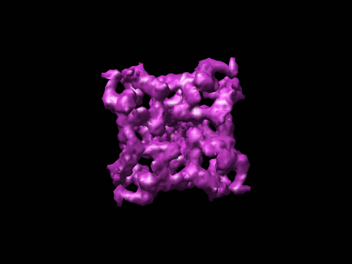
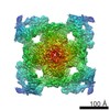
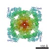
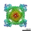
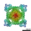
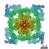
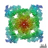
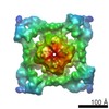
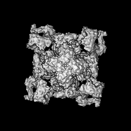
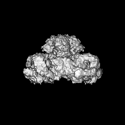
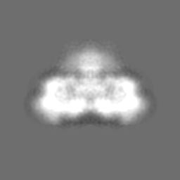
| タイトル | The pore structure of the closed RyR1 channel. | |||||||||
 マップデータ マップデータ | Z-axis of a 3D reconstruction coincides with the 4-fold axis of the channel complex | |||||||||
 試料 試料 |
| |||||||||
| 機能・相同性 | ryanodine-sensitive calcium-release channel activity / RIH domain 機能・相同性情報 機能・相同性情報 | |||||||||
| 生物種 |  | |||||||||
| 手法 | 単粒子再構成法 / クライオ電子顕微鏡法 / 解像度: 9.6 Å | |||||||||
 データ登録者 データ登録者 | Ludtke SJ / Serysheva II / Hamilton SL / Chiu W | |||||||||
 引用 引用 | ジャーナル: Structure / 年: 2005 タイトル: The pore structure of the closed RyR1 channel. 著者: Steven J Ludtke / Irina I Serysheva / Susan L Hamilton / Wah Chiu /  要旨: Using single particle electron cryomicroscopy, several helices in the membrane-spanning region of RyR1, including an inner transmembrane helix, a short pore helix, and a helix parallel to the ...Using single particle electron cryomicroscopy, several helices in the membrane-spanning region of RyR1, including an inner transmembrane helix, a short pore helix, and a helix parallel to the membrane on the cytoplasmic side, have been clearly resolved. Our model places a highly conserved glycine (G4934) at the hinge position of the bent inner helix and two rings of negative charges at the luminal and cytoplasmic mouths of the pore. The kinked inner helix closely resembles the inner helix of the open MthK channel, suggesting that kinking alone does not open RyR1, as proposed for K+ channels. | |||||||||
| 履歴 |
|
- 構造の表示
構造の表示
| ムービー |
ムービービューア |
|---|---|
| 構造ビューア | EMマップ: SurfViewMolmilJmol/JSmol |
| 添付画像 |
- ダウンロードとリンク
-EMDBアーカイブ
| マップデータ | emd_1275.map.gz | 9.1 MB | EMDBマップデータ形式 | |
|---|---|---|---|---|
| ヘッダ (付随情報) | emd-1275-v30.xmlemd-1275.xml | 9.6 KB 9.6 KB | 表示 表示 | EMDBヘッダ |
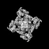
| 画像 |  1275.gif 1275.gif | 37.1 KB | ||
| アーカイブディレクトリ |  http://ftp.pdbj.org/pub/emdb/structures/EMD-1275ftp://ftp.pdbj.org/pub/emdb/structures/EMD-1275 http://ftp.pdbj.org/pub/emdb/structures/EMD-1275ftp://ftp.pdbj.org/pub/emdb/structures/EMD-1275 | HTTPS FTP |
-検証レポート
| 文書・要旨 | emd_1275_validation.pdf.gz | 222.4 KB | 表示 | EMDB検証レポート |
|---|---|---|---|---|
| 文書・詳細版 | emd_1275_full_validation.pdf.gz | 221.5 KB | 表示 | |
| XML形式データ | emd_1275_validation.xml.gz | 6.4 KB | 表示 | |
| アーカイブディレクトリ | https://ftp.pdbj.org/pub/emdb/validation_reports/EMD-1275ftp://ftp.pdbj.org/pub/emdb/validation_reports/EMD-1275 | HTTPS FTP |
-関連構造データ

-リンク
| EMDBのページ | EMDB (EBI/PDBe) / EMDataResource |
|---|
-マップ
| ファイル | ダウンロード / ファイル: emd_1275.map.gz / 形式: CCP4 / 大きさ: 62.5 MB / タイプ: IMAGE STORED AS FLOATING POINT NUMBER (4 BYTES) | ||||||||||||||||||||||||||||||||||||||||||||||||||||||||||||||||||||
|---|---|---|---|---|---|---|---|---|---|---|---|---|---|---|---|---|---|---|---|---|---|---|---|---|---|---|---|---|---|---|---|---|---|---|---|---|---|---|---|---|---|---|---|---|---|---|---|---|---|---|---|---|---|---|---|---|---|---|---|---|---|---|---|---|---|---|---|---|---|
| 注釈 | Z-axis of a 3D reconstruction coincides with the 4-fold axis of the channel complex | ||||||||||||||||||||||||||||||||||||||||||||||||||||||||||||||||||||
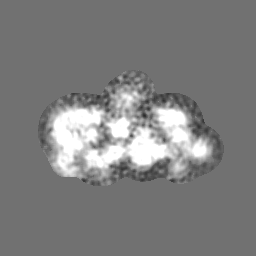
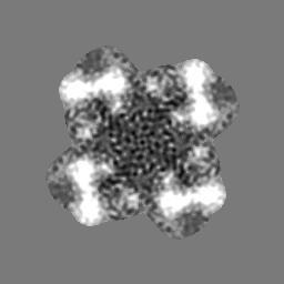
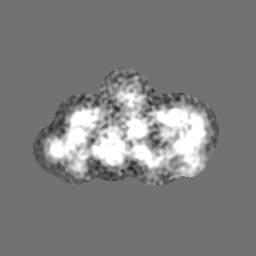
| 投影像・断面図 | 画像のコントロール
画像は Spider により作成 | ||||||||||||||||||||||||||||||||||||||||||||||||||||||||||||||||||||
| ボクセルのサイズ | X=Y=Z: 1.81 Å | ||||||||||||||||||||||||||||||||||||||||||||||||||||||||||||||||||||
| 密度 |
| ||||||||||||||||||||||||||||||||||||||||||||||||||||||||||||||||||||
| 対称性 | 空間群: 1 | ||||||||||||||||||||||||||||||||||||||||||||||||||||||||||||||||||||
| 詳細 | EMDB XML:
CCP4マップ ヘッダ情報:
| ||||||||||||||||||||||||||||||||||||||||||||||||||||||||||||||||||||
 Z (Sec.)
 Y (Row.)
 X (Col.)


-添付データ
- 試料の構成要素
-全体 : ryanodine receptor 1
| 全体 | 名称: ryanodine receptor 1 |
|---|---|
| 要素 |
|
-超分子 #1000: ryanodine receptor 1
| 超分子 | 名称: ryanodine receptor 1 / タイプ: sample / ID: 1000 詳細: The sample was purified after solubilization with detergent from skeletal muscle cells 集合状態: homotetramer / Number unique components: 1 |
|---|---|
| 分子量 | 理論値: 565 KDa |
-分子 #1: Ryanodine receptor type 1
| 分子 | 名称: Ryanodine receptor type 1 / タイプ: protein_or_peptide / ID: 1 / Name.synonym: Skeletal muscle calcium release channel / 詳細: detergent solubilized membrane protein / コピー数: 4 / 集合状態: homotetramer / 組換発現: No |
|---|---|
| 由来(天然) | 生物種: |
| 分子量 | 実験値: 565 KDa |
| 配列 | GO: ryanodine-sensitive calcium-release channel activity / InterPro: RIH domain |
-実験情報
-構造解析
| 手法 | クライオ電子顕微鏡法 |
|---|---|
 解析 解析 | 単粒子再構成法 |
| 試料の集合状態 | particle |
-試料調製
| 濃度 | 2 mg/mL |
|---|---|
| 緩衝液 | pH: 7.4 詳細: 20 mM Mops, 300 mM KCl, 1 mM DTT,0.4% CHAPS,5%sucrose,1mM EGTA |
| グリッド | 詳細: holey carbon 400 mesh Cu grid |
| 凍結 | 凍結剤: ETHANE / チャンバー内温度: 101 K / 装置: HOMEMADE PLUNGER 詳細: Vitrification instrument: Thin carbon film supported by holey carbon film 手法: Blot for 3 second before plunging |
- 電子顕微鏡法
| 顕微鏡 | JEOL 2010F |
|---|---|
| 温度 | 最低: 96 K / 最高: 97 K / 平均: 96 K |
| アライメント法 | Legacy - 非点収差: objective lens astigmatism was corrected at 400,000X |
| 撮影 | カテゴリ: CCD フィルム・検出器のモデル: GENERIC GATAN (4k x 4k) デジタル化 - サンプリング間隔: 1.81 µm / 実像数: 869 / 平均電子線量: 15 e/Å2 / ビット/ピクセル: 8 |
| Tilt angle min | 0 |
| Tilt angle max | 0 |
| 電子線 | 加速電圧: 200 kV / 電子線源:  FIELD EMISSION GUN FIELD EMISSION GUN |
| 電子光学系 | 照射モード: FLOOD BEAM / 撮影モード: BRIGHT FIELD / Cs: 2.0 mm / 最大 デフォーカス(公称値): 4.55 µm / 最小 デフォーカス(公称値): 1.37 µm / 倍率(公称値): 60000 |
| 試料ステージ | 試料ホルダー: side entry / 試料ホルダーモデル: GATAN LIQUID NITROGEN |
-画像解析
| CTF補正 | 詳細: CTF correction of each micrograph |
|---|---|
| 最終 再構成 | 想定した対称性 - 点群: C4 (4回回転対称) / アルゴリズム: OTHER / 解像度のタイプ: BY AUTHOR / 解像度: 9.6 Å / 解像度の算出法: FSC 0.5 CUT-OFF / ソフトウェア - 名称: EMAN / 使用した粒子像数: 28036 |
| 最終 2次元分類 | クラス数: 2294 |
