Journal: Sci Adv / Year: 2021 Title: Structural basis of the activation of the CC chemokine receptor 5 by a chemokine agonist. Authors: Polina Isaikina / Ching-Ju Tsai / Nikolaus Dietz / Filip Pamula / Anne Grahl / Kenneth N Goldie / Ramon Guixà-González / Camila Branco / Marianne Paolini-Bertrand / Nicolas Calo / Fabrice ...Authors: Polina Isaikina / Ching-Ju Tsai / Nikolaus Dietz / Filip Pamula / Anne Grahl / Kenneth N Goldie / Ramon Guixà-González / Camila Branco / Marianne Paolini-Bertrand / Nicolas Calo / Fabrice Cerini / Gebhard F X Schertler / Oliver Hartley / Henning Stahlberg / Timm Maier / Xavier Deupi / Stephan Grzesiek / Abstract: The human CC chemokine receptor 5 (CCR5) is a G protein-coupled receptor (GPCR) that plays a major role in inflammation and is involved in cancer, HIV, and COVID-19. Despite its importance as a drug ...The human CC chemokine receptor 5 (CCR5) is a G protein-coupled receptor (GPCR) that plays a major role in inflammation and is involved in cancer, HIV, and COVID-19. Despite its importance as a drug target, the molecular activation mechanism of CCR5, i.e., how chemokine agonists transduce the activation signal through the receptor, is yet unknown. Here, we report the cryo-EM structure of wild-type CCR5 in an active conformation bound to the chemokine super-agonist [6P4]CCL5 and the heterotrimeric G protein. The structure provides the rationale for the sequence-activity relation of agonist and antagonist chemokines. The N terminus of agonist chemokines pushes onto specific structural motifs at the bottom of the orthosteric pocket that activate the canonical GPCR microswitch network. This activation mechanism differs substantially from other CC chemokine receptors that bind chemokines with shorter N termini in a shallow binding mode involving unique sequence signatures and a specialized activation mechanism.
History
Deposition
Apr 13, 2021
-
Header (metadata) release
Jun 30, 2021
-
Map release
Jun 30, 2021
-
Update
Oct 23, 2024
-
Current status
Oct 23, 2024
Processing site: PDBe / Status: Released
-
Structure visualization
Movie
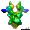
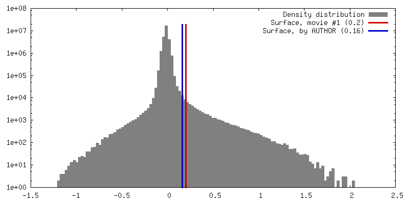
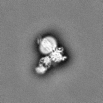
Surface view with section colored by density value
In the structure databanks used in Yorodumi, some data are registered as the other names, "COVID-19 virus" and "2019-nCoV". Here are the details of the virus and the list of structure data.
Jan 31, 2019. EMDB accession codes are about to change! (news from PDBe EMDB page)
EMDB accession codes are about to change! (news from PDBe EMDB page)
The allocation of 4 digits for EMDB accession codes will soon come to an end. Whilst these codes will remain in use, new EMDB accession codes will include an additional digit and will expand incrementally as the available range of codes is exhausted. The current 4-digit format prefixed with “EMD-” (i.e. EMD-XXXX) will advance to a 5-digit format (i.e. EMD-XXXXX), and so on. It is currently estimated that the 4-digit codes will be depleted around Spring 2019, at which point the 5-digit format will come into force.
The EM Navigator/Yorodumi systems omit the EMD- prefix.
Related info.:Q: What is EMD? / ID/Accession-code notation in Yorodumi/EM Navigator
Yorodumi is a browser for structure data from EMDB, PDB, SASBDB, etc.
This page is also the successor to EM Navigator detail page, and also detail information page/front-end page for Omokage search.
The word "yorodu" (or yorozu) is an old Japanese word meaning "ten thousand". "mi" (miru) is to see.
Related info.:EMDB / PDB / SASBDB / Comparison of 3 databanks / Yorodumi Search / Aug 31, 2016. New EM Navigator & Yorodumi / Yorodumi Papers / Jmol/JSmol / Function and homology information / Changes in new EM Navigator and Yorodumi
 Movie
Movie Controller

 Yorodumi
 Open data
 Basic information
 Map data
 Sample
 Keywords
 Function and homology information
 Homo sapiens (human) /
 Authors
 Switzerland, European Union, 4 items
  Citation
 Structure visualization
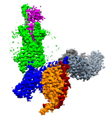
 Downloads & links
 emd_12746.png
 http://ftp.pdbj.org/pub/emdb/structures/EMD-12746


 Z (Sec.)
 Y (Row.)
 X (Col.)


 Sample components

 Spodoptera frugiperda (fall armyworm)
 Processing
 Electron microscopy
 FIELD EMISSION GUN