- EMDB-12575: Structure of ErmDL-Telithromycin-stalled 70S E. coli ribosomal co... -
+
データを開く
IDまたはキーワード:
読み込み中...
-
基本情報
登録情報
データベース: EMDB / ID: EMD-12575
タイトル
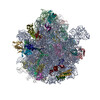
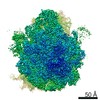
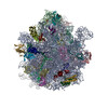
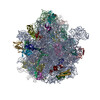
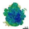
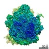
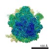
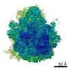
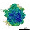
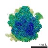
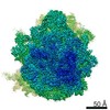
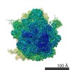
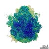
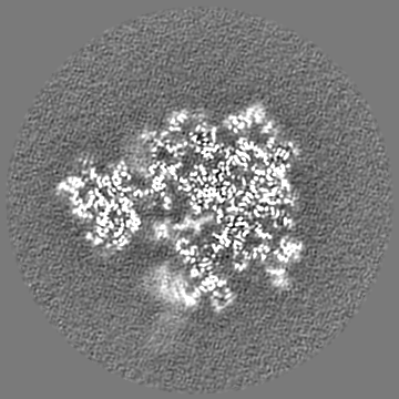
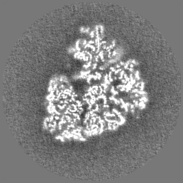
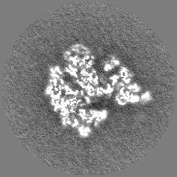
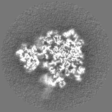
Structure of ErmDL-Telithromycin-stalled 70S E. coli ribosomal complex with A and P-tRNA
マップデータ
main map
試料
複合体: Structure of ErmDL-Erythromycin-stalled 70S E. coli ribosomal complex with P-tRNA
RNA: x 6種
タンパク質・ペプチド: x 50種
リガンド: x 1種
機能・相同性
機能・相同性情報
transcriptional attenuation / endoribonuclease inhibitor activity / RNA-binding transcription regulator activity / negative regulation of cytoplasmic translation / DnaA-L2 complex / translation repressor activity / negative regulation of DNA-templated DNA replication initiation / ribosome assembly / assembly of large subunit precursor of preribosome / cytosolic ribosome assembly ...transcriptional attenuation / endoribonuclease inhibitor activity / RNA-binding transcription regulator activity / negative regulation of cytoplasmic translation / DnaA-L2 complex / translation repressor activity / negative regulation of DNA-templated DNA replication initiation / ribosome assembly / assembly of large subunit precursor of preribosome / cytosolic ribosome assembly / DNA-templated transcription termination / response to radiation / ribosomal large subunit assembly / mRNA 5'-UTR binding / large ribosomal subunit / ribosomal small subunit assembly / small ribosomal subunit / small ribosomal subunit rRNA binding / transferase activity / 5S rRNA binding / large ribosomal subunit rRNA binding / cytosolic small ribosomal subunit / cytosolic large ribosomal subunit / tRNA binding / cytoplasmic translation / rRNA binding / negative regulation of translation / ribosome / structural constituent of ribosome / ribonucleoprotein complex / translation / response to antibiotic / negative regulation of DNA-templated transcription / mRNA binding / DNA binding / RNA binding / zinc ion binding / membrane / cytoplasm / cytosol 類似検索 - 分子機能
Ribosomal protein S21, conserved site / Ribosomal protein S21 signature. / Ribosomal protein L25, short-form / Ribosomal protein S14, bacterial/plastid / Ribosomal protein S21 superfamily / Ribosomal protein S21 / Ribosomal protein S16, conserved site / Ribosomal protein S16 signature. / Ribosomal protein S21 / Ribosomal protein L21, conserved site ...Ribosomal protein S21, conserved site / Ribosomal protein S21 signature. / Ribosomal protein L25, short-form / Ribosomal protein S14, bacterial/plastid / Ribosomal protein S21 superfamily / Ribosomal protein S21 / Ribosomal protein S16, conserved site / Ribosomal protein S16 signature. / Ribosomal protein S21 / Ribosomal protein L21, conserved site / Ribosomal protein L21 signature. / Ribosomal protein L16 signature 1. / Ribosomal protein L6, conserved site / Ribosomal protein L6 signature 1. / Ribosomal protein L16, conserved site / Ribosomal protein L16 signature 2. / Ribosomal protein L9 signature. / Ribosomal protein L9, bacteria/chloroplast / Ribosomal protein L9, C-terminal / Ribosomal protein L9, C-terminal domain / Ribosomal protein L9, C-terminal domain superfamily / Ribosomal L25p family / Ribosomal protein L25 / Ribosomal protein L36 signature. / Ribosomal protein L28/L24 superfamily / Ribosomal protein L25/Gln-tRNA synthetase, N-terminal / Ribosomal protein L25/Gln-tRNA synthetase, anti-codon-binding domain superfamily / Ribosomal protein L9, N-terminal domain superfamily / Ribosomal protein L9 / Ribosomal protein L9, N-terminal / Ribosomal protein L9, N-terminal domain / Ribosomal protein L28 / Ribosomal protein L33, conserved site / Ribosomal protein L33 signature. / Ribosomal protein L5, bacterial-type / Ribosomal protein L18, bacterial-type / Ribosomal protein L6, bacterial-type / Ribosomal protein L19, conserved site / Ribosomal protein L19 signature. / Ribosomal protein L9/RNase H1, N-terminal / Ribosomal protein S6, conserved site / Ribosomal protein S6 signature. / Ribosomal protein L36 / Ribosomal protein L36 superfamily / Ribosomal protein L36 / Ribosomal protein S7, bacterial/organellar-type / Ribosomal protein S11, bacterial-type / Ribosomal protein L27, conserved site / Ribosomal protein S13, bacterial-type / Ribosomal protein L27 signature. / Ribosomal protein S20 / Ribosomal protein S20 superfamily / Ribosomal protein S20 / Ribosomal protein S9, bacterial/plastid / Ribosomal protein S4, bacterial-type / 30S ribosomal protein S17 / Ribosomal protein S5, bacterial-type / Ribosomal protein L14P, bacterial-type / Ribosomal protein L34, conserved site / Ribosomal protein L34 signature. / Ribosomal protein S6, plastid/chloroplast / Ribosomal protein L22, bacterial/chloroplast-type / Ribosomal protein L2, bacterial/organellar-type / Ribosomal protein S2, bacteria/mitochondria/plastid / Ribosomal L28 family / Ribosomal protein L33 / Ribosomal protein L33 / Ribosomal protein L28/L24 / Ribosomal protein L18 / Ribosomal L18 of archaea, bacteria, mitoch. and chloroplast / Ribosomal protein L33 superfamily / Ribosomal protein L30, bacterial-type / : / Ribosomal protein L16 / Ribosomal protein S18, conserved site / Ribosomal protein S18 signature. / L28p-like / Ribosomal protein S16 / Ribosomal protein S16 / Ribosomal protein S16 domain superfamily / Ribosomal protein S15, bacterial-type / Ribosomal protein L21 / Ribosomal protein L27 / Ribosomal L27 protein / Ribosomal protein L19 / Ribosomal protein L19 superfamily / Ribosomal protein L19 / Ribosomal proteins 50S L24/mitochondrial 39S L24 / Ribosomal protein S6 / Ribosomal protein S6 / Ribosomal protein S6 superfamily / Ribosomal protein L21-like / L21-like superfamily / Ribosomal prokaryotic L21 protein / Ribosomal protein S2 signature 2. / Ribosomal L32p protein family / Ribosomal protein S12, bacterial-type / Ribosomal protein L32p / Ribosomal protein L24 / Ribosomal protein L34 類似検索 - ドメイン・相同性
50S ribosomal protein L27 / 50S ribosomal protein L30 / 30S ribosomal protein S14 / 30S ribosomal protein S2 / : / 50S ribosomal protein L6 / 50S ribosomal protein L28 / Small ribosomal subunit protein uS10 / Small ribosomal subunit protein uS11 / 30S ribosomal protein S9 ...50S ribosomal protein L27 / 50S ribosomal protein L30 / 30S ribosomal protein S14 / 30S ribosomal protein S2 / : / 50S ribosomal protein L6 / 50S ribosomal protein L28 / Small ribosomal subunit protein uS10 / Small ribosomal subunit protein uS11 / 30S ribosomal protein S9 / : / : / : / : / : / : / Small ribosomal subunit protein bS16 / Small ribosomal subunit protein bS21 / 30S ribosomal protein S15 / 50S ribosomal protein L18 / 50S ribosomal protein L24 / 50S ribosomal protein L5 / 50S ribosomal protein L32 / 30S ribosomal protein S8 / 30S ribosomal protein S18 / Small ribosomal subunit protein uS12 / 30S ribosomal protein S13 / 50S ribosomal protein L19 / 30S ribosomal protein S20 / Small ribosomal subunit protein uS7 / Large ribosomal subunit protein uL15 / Large ribosomal subunit protein uL29 / Large ribosomal subunit protein bL34 / Large ribosomal subunit protein bL36A / Large ribosomal subunit protein bL9 / Large ribosomal subunit protein uL13 / Large ribosomal subunit protein uL16 / Large ribosomal subunit protein bL21 / Large ribosomal subunit protein uL2 / Large ribosomal subunit protein uL3 / Large ribosomal subunit protein uL4 / Large ribosomal subunit protein uL22 / Large ribosomal subunit protein bL25 / 30S ribosomal protein S17 / 50S ribosomal protein L33 / 30S ribosomal protein S5 / Small ribosomal subunit protein uS4 / Large ribosomal subunit protein uL14 / Small ribosomal subunit protein bS6 類似検索 - 構成要素
ジャーナル: Nat Commun / 年: 2021 タイトル: Structural and mechanistic basis for translation inhibition by macrolide and ketolide antibiotics. 著者: Bertrand Beckert / Elodie C Leroy / Shanmugapriya Sothiselvam / Lars V Bock / Maxim S Svetlov / Michael Graf / Stefan Arenz / Maha Abdelshahid / Britta Seip / Helmut Grubmüller / Alexander S ...著者: Bertrand Beckert / Elodie C Leroy / Shanmugapriya Sothiselvam / Lars V Bock / Maxim S Svetlov / Michael Graf / Stefan Arenz / Maha Abdelshahid / Britta Seip / Helmut Grubmüller / Alexander S Mankin / C Axel Innis / Nora Vázquez-Laslop / Daniel N Wilson / 要旨: Macrolides and ketolides comprise a family of clinically important antibiotics that inhibit protein synthesis by binding within the exit tunnel of the bacterial ribosome. While these antibiotics are ...Macrolides and ketolides comprise a family of clinically important antibiotics that inhibit protein synthesis by binding within the exit tunnel of the bacterial ribosome. While these antibiotics are known to interrupt translation at specific sequence motifs, with ketolides predominantly stalling at Arg/Lys-X-Arg/Lys motifs and macrolides displaying a broader specificity, a structural basis for their context-specific action has been lacking. Here, we present structures of ribosomes arrested during the synthesis of an Arg-Leu-Arg sequence by the macrolide erythromycin (ERY) and the ketolide telithromycin (TEL). Together with deep mutagenesis and molecular dynamics simulations, the structures reveal how ERY and TEL interplay with the Arg-Leu-Arg motif to induce translational arrest and illuminate the basis for the less stringent sequence-specific action of ERY over TEL. Because programmed stalling at the Arg/Lys-X-Arg/Lys motifs is used to activate expression of antibiotic resistance genes, our study also provides important insights for future development of improved macrolide antibiotics.
 ムービー
ムービー コントローラー


 データを開く
 基本情報
基本情報 マップデータ
 試料
試料 機能・相同性情報
 データ登録者
 ドイツ, 2件
  引用
引用

 構造の表示
構造の表示
 ダウンロードとリンク
 emd_12575.png
 http://ftp.pdbj.org/pub/emdb/structures/EMD-12575
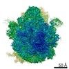


 Z
Z Y
Y X
X


 試料の構成要素
 解析
解析 電子顕微鏡法
 FIELD EMISSION GUN