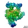
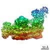
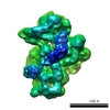
Journal: Mol Cell / Year: 2006 Title: Organization of core spliceosomal components U5 snRNA loop I and U4/U6 Di-snRNP within U4/U6.U5 Tri-snRNP as revealed by electron cryomicroscopy. Authors: Bjoern Sander / Monika M Golas / Evgeny M Makarov / Hero Brahms / Berthold Kastner / Reinhard Lührmann / Holger Stark / Abstract: In eukaryotes, pre-mRNA exons are interrupted by large noncoding introns. Alternative selection of exons and nucleotide-exact removal of introns are performed by the spliceosome, a highly dynamic ...In eukaryotes, pre-mRNA exons are interrupted by large noncoding introns. Alternative selection of exons and nucleotide-exact removal of introns are performed by the spliceosome, a highly dynamic macromolecular machine. U4/U6.U5 tri-snRNP is the largest and most conserved building block of the spliceosome. By 3D electron cryomicroscopy and labeling, the exon-aligning U5 snRNA loop I is localized at the center of the tetrahedrally shaped tri-snRNP reconstructed to approximately 2.1 nm resolution in vitrified ice. Independent 3D reconstructions of its subunits, U4/U6 and U5 snRNPs, show how U4/U6 and U5 combine to form tri-snRNP and, together with labeling experiments, indicate a close proximity of the spliceosomal core components U5 snRNA loop I and U4/U6 at the center of tri-snRNP. We suggest that this central tri-snRNP region may be the site to which the prespliceosomal U2 snRNA has to approach closely during formation of the catalytic core of the spliceosome.
History
Deposition
Aug 17, 2006
-
Header (metadata) release
Aug 17, 2006
-
Map release
Aug 17, 2008
-
Update
Dec 11, 2013
-
Current status
Dec 11, 2013
Processing site: PDBe / Status: Released
-
Structure visualization
Movie
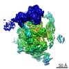
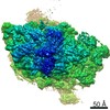
Surface view with section colored by density value
In the structure databanks used in Yorodumi, some data are registered as the other names, "COVID-19 virus" and "2019-nCoV". Here are the details of the virus and the list of structure data.
Jan 31, 2019. EMDB accession codes are about to change! (news from PDBe EMDB page)
EMDB accession codes are about to change! (news from PDBe EMDB page)
The allocation of 4 digits for EMDB accession codes will soon come to an end. Whilst these codes will remain in use, new EMDB accession codes will include an additional digit and will expand incrementally as the available range of codes is exhausted. The current 4-digit format prefixed with “EMD-” (i.e. EMD-XXXX) will advance to a 5-digit format (i.e. EMD-XXXXX), and so on. It is currently estimated that the 4-digit codes will be depleted around Spring 2019, at which point the 5-digit format will come into force.
The EM Navigator/Yorodumi systems omit the EMD- prefix.
Related info.:Q: What is EMD? / ID/Accession-code notation in Yorodumi/EM Navigator
Yorodumi is a browser for structure data from EMDB, PDB, SASBDB, etc.
This page is also the successor to EM Navigator detail page, and also detail information page/front-end page for Omokage search.
The word "yorodu" (or yorozu) is an old Japanese word meaning "ten thousand". "mi" (miru) is to see.
Related info.:EMDB / PDB / SASBDB / Comparison of 3 databanks / Yorodumi Search / Aug 31, 2016. New EM Navigator & Yorodumi / Yorodumi Papers / Jmol/JSmol / Function and homology information / Changes in new EM Navigator and Yorodumi
 Movie
Movie Controller

 Yorodumi
 Open data
 Basic information
 Map data
 Sample
 Homo sapiens (human)
 Authors
 Citation
 Structure visualization
 Movie viewer
 Downloads & links
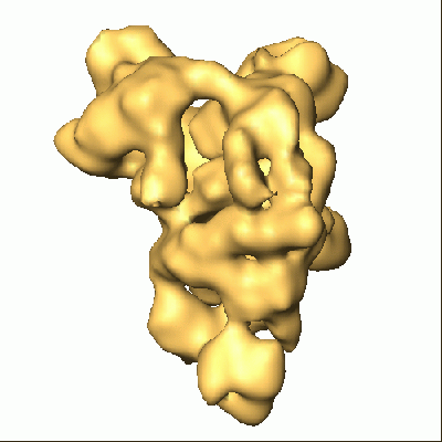 1257.gif
 http://ftp.pdbj.org/pub/emdb/structures/EMD-1257
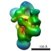


 Z (Sec.)
 Y (Row.)
 X (Col.)


 Sample components
 Processing
 Electron microscopy
 FIELD EMISSION GUN